

Primary pulmonary meningioma: A case report
- PMID: 28489736
- PMCID: PMC5428570
- DOI: 10.1097/MD.0000000000006474
Primary pulmonary meningioma: A case report
Abstract
Rationale: Primary extracranial meningiomas are rare outside the head and neck region.
Patient concerns: A 44-year-old female patient had chest pain for more than 1 year.
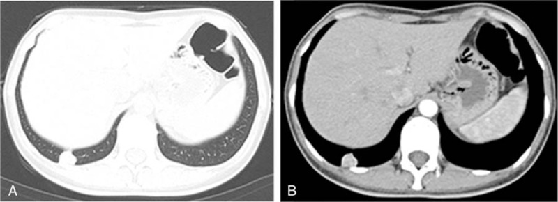
Diagnoses: Preoperative chest computed tomography (CT) scan revealed a nodule in the right lower lobe, 1.8 cm in diameter. Tumor tissues were examined by immunohistochemistry for vimentin and S-100.
Interventions: Histopathologically, the tumor was characterized by whorled nests of spindle-shaped cells accompanied by psammoma bodies. Immunohistochemistry demonstrated tumor cell positivity for vimentin and S-100. This case was diagnosed as a primary pulmonary meningioma. The tumor was removed by a thoracoscopic pulmonary wedge resection.
Outcomes: Postoperative cranial and spinal CT scan did not show any intracranial or spinal mass. No recurrence of the tumor was reported at the time of writing up this case report.
Lessons: A primary pulmonary meningioma should be considered in the differential diagnosis workup of pulmonary nodules.
Conflict of interest statement
The authors have no conflicts of interest to disclose.
Figures

References
-
- Muzumdar DP, Vengsarkar US, Bhatjiwale MG, et al. Diffuse calvarial meningioma: a case report. J Postgrad Med 2001;47:116–8. - PubMed
-
- Wu ZB, Yang GH. People's Medical Publishing House Chinese Surgical Pathology [Book in Chinese] 2002:2544-5.
-
- Kemnitz PSH, Heinrich P. Meningiom a of lung: first report with light and electronmicroscopic findings. Ultrastruct Pathol 1982;3:359–65. - PubMed
-
- Kim YY, Hong YK, Kie JH, et al. Primary pulmonary meningioma: an unusual cause of a nodule with strong and homogeneous enhancement. Clin Imaging 2016;40:170–3. - PubMed
Publication types
MeSH terms
LinkOut - more resources
Full Text Sources
Other Literature Sources
Medical
