. 2017 May 10;12(5):e0177365.
doi: 10.1371/journal.pone.0177365. eCollection 2017.

Bystander activation of irrelevant CD4+ T cells following antigen-specific vaccination occurs in the presence and absence of adjuvant

Affiliations

Bystander activation of irrelevant CD4+ T cells following antigen-specific vaccination occurs in the presence and absence of adjuvant

Susan van Aalst et al. PLoS One. .

Abstract

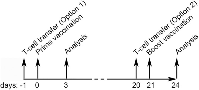
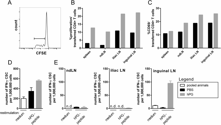
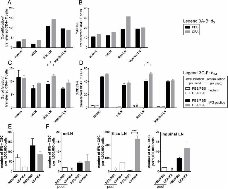
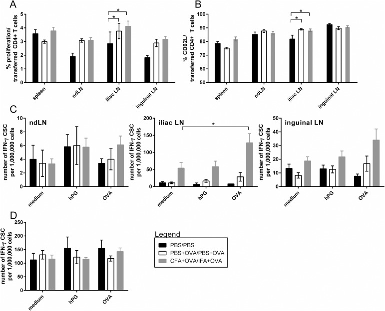
Autoimmune and other chronic inflammatory diseases (AID) are prevalent diseases which can severely impact the quality of life of those that suffer from the disease. In most cases, the etiology of these conditions have remained unclear. Immune responses that take place e.g. during natural infection or after vaccination are often linked with the development or exacerbation of AID. It is highly debated if vaccines induce or aggravate AID and in particular adjuvants are mentioned as potential cause. Since vaccines are given on a large scale to healthy individuals but also to elderly and immunocompromised individuals, more research is warranted. Non-specific induction of naïve or memory autoreactive T cells via bystander activation is one of the proposed mechanisms of how vaccination might be involved in AID. During bystander activation, T cells unrelated to the antigen presented can be activated without (strong) T cell receptor (TCR) ligation, but via signals derived from the ongoing response directed against the vaccine-antigen or adjuvant at hand. In this study we have set up a TCR transgenic T cell transfer mouse model by which we were able to measure local bystander activation of transferred and labeled CD4+ T cells. Intramuscular injection with the highly immunogenic Complete Freund's Adjuvant (CFA) led to local in vivo proliferation and activation of intravenously transferred CD4+ T cells in the iliac lymph node. This local bystander activation was also observed after CFA prime and Incomplete Freund's Adjuvant (IFA) boost injection. Furthermore, we showed that an antigen specific response is sufficient for the induction of a bystander activation response and the general, immune stimulating effect of CFA or IFA does not appear to increase this effect. In other words, no evidence was obtained that adjuvation of antigen specific responses is essential for bystander activation.


Conflict of interest statement

Competing Interests: The authors have declared that no competing interests exist.

Figures

Fig 1
Fig 1. Set up of in vivo transfer studies.
CD4+ T cells were isolated from TCR Tg mice which have CD4+ T cells specific for hPG peptide. The (CD90.1+)CD4+ T cells were CFSE-labeled and i.v. transferred to donor mice at d-1 (approach 1) or d20 (approach 2). At d0 acceptor mice received an i.m. injection (50 μl) in the left quadriceps with CFA or PBS, sometimes supplemented with hPG-peptide or OVA. Animals were sacrificed at d3 or d24. Animals sacrificed on d24 received a booster shot (right quadriceps, d21) with IFA or PBS, sometimes supplemented with OVA.
Fig 2
Fig 2. Injection with hPG peptide results in proliferation and activation of transferred hPG-T cells at d3.
Animals received an i.v. transfer of CFSE-labeled hPG specific CD4+ T cells at d-1 and an i.m. injection with PBS or hPG peptide in PBS at d0 before analysis at d3. (A) Representative FACS-histogram and -gating strategy of CFSE-labeled hPG-specific donor CD4+ T cells in the iliac LN after hPG peptide injection. (B,C) Percentage of (B) proliferation of and (C) CD69 expression on CFSE-labeled CD4+ T cells in spleen and LN. (D,E) IFN-γ-ELISpot assay with 48h ex vivo hPG peptide restimulation of (D) splenocytes and (E) LNs. The ELISpot was performed in duplo (LN) and triplo (spleen). 1 animal per group was used. This experiment was performed twice. ND: not determined, hPG: hPG peptide, CSC: cytokine secreting cell, med: medium, ndLN: non-draining LN.
Fig 3
Fig 3. CFA prime and CFA/IFA prime-boost leads to local activation and proliferation of transferred hPG-T cells.
(A, B) Animals received an i.v. transfer of CFSE-labeled hPG specific CD4+ T cells at d-1 and an i.m. injection with PBS or CFA at d0 before analysis at d3. Percentage of (A) proliferation of and (B) CD69 expressing cells in CFSE-labeled CD4+ cell population in spleen and LN. (C-F) Animals received an i.v. transfer of CFSE-labeled hPG specific CD4+CD90.1+ T cells at d-1 and an i.m. prime with PBS or CFA at d0, followed by a boost with PBS or IFA at d21. (C,D) At d24 cells were restimulated with hPG peptide for 24h. Indicated are the percentage of (C) proliferation of and (D) CD69 expressing cells in CFSE-labeled CD90.1+CD4+ cells in spleen and LN after ex vivo hPG restimulation. (E,F) At d24 (E) splenocytes and (F) LNs were used for an IFN-y-ELISpot assay with 48h ex vivo hPG-peptide restimulation. The ELISpot was performed in duplo (LN) and triplo (spleen).Results were obtained with N = 3–4. In some cases, LN cells were pooled per group for the medium stimulus (pool). Means+SEM are shown. Differences between two groups were determined with an unpaired two-tailed student’s t-test. P < 0.05 was considered significant. ND: not determined, CSC: cytokine secreting cell, ndLN: non-draining LN.
Fig 4
Fig 4. Antigen-specific boost leads to local proliferation of transferred hPG-specific T cells.
Animals received a prime at d0 (PBS, OVA+PBS or OVA+CFA) followed by an i.v. transfer of labeled CD90.1+CD4+ T cells at d20 and a boost at d21 (PBS, OVA+PBS or OVA+IFA) before analysis at d24. (A,B) Percentage of (A) proliferation of and (B) CD62L expression in CFSE-labeled CD90.1+CD4+ cells in spleen and LN. (C,D) IFN-γ-ELISpot assay with 48h ex vivo hPG-peptide restimulation of (D) splenocytes and (C) LNs. The ELISpot was performed in duplo (LN) and triplo (spleen). Data shown are the means+SEM of two independent experiments (each 2–5 animals per group). Differences between two groups were determined with an unpaired two-tailed student’s t-test. Differences between the three groups were determined with a one-way ANOVA (two-tailed) with Dunnett’s multiple comparison test. P < 0.05 was considered significant. CSC: cytokine secreting cell, ndLN: non-draining LN.

References

    1. Sharma S, Thomas PG. The two faces of heterologous immunity: Protection or immunopathology. J Leukocyte Biol. 2014;95: 405–416. doi: 10.1189/jlb.0713386 - DOI - PMC - PubMed
    1. Münz C., Lünemann J.D., Getts M.T., Miller S.D. Antiviral immune responses: Triggers of or triggered by autoimmunity? Nat Rev Immunol. 2009;9: 246–258. doi: 10.1038/nri2527 - DOI - PMC - PubMed
    1. Tough DF, Sprent J. Bystander stimulation of T cells in vivo by cytokines. Vet Immunol Immunopathol. 1998;63: 123–129. - PubMed
    1. Bangs SC, McMichael AJ, Xu X. Bystander T cell activation–implications for HIV infection and other diseases. Trends Immunol. 2006;27: 518–524. doi: 10.1016/j.it.2006.09.006 - DOI - PubMed
    1. Su L, Kidd B, Han A, Kotzin J, Davis M. Virus-Specific CD4+ Memory-Phenotype T Cells Are Abundant in Unexposed Adults. Immunity. 2013;38: 373–383. doi: 10.1016/j.immuni.2012.10.021 - DOI - PMC - PubMed

MeSH terms