. 2017 May 11;376(19):1835-1848.
doi: 10.1056/NEJMoa1614814.

Cancer-Associated Mutations in Endometriosis without Cancer

Affiliations

Cancer-Associated Mutations in Endometriosis without Cancer

Michael S Anglesio et al. N Engl J Med. .

Abstract

Background: Endometriosis, defined as the presence of ectopic endometrial stroma and epithelium, affects approximately 10% of reproductive-age women and can cause pelvic pain and infertility. Endometriotic lesions are considered to be benign inflammatory lesions but have cancerlike features such as local invasion and resistance to apoptosis.

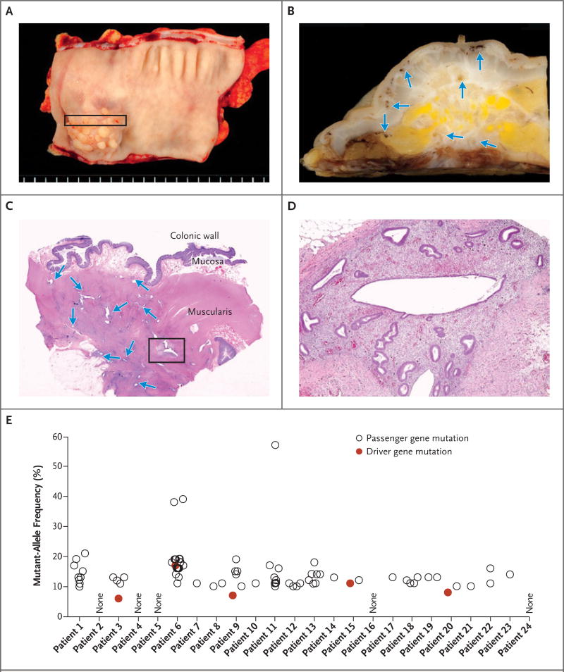
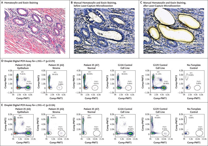
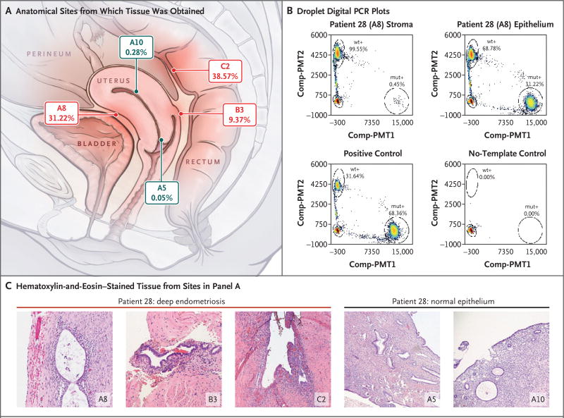
Methods: We analyzed deeply infiltrating endometriotic lesions from 27 patients by means of exomewide sequencing (24 patients) or cancer-driver targeted sequencing (3 patients). Mutations were validated with the use of digital genomic methods in microdissected epithelium and stroma. Epithelial and stromal components of lesions from an additional 12 patients were analyzed by means of a droplet digital polymerase-chain-reaction (PCR) assay for recurrent activating KRAS mutations.

Results: Exome sequencing revealed somatic mutations in 19 of 24 patients (79%). Five patients harbored known cancer driver mutations in ARID1A, PIK3CA, KRAS, or PPP2R1A, which were validated by Safe-Sequencing System or immunohistochemical analysis. The likelihood of driver genes being affected at this rate in the absence of selection was estimated at P=0.001 (binomial test). Targeted sequencing and a droplet digital PCR assay identified KRAS mutations in 2 of 3 patients and 3 of 12 patients, respectively, with mutations in the epithelium but not the stroma. One patient harbored two different KRAS mutations, c.35G→T and c.35G→C, and another carried identical KRAS c.35G→A mutations in three distinct lesions.

Conclusions: We found that lesions in deep infiltrating endometriosis, which are associated with virtually no risk of malignant transformation, harbor somatic cancer driver mutations. Ten of 39 deep infiltrating lesions (26%) carried driver mutations; all the tested somatic mutations appeared to be confined to the epithelial compartment of endometriotic lesions.


Figures

Figure 1
Figure 1. An Example of Deeply Infiltrative Endometriosis in the Colon
In Panel A, a segment of involved colon shows a papillary lesion projecting into the lumen. Panel B shows the cross section of the colonic wall that is indicated by the rectangle in Panel A; arrows indicate endometriotic lesions. In Panel C, a section stained with hematoxylin and eosin shows multiple, discrete endometriotic foci (arrows) infiltrating into the muscle layer of the colon. The box indicates the approximate region that is core-targeted for molecular analysis. In Panel D, a higher magnification shows the characteristic morphologic features of endometriosis, with both glandular and stromal components. Results for all 24 exome-sequenced patients are shown in Panel E; not all patients’ samples yielded detectable somatic mutations. Both “driver” and “passenger” mutations are indicated (Tables S1 and S2 in Supplementary Appendix 2). As opposed to driver mutations, passenger mutations are defined as somatic mutations that are not known or presumed to directly contribute to cancer initiation or progression.
Figure 2
Figure 2. Immunohistochemistry of ARID1A (BAF250A) in Deep Infiltrating Endometriosis
ARID1A immunoreactivity was detected in all stromal (St) cells and epithelial (Epi) cells within an endometriotic lesion containing wild-type ARID1A (Panel A). In Patient 20, harboring an ARID1A inactivating mutation, loss of ARID1A immunoreactivity was observed in a subset of epithelial cells (arrows indicate examples), but immunoreactivity was preserved in a much larger fraction of the adjacent stromal cells within the same lesion (Panel B). The mutant-allele fraction of the ARID1A mutation in this patient was 8%.
Figure 3
Figure 3. Confirmation of Activating, Somatic KRAS Mutations in the Glandular Epithelium but not Stromal Compartments of Deep Endometriotic Lesions
Panel A shows a photomicrograph of endometriotic tissue from Patient 25, with standard hematoxylin and eosin staining. Panels B and C show manually stained, non–cover-slipped sections, also from Patient 25, that were prepared for laser-capture microdissection. Panels D and E show droplet digital polymerase-chain- reaction (PCR) plots illustrating the presence of both c.35G→T (p.G12V) and c.35G→C (p.G12A) KRAS mutations at different allelic frequencies but exclusive to the glandular epithelium in Patient 25. The allelic frequencies represent the percentage of droplets that were positive for the mutant allele (mut+) or positive for the wild-type allele (wt+). Control cell lines and no-template controls (including all reaction components except a DNA template) are also shown. Comp-PMT1 denotes spectrally compensated photomultiplier tube 1 (the dye channel used for the mutant assay), and Comp-PMT2 spectrally compensated photo-multiplier tube 2 (the dye channel used for the wild-type assay).
Figure 4
Figure 4. Co-Occurring and Anatomically Distinct Deep Infiltrating Endometriosis Lesions That Harbor Identical KRAS Mutations
Panel A shows an anatomical diagram outlining the position of three distinct deep infiltrating endometriosis lesions (in red) and normal sampling of eutopic endometrial and endocervical epithelium (in gray), all from Patient 28. The allelic frequency of the epithelial-restricted c.35G→A (p.G12D) KRAS mutation from droplet digital PCR experiments is also shown below each block identifier. Panel B shows droplet digital PCR plots confirming the KRAS mutation in the epithelial, but not stromal, component of the A8 endometriotic lesion, with the mutant-droplet-positive fraction (allelic frequency). Controls are also shown. Panel C shows hematoxylin-and-eosin photomicrographs of each location shown in Panel A, including endometriotic tissue taken from the three distinct lesions: in the anterior serosal surface of the uterus (A8) as well as the vaginal (B3) and rectal (C2) surfaces of the rectouterine pouch.

Comment in

Similar articles

Cited by

References

    1. Giudice LC. Endometriosis. N Engl J Med. 2010;362:2389–98. - PMC - PubMed
    1. Reese KA, Reddy S, Rock JA. Endometriosis in an adolescent population: the Emory experience. J Pediatr Adolesc Gynecol. 1996;9:125–8. - PubMed
    1. Bulun SE. Endometriosis. N Engl J Med. 2009;360:268–79. - PubMed
    1. Leyland N, Casper R, Laberge P, Singh SS. Endometriosis: diagnosis and management. J Obstet Gynaecol Can. 2010;32(Suppl 2):S1–S32. - PubMed
    1. Tosti C, Pinzauti S, Santulli P, Chapron C, Petraglia F. Pathogenetic mechanisms of deep infiltrating endometriosis. Reprod Sci. 2015;22:1053–9. - PubMed

Publication types

MeSH terms