. 2017 Jan 25;37(4):1039-1054.
doi: 10.1523/JNEUROSCI.1647-16.2016.

Opposing Effects of Valproic Acid Treatment Mediated by Histone Deacetylase Inhibitor Activity in Four Transgenic X. laevis Models of Retinitis Pigmentosa

Affiliations

Opposing Effects of Valproic Acid Treatment Mediated by Histone Deacetylase Inhibitor Activity in Four Transgenic X. laevis Models of Retinitis Pigmentosa

Ruanne Y J Vent-Schmidt et al. J Neurosci. .

Abstract

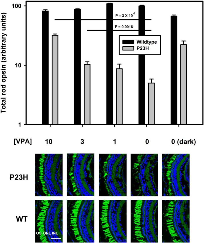
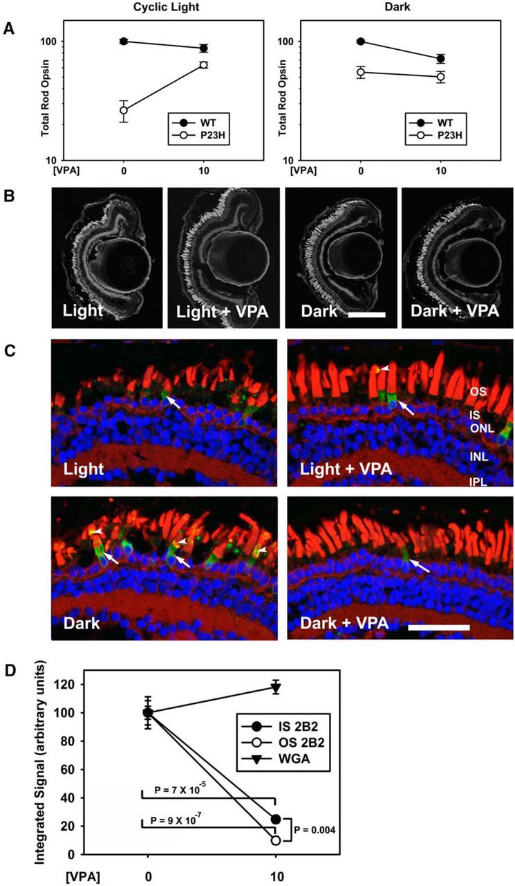
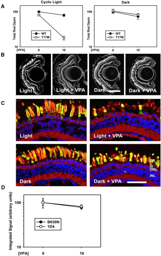
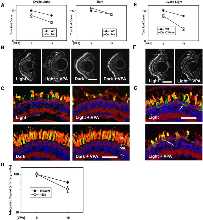
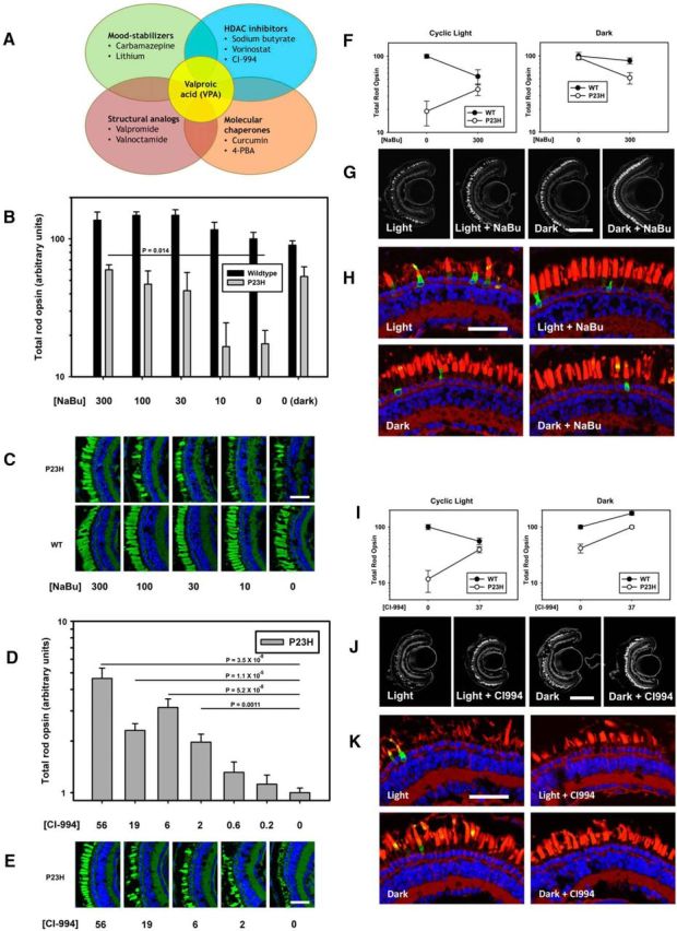
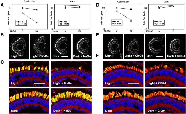
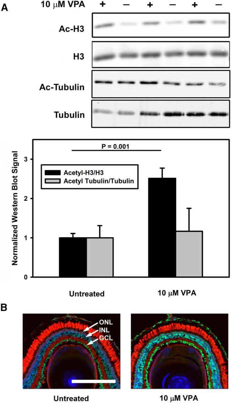
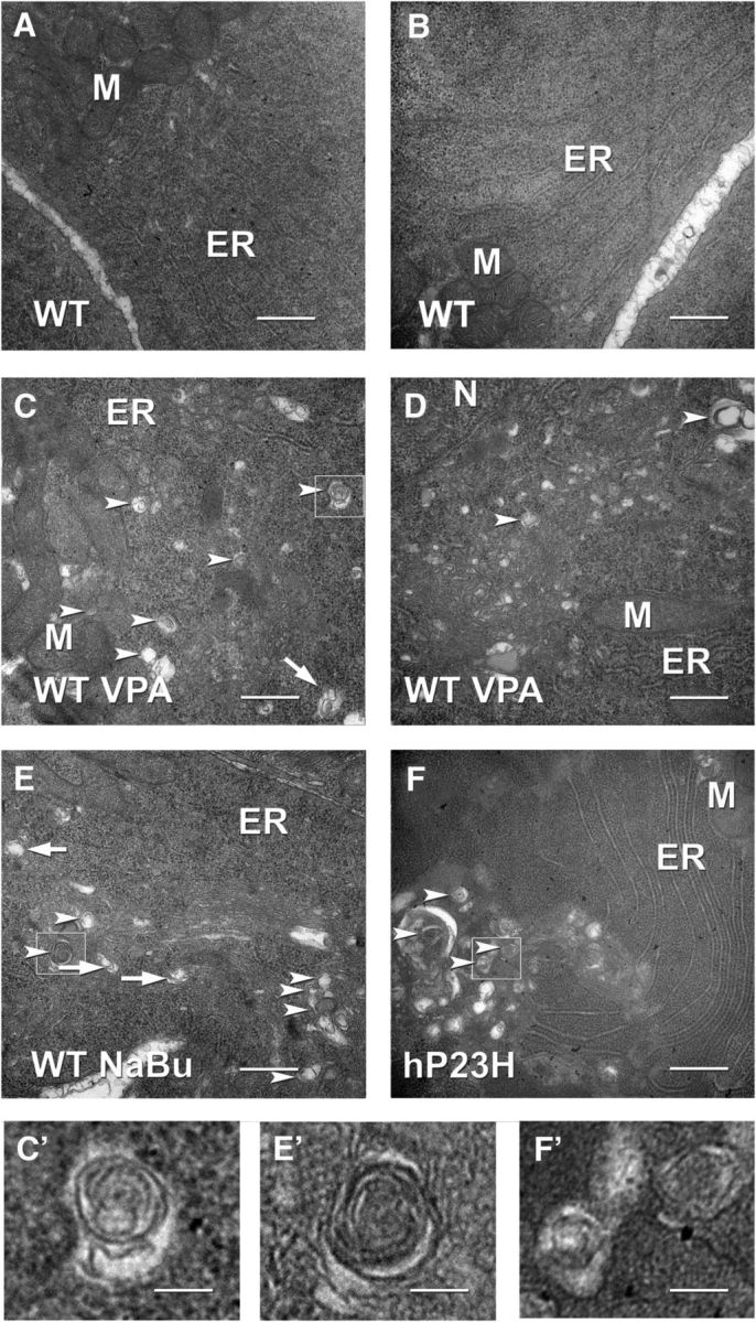
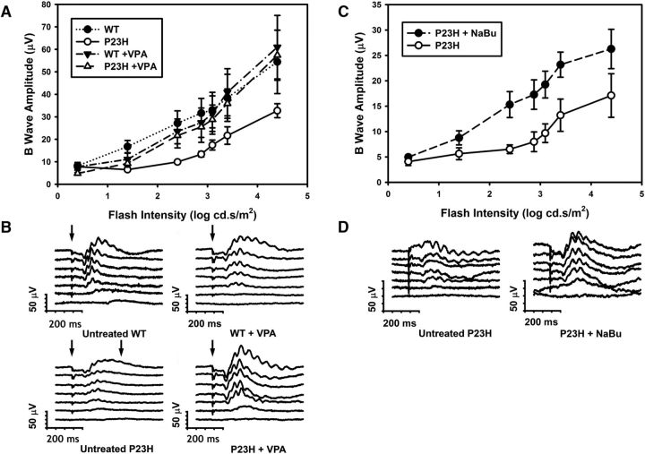
Retinitis pigmentosa (RP) is an inherited retinal degeneration (RD) that leads to blindness for which no treatment is available. RP is frequently caused by mutations in Rhodopsin; in some animal models, RD is exacerbated by light. Valproic acid (VPA) is a proposed treatment for RP and other neurodegenerative disorders, with a phase II trial for RP under way. However, the therapeutic mechanism is unclear, with minimal research supporting its use in RP. We investigated the effects of VPA on Xenopus laevis models of RP expressing human P23H, T17M, T4K, and Q344ter rhodopsins, which are associated with RP in humans. VPA ameliorated RD associated with P23H rhodopsin and promoted clearing of mutant rhodopsin from photoreceptors. The effect was equal to that of dark rearing, with no additive effect observed. Rescue of visual function was confirmed by electroretinography. In contrast, VPA exacerbated RD caused by T17M rhodopsin in light, but had no effect in darkness. Effects in T4K and Q344ter rhodopsin models were also negative. These effects of VPA were paralleled by treatment with three additional histone deacetylase (HDAC) inhibitors, but not other antipsychotics, chemical chaperones, or VPA structural analogues. In WT retinas, VPA treatment increased histone H3 acetylation. In addition, electron microscopy showed increased autophagosomes in rod inner segments with HDAC inhibitor (HDACi) treatment, potentially linking the therapeutic effects in P23H rhodopsin animals and negative effects in other models with autophagy. Our results suggest that the success or failure of VPA treatment is dependent on genotype and that HDACi treatment is contraindicated for some RP cases.SIGNIFICANCE STATEMENT Retinitis pigmentosa (RP) is an inherited, degenerative retinal disease that leads to blindness for which no therapy is available. We determined that valproic acid (VPA), currently undergoing a phase II trial for RP, has both beneficial and detrimental effects in animal models of RP depending on the underlying disease mechanism and that both effects are due to histone deacetylase (HDAC) inhibition possibly linked to autophagy regulation. Off-label use of VPA and other HDAC inhibitors for the treatment of RP should be limited to the research setting until this effect is understood and can be predicted. Our study suggests that, unless genotype is accounted for, clinical trials for RP treatments may give negative results due to multiple disease mechanisms with differential responses to therapeutic interventions.

Keywords: HDAC inhibitor; autophagy; retinal degeneration; retinitis pigmentosa; rhodopsin; valproic acid.


Figures

Figure 1.
Figure 1.
Effect of VPA on RD in transgenic X. laevis expressing P23H rod opsin. Top, Dot blot analysis of total rod opsin from whole eye extracts of P23H X. laevis (gray bars) and their WT siblings (black bars). VPA ameliorates RD significantly in a dose-dependent manner (ANOVA, p = 1.5 × 10⁻¹²). At 10 μm, the effect of VPA is equivalent to the effect of dark rearing. WT animals are unaffected by VPA (ANOVA, p = 0.363). p-values shown on chart are Dunnet's multiple comparisons. n = 10–17 animals per condition. Error bars indicate SEM. Bottom, Representative cryosections of contralateral eyes confirm that effects of VPA in dot blot assays are due to reduced RD in P23H-transgenic animals. OSs were stained with WGA (green) and Hoechst dye (blue). VPA treatment resulted in greater density of OSs in P23H X.laevis. WT animals were unaffected. Scale bar, 50 μm.
Figure 2.
Figure 2.
VPA ameliorates light-induced RD in a P23H rod opsin model. A, Dot blot analysis of P23H X. laevis and their WT siblings treated with 10 μm VPA in cyclic light and dark-reared conditions. In cyclic light, the effect of genotype and treatment were significant (two-way ANOVA, pg = 6 × 10⁻⁹, pt = 2 × 10⁻⁵) and VPA treatment significantly modified the effect of genotype (pi = 2 × 10⁻⁵). In the dark, the effect of genotype was significant (pg = 4 × 10⁻⁵), the effect of treatment was minimally significant (pt = 0.047), and treatment did not significantly modify the effect of genotype (p = 0.22). n = 5–13 animals per group. Error bars indicate SEM. B, Representative low-magnification confocal micrographs of cryosections from transgenic retinas expressing P23H rod opsin stained with WGA. Scale bar, 200 μm. C, Representative high-magnification confocal micrographs of transgenic retinas expressing P23H rod opsin stained with 2B2 anti-mammalian rhodopsin (green), WGA (red), and Hoechst dye (blue). Dark rearing promotes OS localization of P23H rhodopsin (arrowheads). VPA treatment does not alter P23H rhodopsin localization. ONL, Outer nuclear layer; INL, inner nuclear layer; IPL, inner plexiform layer. Scale bar, 50 μm. D, OS WGA and whole-retina 2B2 (P23H rhodopsin) signals were quantified in confocal microscopy images of dark reared transgenic animals. 2B2 signal was markedly reduced in both IS and OS of treated animals, whereas WGA signal was unchanged (two-way ANOVA pi = 3.5 × 10⁻⁹, t tests p = 7 × 10⁻⁵, p = 9 × 10⁻⁷, p = 0.004).
Figure 3.
Figure 3.
VPA exacerbates light-induced RD in a T17M rod opsin model. A, Dot blot analysis of VPA-treated T17M X. laevis and their WT siblings in cyclic light and dark-reared conditions. In cyclic light, the effects of genotype and treatment were significant (pg = 1.7 × 10⁻⁹, pt = 1.8 × 10⁻⁹) and VPA treatment modified the effect of genotype significantly (pi = 1.2 × 10⁻⁶). In the dark, there was no significant effect of genotype and no interaction between VPA and genotype (pg = 0.56, pt = 0.029, pi = 0.67). n = 6–16 animals per group. Error bars indicate SEM. B, Representative low-magnification confocal micrographs of cryosections from transgenic retinas expressing T17M rod opsin stained with WGA. Scale bar, 200 μm. C, Representative high-magnification confocal micrographs of transgenic retinas expressing P23H rod opsin stained with 2B2 anti-mammalian rhodopsin (green), WGA (red), and Hoechst dye (blue). Neither dark rearing nor VPA treatment altered the distribution of T17M rod opsin significantly. ONL, Outer nuclear layer; INL, inner nuclear layer; IPL, inner plexiform layer. Scale bar, 50 μm. D, Total rod opsin (B630N) and T17M rhodopsin (1D4) signals were quantified in dot blot analyses of dark-reared animals. Two-way ANOVA analysis showed no significant effects of treatment on antibody signals, indicating that VPA treatment did not alter T17M rhodopsin expression levels. n = 11–13 animals for each condition.
Figure 4.
Figure 4.
VPA has negative effects in a T4K rod opsin model and exacerbates RD in a Q344ter rod opsin model. AD, Effects of VPA in a T4K rod opsin model. A, In cyclic light, effects of genotype and treatment were significant (pg = 5.8 × 10⁻⁵, pt = 1.2 × 10⁻⁴) and negative, but treatment did not modify the effect of genotype (pi = 0.45). In the dark, there was no effect of genotype (p = 0.111) and no interaction between treatment and genotype (p = 0.133). n = 6–11 animals per group. B, Representative low-magnification confocal micrographs of transgenic retinas expressing T4K rod opsin stained with WGA. C, Representative high-magnification confocal micrographs of transgenic retinas expressing T4K rod opsin stained with 2B2 anti-mammalian rhodopsin (green), WGA (red), and Hoechst dye (blue). VPA treatment did not alter T4K rod opsin distribution. D, Total rod opsin (B630N) and T4K rhodopsin (1D4) signals were quantified in dark reared animals. Two-way ANOVA analysis showed that VPA treatment reduced the antibody signals (p = 1.0 × 10⁻⁴), but the effect was not significantly different between total and transgenic rhodopsins (p = 0.099). n = 6–13 animals for each condition. EG, Effects of VPA in a Q344ter rod opsin model. E, In cyclic light, the effects of genotype and treatment were significant (pg = 2 × 10⁻⁷, pt = 1 × 10⁻⁶) and treatment modified the effect of genotype significantly (pi = 0.016). n = 8–11 animals for each condition. F, Representative low-magnification confocal micrographs of transgenic retinas expressing Q344ter rod opsin stained with WGA. Scale bar, 200 μm. G, Representative high-magnification confocal micrographs of transgenic retinas expressing Q344ter rod opsin stained with 2B2 anti-mammalian rhodopsin (green), WGA (red), and Hoechst dye (blue). VPA treatment did not alter the distribution of Q344ter rod opsin. Scale bar, 50 μm. Error bars indicate SEM.
Figure 5.
Figure 5.
The HDACi's sodium butyrate (NaBu) and CI-994 reproduce the effects of VPA. A, Venn diagram showing overlapping pharmacological activities of compounds examined. B, Effect of NaBu on total rod opsin levels. NaBu ameliorates RD in P23H X. laevis in a dose-dependent manner (ANOVA, p = 0.0022). Treatment with 300 μm sodium butyrate is equivalent to dark rearing. WT animals are unaffected (ANOVA, p = 0.10). p-values shown are Dunnet's multiple comparisons. n = 7–10 animals per group. C, Representative cryosections of contralateral eyes confirm that effects of NaBu in dot blot assays are due to reduced RD in P23H-transgenic animals. OSs were stained with WGA (green) and Hoechst dye (blue). NaBu treatment resulted in greater density of OSs in P23H X. laevis. WT animals were unaffected. D, Dot blot analysis of total rod opsin from whole-eye extracts of P23H X. laevis treated with varying concentrations of CI-994. CI-994 increased total rhodopsin significantly and in a dose-dependent manner (ANOVA, p = 4.5 × 10⁻¹³). p-values shown on chart are Dunnet's multiple comparisons. n = 7–10 animals per group. E, Representative cryosections of contralateral eyes confirm that effects of CI-994 in dot blot assays are due to reduced RD in P23H-transgenic animals. OSs were stained with WGA (green) and Hoechst dye (blue). CI-994 treatment resulted in greater density of OSs in P23H X. laevis. Scale bar, 50 um. FK, Same as described in Figure 2, A–C. FH, Effects of NaBu on light-exacerbated RD in P23H animals were identical to the effects of VPA (two-way ANOVA, cyclic light: pg = 3.7 × 10⁻¹⁵, pt = 1.6 × 10⁻⁹, pi = 4.3 × 10⁻⁶. Two-way ANOVA, dark: pg = 0.03, pt = 0.005, pi = 0.11), n = 5–14 animals per group. IK, Effects of CI-994 on light-exacerbated RD were essentially identical to the effects of VPA (two-way ANOVA, cyclic light: pg = 1.9 × 10⁻⁷, pt = 0.038, pi = 5.0 × 10⁻⁶; two-way ANOVA, dark: pg = 2.2 × 10⁻¹⁰, pt = 1.6 × 10⁻¹⁰, pi = 0.036.). N = 8–12 animals per group. Error bars indicate SEM.
Figure 6.
Figure 6.
Effects of VPA on T17M-transgenic retinas are reproduced by the HDACi's sodium butyrate (NaBu) and CI-994. AC, Effects of NaBu on light exacerbated RD in T17M animals were essentially identical to the effects of VPA. Panels are as described in Figure 3, A–C (two-way ANOVA, cyclic light: pg = 3.7 × 10⁻¹⁵, pt = 1.6 × 10⁻⁹, pi = 4.2 × 10⁻⁶; two-way ANOVA, dark: pg = 0.002, pt = 0.29, pi = 0.77). n = 8–14 animals per group. DF, Effects of CI-994 on light-exacerbated RD in T17M animals were essentially identical to the effects of VPA. Panels are as described in Figure 3, A–C (two-way ANOVA, cyclic light: pg = 6.1 × 10⁻¹¹, pt = 5.2 × 10⁻⁸, pi = 2.6 × 10⁻⁵; two-way ANOVA, dark: pg = 0.156, pt = 0.013, pi = 0.538). n = 8–13 animals per group.
Figure 7.
Figure 7.
VPA treatment increases histone H3 acetylation in WT X. laevis eyes. A, Western blots of eye extracts probed with anti-acetyl H3, anti-H3, anti-acetyl tubulin, and anti-tubulin (top) were quantified (bottom) and showed an increase in relative levels of H3 acetylation on treatment with 10 μm VPA (p = 0.001, t test), but no significant change in relative levels of tubulin acetylation. n = 6–7 animals per condition. Error bars indicate SEM. B, Immunolabeling with anti-acetyl H3 (green) shows that the effect observed in A is due to increased anti-acetyl H3 labeling in all retinal layers, including photoreceptors. Blue indicates Hoechst 33342; red, WGA; ONL, outer nuclear layer; INL, inner nuclear layer; IPL, inner plexiform layer.
Figure 8.
Figure 8.
Transmission electron microscopy of X. laevis rod photoreceptors. A, B, Untreated WT. C, D, VPA-treated WT. Structures indicated by arrowheads are consistent with autophagosomes or autolysosomes. Structures indicated by arrows are consistent with newly forming autophagosomes (phagophores). Small vesicular structures morphologically consistent with autophagosomes and autolysosomes increased with VPA treatment consistent with an increase in autophagy. E, Vesicular structures also increased in rods treated with sodium butyrate. F, Vesicular structures were increased in photoreceptors expressing P23H rhodopsin, consistent with previous studies suggesting induction of autophagy during retinal degeneration. Larger vesicular structures were also apparent in these samples. C′, E′, F′, Boxed structures from C, E, and F shown at higher magnification. M, Mitochondria; ER, endoplasmic reticulum; N, nucleus. Scale bar, 500 nm.
Figure 9.
Figure 9.
Electroretinography (ERG) of X. laevis tadpoles treated with HDACi's. A, B-wave amplitudes obtained from VPA-treated and untreated WT and P23H X. laevis tadpoles in response to flashes of increasing intensities. Two-way ANOVA shows a significant effect of intensity and group (p = 7 × 10⁻¹⁷, p = 4.1 × 10⁻⁴). n = 5–7 animals per condition. Multiple comparisons (Tukey's test) showed that P23H untreated responses were significantly different from all other groups (p < 0.019) and no other differences between groups were significant. B, Representative ERG traces from each condition in A. C, B-wave amplitudes obtained from sodium butyrate (NaBu)-treated and untreated P23H X. laevis tadpoles in response to increasing flash intensities. Two-way ANOVA showed a significant effect of intensity and group (p = 1.1 × 10⁻⁶, p = 0.012). Responses from treated P23H X. laevis improved significantly. D, Representative ERG traces from each condition in C. n = 4–8 animals per condition. Error bars indicate SEM.

Comment in

Similar articles

Cited by

References

    1. Adamus G, Zam ZS, Arendt A, Palczewski K, McDowell JH, Hargrave PA. Anti-rhodopsin monoclonal antibodies of defined specificity: characterization and application. Vis Res. 1991;31:17–31. doi: 10.1016/0042-6989(91)90069-H. - DOI - PubMed
    1. Beckers T, Burkhardt C, Wieland H, Gimmnich P, Ciossek T, Maier T, Sanders K. Distinct pharmacological properties of second generation HDAC inhibitors with the benzamide or hydroxamate head group. Int J Cancer. 2007;121:1138–1148. doi: 10.1002/ijc.22751. - DOI - PubMed
    1. Berson EL. Retinitis pigmentosa. The Friedenwald Lecture. Invest Ophthalmol Vis Sci. 1993;34:1659–1676. - PubMed
    1. Bhalla S, Joshi D, Bhullar S, Kasuga D, Park Y, Kay CN. Long-term follow-up for efficacy and safety of treatment of retinitis pigmentosa with valproic acid. Br J Ophthalmol. 2013;97:895–899. doi: 10.1136/bjophthalmol-2013-303084. - DOI - PubMed
    1. Bogéa TH, Wen RH, Moritz OL. Light induces ultrastructural changes in rod outer and inner segments, including autophagy, in a transgenic Xenopus laevis P23H rhodopsin model of retinitis pigmentosa autophagy in P23H light-induced retinal degeneration. Invest Ophthalmol Vis Sci. 2015;56:7947–7955. doi: 10.1167/iovs.15-16799. - DOI - PMC - PubMed

Publication types

MeSH terms

LinkOut - more resources