

Giant Ganglioneuroma of Thoracic Spine: A Case Report and Review of Literature
- PMID: 28490166
- PMCID: PMC5426442
- DOI: 10.3340/jkns.2015.0708.010
Giant Ganglioneuroma of Thoracic Spine: A Case Report and Review of Literature
Abstract
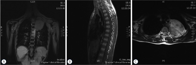
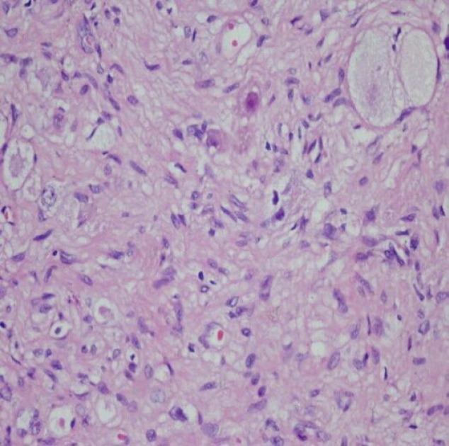
Ganglioneuroma (GN) is a rare benign tumor of neural crest origin usually found in the abdomen, but may occasionally present at uncommon sites including the cervical, lumbar, or sacral spine. However, GNs of thoracic spine are extremely rare. In this report, we describe a 12-year-old girl with giant GN in the thoracic spine, who underwent successful resection (T1-4 level) of the tumor. Histopathological examination confirmed the diagnosis. GN should be considered in the differential diagnosis of any paraspinal mass. A high index of suspicion and correlation of clinico-radiological findings is necessary in differentiating a large benign tumor from a malignant growth. Complete surgical excision is the treatment of choice; however tumor size and location need to be considered for the surgical approach (one-step or multiple surgeries). Close follow-up after surgery is mandatory.
Keywords: Ganglioneuroma; Intradural tumor; Surgery; Thoracic spine.
Figures
Similar articles
-
Total resection of a giant retroperitoneal and mediastinal ganglioneuroma-case report and systematic review of the literature.World J Surg Oncol. 2020 Sep 18;18(1):248. doi: 10.1186/s12957-020-02016-1. World J Surg Oncol. 2020. PMID: 32948207 Free PMC article.
-
A giant lobular thoracic ganglioneuroma cause skeletal erosion: A case report and literature review.Medicine (Baltimore). 2023 Jun 9;102(23):e33891. doi: 10.1097/MD.0000000000033891. Medicine (Baltimore). 2023. PMID: 37335670 Free PMC article. Review.
-
L1 Nerve Root Ganglioneuroma: Surgical Tip on excising this Well-Differentiated and Rarely Appreciated Tumor.J Orthop Case Rep. 2022 Jul;12(7):27-29. doi: 10.13107/jocr.2022.v12.i07.2902. J Orthop Case Rep. 2022. PMID: 36659884 Free PMC article.
-
Multiple cervical ganglioneuroma: A case report and review of the literature.Oncol Lett. 2012 Sep;4(3):509-512. doi: 10.3892/ol.2012.767. Epub 2012 Jun 21. Oncol Lett. 2012. PMID: 23741250 Free PMC article.
-
Giant Ganglioneuroma of the Lumbar Spine: A Rare Cause of Radiculopathy.Case Rep Surg. 2024 May 23;2024:9477892. doi: 10.1155/2024/9477892. eCollection 2024. Case Rep Surg. 2024. PMID: 38883268 Free PMC article.
Cited by
-
Adolescent thoracic scoliosis due to giant ganglioneuroma: a two-case report and literature review.Nagoya J Med Sci. 2024 Nov;86(4):711-719. doi: 10.18999/nagjms.86.4.711. Nagoya J Med Sci. 2024. PMID: 39780926 Free PMC article. Review.
-
Total resection of a giant retroperitoneal and mediastinal ganglioneuroma-case report and systematic review of the literature.World J Surg Oncol. 2020 Sep 18;18(1):248. doi: 10.1186/s12957-020-02016-1. World J Surg Oncol. 2020. PMID: 32948207 Free PMC article.
-
A giant lobular thoracic ganglioneuroma cause skeletal erosion: A case report and literature review.Medicine (Baltimore). 2023 Jun 9;102(23):e33891. doi: 10.1097/MD.0000000000033891. Medicine (Baltimore). 2023. PMID: 37335670 Free PMC article. Review.
-
Mediastinal ganglioneuroma with osseous invasion simulating malignant transformation of osteochondroma on CT imaging.Radiol Case Rep. 2020 Oct 28;16(1):18-21. doi: 10.1016/j.radcr.2020.10.010. eCollection 2021 Jan. Radiol Case Rep. 2020. PMID: 33144905 Free PMC article.
-
Resection of Bilateral Symmetrical Multiple Level Cervical Ganglioneuroma in a 43-Year-Old Man, a Probable Case of Neurofibromatosis Type-1: Report of a Case and Review of Literature.Case Rep Surg. 2022 Jul 15;2022:4547572. doi: 10.1155/2022/4547572. eCollection 2022. Case Rep Surg. 2022. PMID: 35873198 Free PMC article.
References
-
- Kara T, Oztunali C. Radiologic findings of thoracic scoliosis due to giant ganglioneuroma. Clin Imaging. 2013;37:767–768. - PubMed
-
- Kyoshima K, Sakai K, Kanaji M, Oikawa S, Kobayashi S, Sato A, et al. Symmetric dumbbell ganglioneuromas of bilateral C2 and C3 roots with intradural extension associated with von Recklinghausen’s disease: case report. Surg Neurol. 2004;61:468–473. discussion 473. - PubMed
-
- Pang BC, Tchoyoson Lim CC, Tan KK. Giant spinal ganglioneuroma. J Clin Neurosci. 2005;12:967–972. - PubMed
Publication types
LinkOut - more resources
Full Text Sources
Other Literature Sources
Research Materials
