Review
. 2017 Jul;30(3):709-746.
doi: 10.1128/CMR.00013-17.

Bartonella Species, an Emerging Cause of Blood-Culture-Negative Endocarditis

Affiliations
Review

Bartonella Species, an Emerging Cause of Blood-Culture-Negative Endocarditis

Udoka Okaro et al. Clin Microbiol Rev. 2017 Jul.

Abstract

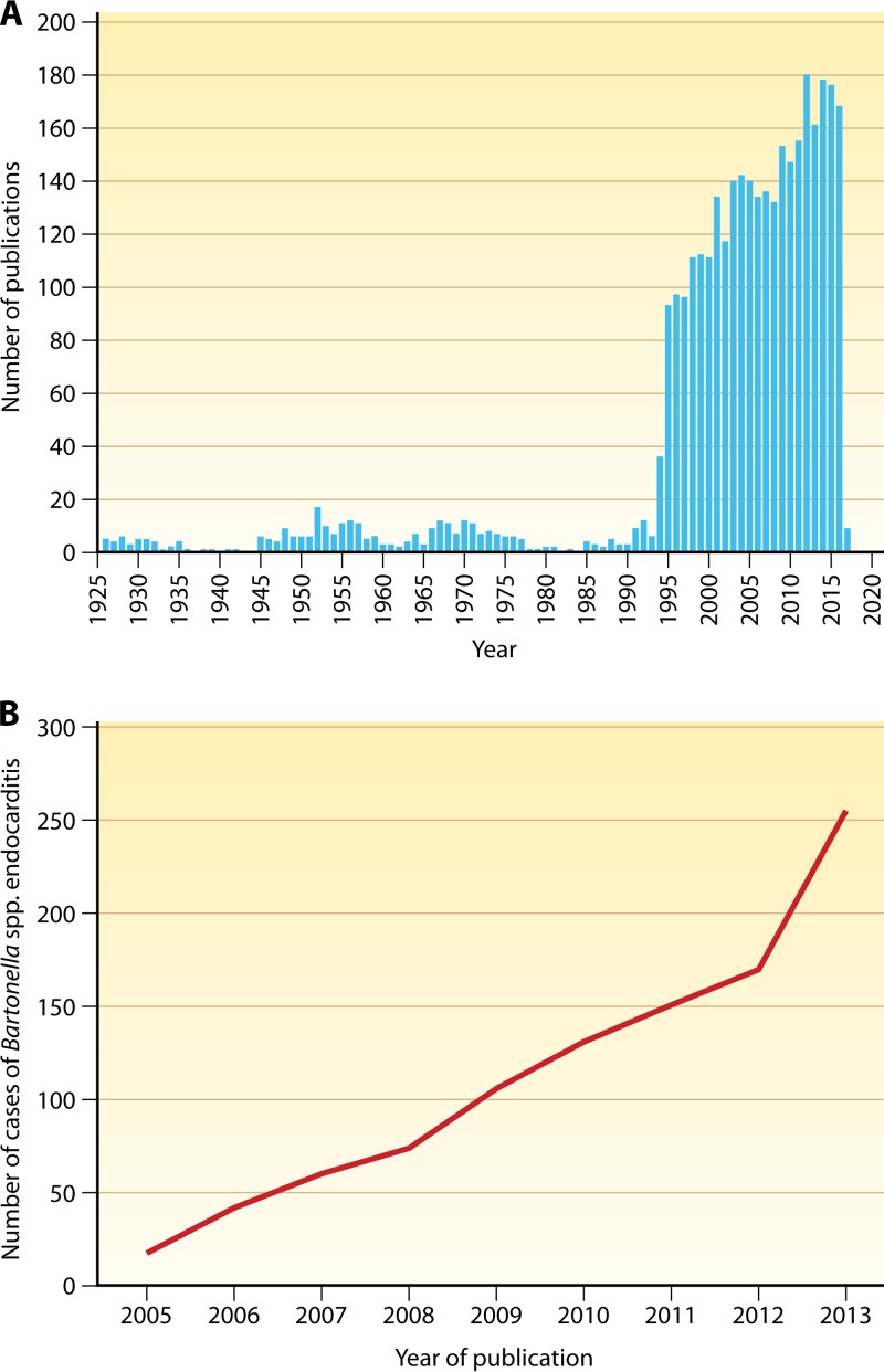
Since the reclassification of the genus Bartonella in 1993, the number of species has grown from 1 to 45 currently designated members. Likewise, the association of different Bartonella species with human disease continues to grow, as does the range of clinical presentations associated with these bacteria. Among these, blood-culture-negative endocarditis stands out as a common, often undiagnosed, clinical presentation of infection with several different Bartonella species. The limitations of laboratory tests resulting in this underdiagnosis of Bartonella endocarditis are discussed. The varied clinical picture of Bartonella infection and a review of clinical aspects of endocarditis caused by Bartonella are presented. We also summarize the current knowledge of the molecular basis of Bartonella pathogenesis, focusing on surface adhesins in the two Bartonella species that most commonly cause endocarditis, B. henselae and B. quintana. We discuss evidence that surface adhesins are important factors for autoaggregation and biofilm formation by Bartonella species. Finally, we propose that biofilm formation is a critical step in the formation of vegetative masses during Bartonella-mediated endocarditis and represents a potential reservoir for persistence by these bacteria.

Keywords: Bartonella; biofilm; blood-culture-negative endocarditis; emerging infections; trimeric autotransporter adhesins.


Figures

FIG 1
FIG 1
(A) Number of publications on Bartonella in PubMed. Source: https://www.ncbi.nlm.nih.gov/pubmed/?term=bartonella. (B) Increase in reported Bartonella endocarditis cases. (Adapted from reference with permission.)
FIG 2
FIG 2
Colony morphology of low-passage-number Houston-1 type strain of B. henselae (ATCC 49882). A highly adherent colony phenotype was observed in this isolate which has subsequently been attributed to expression of badA. (Reproduced from reference .)
FIG 3
FIG 3
Transmission electron micrograph of the bacteriophage-like particles of B. henselae stained with uranyl acetate. White bar, 50 nm.
FIG 4
FIG 4
(A) Transesophageal echocardiogram from a patient with BCNE caused by B. henselae. Bicuspid aortic valve with left coronary leaflet almost entirely replaced by a large vegetation (arrow). (B) Giemsa stain of the patient in panel A showing extensive fibrosis and coccobacilli on the aortic valve that were confirmed to be B. henselae. (Both panels reproduced from reference with permission from Elsevier.)
FIG 5
FIG 5
Transmission electron micrograph of a B. henselae invasome after internalization into an endothelial cell. Magnification, ×12,000.
FIG 6
FIG 6
Paracrine angiogenic loop model for B. henselae. The role of BadA, VirB, and the cognate effectors (Beps) in inducing the angiogenic host response that is unique to Bartonella species is shown. (Adapted from reference .)
FIG 7
FIG 7
Expression and surface localization of BadA in B. henselae. Houston-1 (A) and Marseille (B) strains were reacted with rabbit anti-BadA antibody, followed by goat anti-rabbit IgG conjugated to 10-nm colloidal gold particles. Cells were washed, suspended in phosphate-buffered saline, transferred onto a copper-coated grid, air dried, and imaged using a JEOL JEM 1400 microscope. Surface localization of BadA can be seen in both the Houston-1 and Marseille strains but not the isogenic badA deletion mutants (Houston-1 ΔbadA mutant [C] and Marseille ΔbadA mutant [D]). The markerless, nonpolar in-frame Houston-1 deletion mutant was constructed as previously described (325). The Marseille deletion mutant was constructed by the same approach (unpublished data). Rabbit anti-BadA antibody was raised to the stalk region of the BadA protein (77) and was generously provided by Volkhard Kempf.
FIG 8
FIG 8
Biofilm formation by B. henselae. Scanning electron microscopic images of the adherent cells for both the Houston-1 and Marseille wild-type (WT) strains compared to reduced adherence, autoaggregation, and biofilm formation for the isogenic mutants in which the badA gene is deleted (ΔbadA). Bacterial cells (105) were inoculated onto a coverslip in a six-well plate and grown for 24 h in Schneider's liquid medium at 37°C and 5% CO2. Cells were fixed with 2% paraformaldehyde plus 2% glutaraldehyde and 0.15% alcian blue (to preserve the polysaccharide moieties found in the EPS of biofilms [396, 397]) in 0.2 M sodium cacodylate buffer, pH 7.2. Samples were washed, postfixed for 90 min in 1% OsO4, and dehydrated. Samples were air dried overnight; the coverslip was mounted on adhesive carbon film, coated for 20 s with Au/Pd (60:40) at 16.40 g/cm and 25 mA, and examined using a JEOL JSM6490LV microscope operated at 3-kV low vacuum; and secondary images were collected as JPEG files. The ΔbadA mutants were the same strains as those described in the legend to Fig. 7.
None
None
None
None

References

    1. Anstead GM. 2016. The centenary of the discovery of trench fever, an emerging infectious disease of World War 1. Lancet Infect Dis 16:e164–e172. doi:10.1016/S1473-3099(16)30003-2. - DOI - PMC - PubMed
    1. Murray LM. 1918. The common factor in disordered action of the heart. Br Med J 2:650–652. doi:10.1136/bmj.2.3024.650. - DOI - PMC - PubMed
    1. Venning JA. 1919. The etiology of disordered action of the heart: a report on 7,803 cases. Br Med J 2:337–339. doi:10.1136/bmj.2.3063.337. - DOI - PMC - PubMed
    1. Roden JA, Wells DH, Chomel BB, Kasten RW, Koehler JE. 2012. Hemin binding protein C is found in outer membrane vesicles and protects Bartonella henselae against toxic concentrations of hemin. Infect Immun 80:929–942. doi:10.1128/IAI.05769-11. - DOI - PMC - PubMed
    1. Schulte Fischedick FB, Stuckey MJ, Aguilar-Setien A, Moreno-Sandoval H, Galvez-Romero G, Salas-Rojas M, Arechiga-Ceballos N, Overgaauw PA, Kasten RW, Chomel BB. 2016. Identification of Bartonella species isolated from rodents from Yucatan, Mexico, and isolation of Bartonella vinsonii subsp. yucatanensis subsp nov. Vector Borne Zoonotic Dis 16:636–642. doi:10.1089/vbz.2016.1981. - DOI - PubMed

MeSH terms

LinkOut - more resources