. 2017 Jun 30;292(26):10801-10812.
doi: 10.1074/jbc.M116.771469. Epub 2017 May 10.

Inflammatory cytokines down-regulate the barrier-protective prostasin-matriptase proteolytic cascade early in experimental colitis

Affiliations

Inflammatory cytokines down-regulate the barrier-protective prostasin-matriptase proteolytic cascade early in experimental colitis

Marguerite S Buzza et al. J Biol Chem. .

Abstract

Compromised gastrointestinal barrier function is strongly associated with the progressive and destructive pathologies of the two main forms of irritable bowel disease (IBD), ulcerative colitis (UC), and Crohn's disease (CD). Matriptase is a membrane-anchored serine protease encoded by suppression of tumorigenicity-14 (ST14) gene, which is critical for epithelial barrier development and homeostasis. Matriptase barrier-protective activity is linked with the glycosylphosphatidylinositol (GPI)-anchored serine protease prostasin, which is a co-factor for matriptase zymogen activation. Here we show that mRNA and protein expression of both matriptase and prostasin are rapidly down-regulated in the initiating inflammatory phases of dextran sulfate sodium (DSS)-induced experimental colitis in mice, and, significantly, the loss of these proteases precedes the appearance of clinical symptoms, suggesting their loss may contribute to disease susceptibility. We used heterozygous St14 hypomorphic mice expressing a promoter-linked β-gal reporter to show that inflammatory colitis suppresses the activity of the St14 gene promoter. Studies in colonic T84 cell monolayers revealed that barrier disruption by the colitis-associated Th2-type cytokines, IL-4 and IL-13, down-regulates matriptase as well as prostasin through phosphorylation of the transcriptional regulator STAT6 and that inhibition of STAT6 with suberoylanilide hydroxamic acid (SAHA) restores protease expression and reverses cytokine-induced barrier dysfunction. Both matriptase and prostasin are significantly down-regulated in colonic tissues from human subjects with active ulcerative colitis or Crohn's disease, implicating the loss of this barrier-protective protease pathway in the pathogenesis of irritable bowel disease.

Keywords: cell junction; colitis; cytokine; inflammatory bowel disease (IBD); intestinal epithelium; matriptase; permeability; prostasin; protease; serine protease.


Conflict of interest statement

The authors declare that they have no conflicts of interest with the contents of this article

Figures

Figure 1.
Figure 1.
Clinical symptoms induced during the initiating phase of murine colitis do not become significant until 5 days of DSS treatment in littermate control and St14 hypomorph mice. St14 hypomorph (Hypo) mice and littermate control (Ctl) mice were treated with 2% DSS in drinking water or water alone. A, body weight was monitored daily and expressed relative to day 0, mean ± S.D. (n = 4–6 per group). B, colon lengths measured in littermate controls and St14 hypomorph mice after sacrifice at the indicated times after DSS treatment (Ctl, n = 7–8/group; Hypo, n = 4/group). Colon lengths are significantly shorter after 5 days' DSS treatment compared with mice treated with water alone. **, p < 0.01, unpaired t test. C, spleen weights (expressed as % body weight, mean ± S.D.) are significantly higher after 5 days' DSS treatment in both groups. *, p < 0.05, Mann-Whitney U test (Ctl, n = 7–8/group; Hypo, n = 4/group). D, clinical disease scores mean ± S.D. (Ctl, n = 7–8/group; Hypo, n = 4/group). Both groups do not show significant clinical disease until day 5. ***, p < 0.001, *, p < 0.05, Mann-Whitney U test. E, representative H&E-stained images of distal colons from St14 hypomorph and littermate control mice at the indicated times of DSS treatment. The majority of tissue after 1.5 days' treatment appears normal with some patchy damage (middle panels, 20×, 40×), but extensive damage was evident at day 5 with immune cell infiltrate and loss of colonic villi. Scale bars: 20× = 200 μm, 40× = 100 μm.
Figure 2.
Figure 2.
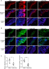
Matriptase and prostasin proteins are significantly down-regulated during murine colitis. A and B, representative immunostaining of distal colon sections from untreated control mice or after 5 days of DSS treatment. Images from different treatment groups were captured at equal exposure times and are shown at 200× or 600×. In untreated mice both proteases are most highly expressed at villous tips, matriptase concentrates at epithelial cell junctions (A, water, 600×, arrow), whereas prostasin displays a more diffuse and apical distribution (B, water, 600×, arrow). DSS treatment causes a reduction in staining intensity for both proteases, with evidence of leukocyte infiltrate (L) in some colonic segments of DSS-treated mice. β-tubulin staining of similar regions of colonic sections shows the epithelial cells, where matriptase and prostasin expression is reduced. C, quantitation of signal intensities of matriptase and prostasin in colons sections from untreated or DSS-treated mice (n = 4–6 mice/group). Graphs show mean ± S.D. *, p < 0.05, ***, p < 0.001, unpaired t test.
Figure 3.
Figure 3.
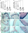
Matriptase and prostasin mRNA are down-regulated early during the initiating phase of murine colitis. A and B, qPCR analysis of mRNA isolated from control mice after treatment with 2% DSS for 1.5 or 5 days for matriptase (A) and prostasin (B). Signals were first normalized to GAPDH to account for cDNA content, and then normalized to the epithelial marker EpCAM. mRNA for both proteases is significantly down-regulated at both 1.5 days and 5 days of DSS treatment (n = 6–8 mice/group). Graphs show individual mice in each treatment group, and mean ± S.D. *, p < 0.05, **, p < 0.01, unpaired t test. C, X-gal staining (blue) of proximal colons from littermate control mice treated with water alone compared with DSS-treated mice. Representative images from 6–8 mice/group. Scale bars: 4× = 1000 μm, 10× = 400 μm, 40× = 100 μm.
Figure 4.
Figure 4.
Cytokines that are implicated in UC down-regulate matriptase and prostasin during disruption of polarized T84 epithelial barriers. T84 cells were plated onto Transwell filters and allowed to develop barrier function for 6 days as assessed by TEER, and were then treated basolaterally with 10 ng/ml of the indicated cytokines for 5 days (arrow below A). A, addition of both combinations of cytokines decreased TEER over time, mean ± S.E. from triplicate wells. Data are representative of two experiments. B, measurement of the paracellular permeability of T84 monolayers to 4 kDa FITC-dextran assessed after 5 days of cytokine treatment (day 11 in A), shows a significant disruption of epithelial barrier function by both combinations of cytokines, mean ± S.E. from quadruplicate wells. ***, p < 0.001, unpaired t test. C and D, immunoblot analysis of whole cell lysates prepared on day 5 of cytokine treatment (day 11 in A) probed with the indicated antibodies. Data show decreased matriptase and prostasin expression, elevated claudin-2, increased STAT6 activation (p-STAT6, phospho-STAT6) induced by IL-4 and IL-13, and increased apoptosis induced by TNFα and IFNγ. Data are representative of two independent experiments.
Figure 5.
Figure 5.
IL-4/IL-13 induce time-dependent down-regulation of matriptase and prostasin. T84 monolayers were allowed to develop barrier function for 10 days, at which time they were treated basolaterally with 10 ng/ml IL-4 and IL-13 (arrow) for 5 days. A, measurement of TEER shows that cytokine-induced TEER loss begins at ∼6 h of treatment (inset). Graphs show mean ± S.E. from triplicate wells. Data are representative of two experiments. B, immunoblot analysis of whole cell lysates prepared at the indicated times after cytokine addition show both matriptase and prostasin protein levels start to decrease after 24 h of treatment. Matriptase expression remains suppressed over 72 h, whereas prostasin expression returns at 72 h. C, qPCR analysis of matriptase, prostasin, and claudin-2 mRNA expression in IL-4/IL-13 treated T84 monolayers compared with untreated monolayers. Signals were normalized to GAPDH and expressed relative to untreated at 3 h. Matriptase mRNA is significantly lower after 24 h of treatment (p < 0.001) and remains suppressed over 72 h, whereas prostasin mRNA is significantly lost by 6 h (p < 0.05) but begins to return at 24 h (n.s). Data are representative of two independent experiments, from triplicate wells each.
Figure 6.
Figure 6.
The STAT6 inhibitor SAHA prevents the loss of barrier function and down-regulation of matriptase and prostasin mRNA induced by IL-4/IL-13. Polarized T84 monolayers on Transwell filters were treated basolaterally with 10 ng/ml each IL-4 and IL-13 with or without pretreatment with 5 μm SAHA for 1 h. A, analysis of TEER and matriptase, prostasin, and claudin-2 mRNA expression at 6 and 24 h after cytokine addition. Both TEER and matriptase mRNA expression are significantly decreased by 24 h of treatment with IL-4/IL-13 (p < 0.05), which is abrogated in the presence of SAHA. The significant loss of prostasin mRNA that occurs at 6 h in the presence of IL-4/IL-13 (p < 0.001) is also inhibited by SAHA. Signals are normalized to 18S rRNA because we found GAPDH mRNA to be regulated by SAHA. Data are representative of three independent experiments. Mean ± S.E. B, immunoblots of whole cell lysates at 24 h (top) and densitometry analysis of matriptase protein expression (bottom). The data show a decrease in IL-4/IL-13 induced STAT6 phosphorylation, the prevention of loss of matriptase protein, and the failure to upregulate claudin-2 in the presence of cytokines by SAHA. The bottom panel shows a significant loss of matriptase protein that is recovered in the presence of SAHA as quantified by densitometry. Matriptase protein expression is expressed relative to vehicle-treated cells after normalizing to β-tubulin at 24 h of treatment. Graph shows mean ± S.E. from three independent experiments. **, p < 0.01, unpaired t test.
Figure 7.
Figure 7.
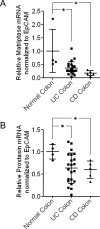
Matriptase and prostasin are significantly decreased in colonic epithelium of human IBD patients. A and B, the TissueScan Crohn's and Colitis Tissue qPCR Panel II was analyzed for matriptase (A) and prostasin (B) expression by qPCR. Signals were normalized to β-actin mRNA and then normalized to EpCAM mRNA to account for variation in epithelial content of individual samples. Graphs show mean ± S.D. *, p < 0.05, unpaired t test.

Similar articles

Cited by

References

    1. Xavier R. J., and Podolsky D. K. (2007) Unravelling the pathogenesis of inflammatory bowel disease. Nature 448, 427–434 - PubMed
    1. Michielan A., and D'Incà R. (2015) Intestinal permeability in inflammatory bowel disease: Pathogenesis, clinical evaluation, and therapy of leaky gut. Mediators Inflamm. 2015, 628157. - PMC - PubMed
    1. Hollander D., Vadheim C. M., Brettholz E., Petersen G. M., Delahunty T., and Rotter J. I. (1986) Increased intestinal permeability in patients with Crohn's disease and their relatives. A possible etiologic factor. Ann. Intern. Med. 105, 883–885 - PubMed
    1. Katz K. D., Hollander D., Vadheim C. M., McElree C., Delahunty T., Dadufalza V. D., Krugliak P., and Rotter J. I. (1989) Intestinal permeability in patients with Crohn's disease and their healthy relatives. Gastroenterology 97, 927–931 - PubMed
    1. Irvine E. J., and Marshall J. K. (2000) Increased intestinal permeability precedes the onset of Crohn's disease in a subject with familial risk. Gastroenterology 119, 1740–1744 - PubMed

MeSH terms