. 2017 Jun 30;292(26):10912-10925.
doi: 10.1074/jbc.M116.773077. Epub 2017 May 10.

The Cox1 C-terminal domain is a central regulator of cytochrome c oxidase biogenesis in yeast mitochondria

Affiliations

The Cox1 C-terminal domain is a central regulator of cytochrome c oxidase biogenesis in yeast mitochondria

Rodolfo García-Villegas et al. J Biol Chem. .

Abstract

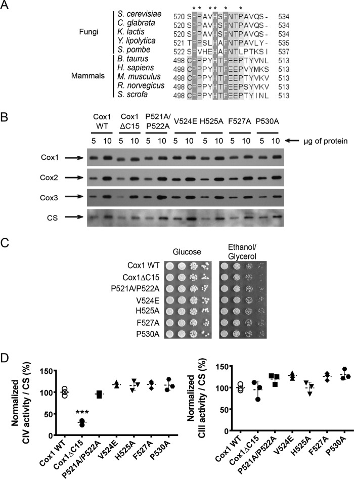
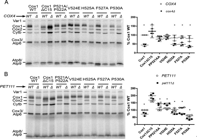
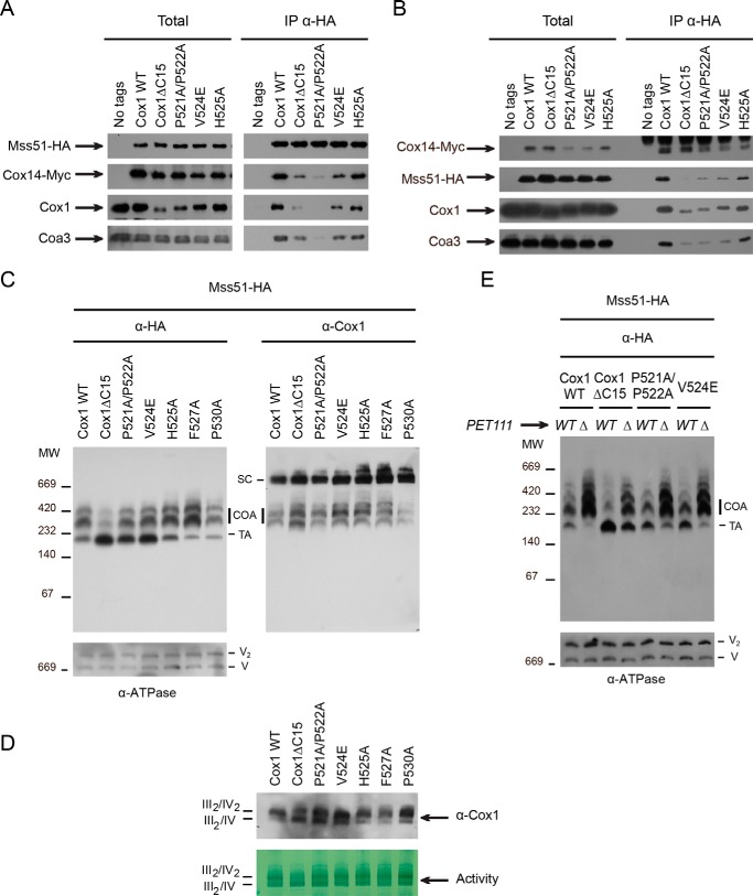
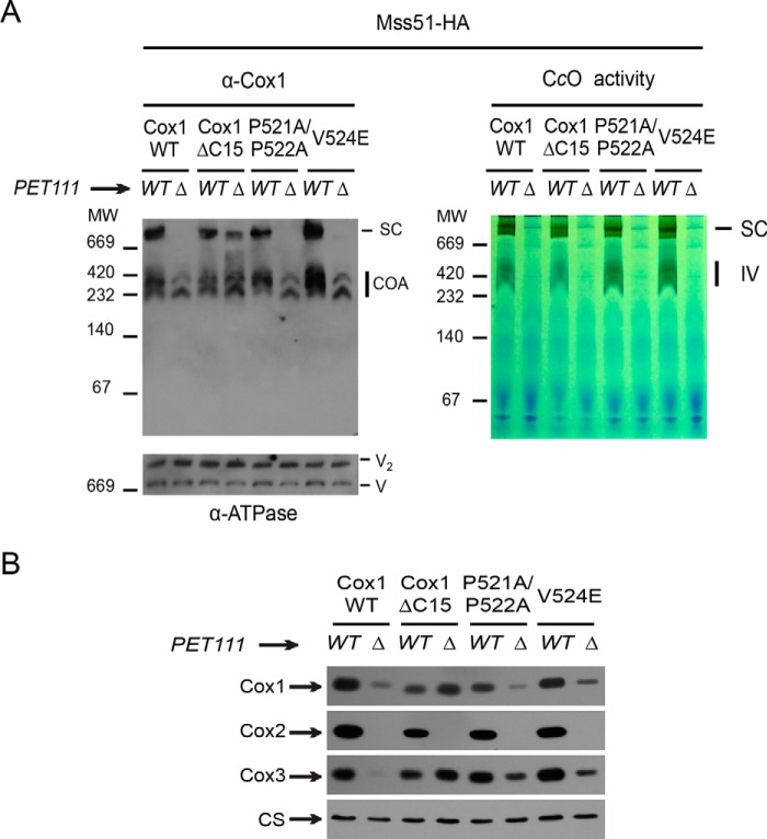
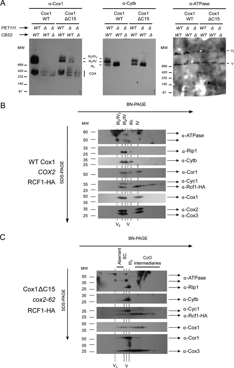
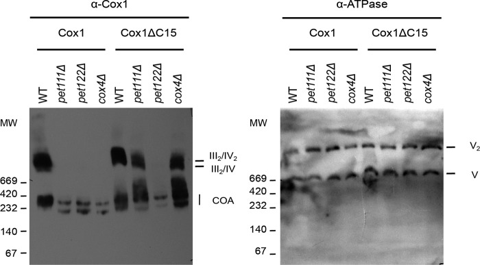
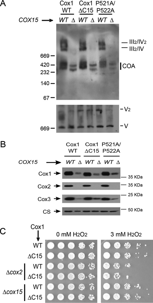
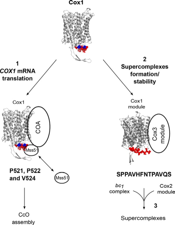
Cytochrome c oxidase (CcO) is the last electron acceptor in the respiratory chain. The CcO core is formed by mitochondrial DNA-encoded Cox1, Cox2, and Cox3 subunits. Cox1 synthesis is highly regulated; for example, if CcO assembly is blocked, Cox1 synthesis decreases. Mss51 activates translation of COX1 mRNA and interacts with Cox1 protein in high-molecular-weight complexes (COA complexes) to form the Cox1 intermediary assembly module. Thus, Mss51 coordinates both Cox1 synthesis and assembly. We previously reported that the last 15 residues of the Cox1 C terminus regulate Cox1 synthesis by modulating an interaction of Mss51 with Cox14, another component of the COA complexes. Here, using site-directed mutagenesis of the mitochondrial COX1 gene from Saccharomyces cerevisiae, we demonstrate that mutations P521A/P522A and V524E disrupt the regulatory role of the Cox1 C terminus. These mutations, as well as C terminus deletion (Cox1ΔC15), reduced binding of Mss51 and Cox14 to COA complexes. Mss51 was enriched in a translationally active form that maintains full Cox1 synthesis even if CcO assembly is blocked in these mutants. Moreover, Cox1ΔC15, but not Cox1-P521A/P522A and Cox1-V524E, promoted formation of aberrant supercomplexes in CcO assembly mutants lacking Cox2 or Cox4 subunits. The aberrant supercomplex formation depended on the presence of cytochrome b and Cox3, supporting the idea that supercomplex assembly factors associate with Cox3 and demonstrating that supercomplexes can be formed even if CcO is inactive and not fully assembled. Our results indicate that the Cox1 C-terminal end is a key regulator of CcO biogenesis and that it is important for supercomplex formation/stability.

Keywords: Cox1; Mss51; cytochrome c oxidase (complex IV); mitochondria; mitochondrial DNA (mtDNA); supercomplex; translation; yeast.


Conflict of interest statement

The authors declare that they have no conflicts of interest with the contents of this article

Figures

Figure 1.
Figure 1.
Cox1 C-terminal end mutations do not affect CcO activity. A, alignment of the last 15 residues from S. cerevisiae Cox1 with fungal and mammal Cox1 homologues. The alignment was obtained using the Jalview version 2.8.0b1 software (56). Dark gray boxes, highly conserved residues; light gray boxes, partially conserved residues. Asterisks show the amino acids that were mutated in the present study. Numbers indicate position of the last 15 residues of Cox1 in each species. B, mitochondria from wild type and the Cox1 mutants were separated by SDS-PAGE and analyzed by Western blotting using the indicated antibodies. Citrate synthase (CS) was used as loading control. C, 10-fold serial dilutions from wild type and Cox1 mutants were spotted in complete medium with either glucose or ethanol/glycerol. The plates were incubated for 3 days at 30 °C. D, spectrophotometric measurements of complex IV and complex III activities from mitochondria carrying the indicated Cox1 mutations were normalized to the activity of CS. Statistical significance was determined by one-way analysis of variance and Dunnett's test for multiple comparisons with the WT values (***, p < 0.001; n = 3 independent assays) using Prism version 6 software. Error bars, S.D.
Figure 2.
Figure 2.
Mutations Cox1ΔC15, Cox1-P521A/P522A, and Cox1-V524E are important for assembly feedback regulation of Cox1 synthesis. A and B, cells carrying either wild-type or mutated Cox1 were pulse-labeled with [35S]methionine in the presence of cycloheximide. The Cox1 variants were combined with either cox4Δ (A) or pet111Δ (B) mutations, as indicated. Translation products were separated by SDS-PAGE and analyzed by autoradiography. Cox1 labeling intensity is shown at the right side of each panel. It was quantified using the ImageJ software and normalized to the Cox3/Atp6 (ATPase subunit 6) signal. Labeling intensity was expressed as a percentage of the wild-type Cox1 signal. Error bars, S.D. from three independent experiments. We also compared the signals of Cytb and Var1 (ribosomal protein) to the Cox3/Atp6 signal, showing that the observed pattern in A and B is specific for Cox1 (data not shown). The relevant significant differences between strains (*) were determined by Student's t test. A p value of < 0.01 was considered statistically significant.
Figure 3.
Figure 3.
The interaction between Mss51 and the COA complexes decreased in the Cox1-P521A/P522A, Cox1-V524E, and Cox1ΔC15 mutants. A, mitochondria (500 μg of proteins) from wild type and the Cox1 mutants were solubilized with dodecyl maltoside and incubated with an antibody against HA epitope to immunoprecipitate Mss51-HA. A strain without HA tag in Mss51 was used as a negative control. The total and immunoprecipitated (IP) fractions were separated by SDS-PAGE and transferred to a PVDF membrane for Western blotting analysis with the indicated antibodies. Total fractions represent 3% of the mitochondrial extract, whereas all immunoprecipitated protein was loaded onto the gel. B, mitochondrial proteins from wild type or Cox1 mutants were analyzed as in A, but an antibody against Myc was used to immunoprecipitate Cox14-Myc. C, digitonin-solubilized mitochondria from WT and Cox1 mutants were separated by BN-PAGE (5–13%) and transferred to a PVDF membrane for Western blotting analysis with antibodies against HA epitope (α-HA) or Cox1 (α-Cox1). An antibody against ATP synthase was used as loading control (V and V2). Bands corresponding to supercomplexes (SC), COA complexes containing Cox1, and the translational active form of Mss51 (TA) are indicated. All strains carried the Mss51-HA variant. D, mitochondria from C were separated by a BN-PAGE (4–12%) to resolve supercomplexes III2/IV2 and III2/IV. The separated proteins were analyzed by Western blotting using antibody against Cox1 and by CcO in-gel activity. E, digitonin-solubilized mitochondria from WT and Cox1 mutants bearing wild-type PET111 or a pet111Δ mutation were separated by BN-PAGE and analyzed as in C.
Figure 4.
Figure 4.
In the absence of Pet111, Cox1ΔC15 led to formation of a supercomplex-like band in BN-PAGE. A, digitonin-solubilized mitochondria from wild type and Cox1 mutants carrying either wild-type PET111 (WT) or a pet111Δ mutation (Δ) were separated by BN-PAGE (5–13%) as in Fig. 3. In parallel, a second BN gel was stained for CcO activity using diaminobenzidine and cytochrome c (right). The monomeric form of active CcO is indicated (IV). B, mitochondria from A were separated by SDS-PAGE and analyzed by Western blotting using the indicated antibodies. CS was used as a loading control.
Figure 5.
Figure 5.
Cox1ΔC15-induced formation of non-functional, bc1 complex-dependent supercomplexes in the absence of Cox2. A, digitonin-solubilized mitochondria of WT, pet111Δ, or pet111Δ/cbs2Δ mutants containing Cox1 or Cox1ΔC15 were separated by BN-PAGE (4–12%) and analyzed by Western blotting using antibodies against Cox1 (α-Cox1), cytochrome b (α-Cytb), or ATP synthase. Supercomplexes III2/IV2 and III2/IV are indicated. B, mitochondria from cells carrying wild-type Cox1, COX2, and a plasmid encoding Rcf1 fused to His6 and HA3 (9) were solubilized with digitonin and separated by BN-PAGE (4–12%). A lane of this gel was further resolved by 2D Tricine-SDS-PAGE (12%) and analyzed by Western blotting with the indicated antibodies. C, mitochondria of cox2-62 cells containing Cox1ΔC15 and Rcf1-His6-HA3 were analyzed as in B.
Figure 6.
Figure 6.
Supercomplex-like bands induced by Cox1ΔC15 are absent in pet122Δ (and thereof Cox3 deletion) but present in cox4Δ mutants. Digitonin-solubilized mitochondria of pet111Δ, pet122Δ, and cox4Δ cells containing either Cox1 or Cox1ΔC15 were separated by BN-PAGE (4–12%) and analyzed by immunoblotting with a Cox1 antibody. ATPase antibodies were used to detect complex V as loading control.
Figure 7.
Figure 7.
Formation of aberrant supercomplexes requires Cox1 hemylation. A, mitochondria from cells carrying wild-type Cox1, Cox1ΔC15, or Cox1-P521A/P522A and either cox15Δ or wild-type COX15 were separated on BN-PAGE and analyzed as in Fig. 6. B, mitochondria from A were separated by SDS-PAGE and analyzed by Western blotting using the indicated antibodies. CS was used as a loading control. C, cells carrying either the wild-type Cox1 or Cox1ΔC15 and either cox2Δ (allele cox2-62 (36)) or cox15Δ were incubated with 3 mm H2O2 or were mock-treated. 10-fold serial dilutions were grown on YPD plates for 3 days at 30 °C.
Figure 8.
Figure 8.
Model depicting the participation of the Cox1 C-terminal end in CcO biogenesis and supercomplex accumulation. A model of S. cerevisiae Cox1 was constructed with SWISS-MODEL (57) based on the crystallographic structure of bovine Cox1 (58). The last 15 residues of Cox1 are indicated in red. Pro-521, Pro-522, and Val-524 are indicated in blue. The last 15 Cox1 residues, and mainly Pro-521/Pro-522 and Val-524, regulate Cox1 synthesis by promoting association of Mss51 with the COA complexes (1). In addition, the Cox1 15-residue C terminus (with sequence SPPAVHFNTPAVQS), rather than Pro-521/Pro-522 and V524E is important to modulate accumulation of respiratory chain supercomplexes (2). According to our results, the Cox1 and Cox3 assembly modules can associate with bc1 complex before the integration of the Cox2 module to form fully functional supercomplexes (3).

Similar articles

Cited by

References

    1. Mick D. U., Fox T. D., and Rehling P. (2011) Inventory control: cytochrome c oxidase assembly regulates mitochondrial translation. Nat. Rev. Mol. Cell Biol. 12, 14–20 - PMC - PubMed
    1. Soto I. C., Fontanesi F., Liu J., and Barrientos A. (2012) Biogenesis and assembly of eukaryotic cytochrome c oxidase catalytic core. Biochim. Biophys. Acta 1817, 883–897 - PMC - PubMed
    1. McStay G. P., Su C. H., Thomas S. M., Xu J. T., and Tzagoloff A. (2013) Characterization of Cox1p assembly intermediates in Saccharomyces cerevisiae. J. Biol. Chem. 288, 26546–26556 - PMC - PubMed
    1. McStay G. P., Su C. H., and Tzagoloff A. (2013) Modular assembly of yeast cytochrome oxidase. Mol. Biol. Cell 24, 440–452 - PMC - PubMed
    1. McStay G. P., Su C. H., and Tzagoloff A. (2013) Stabilization of Cox1p intermediates by the Cox14p-Coa3p complex. FEBS Lett. 587, 943–949 - PMC - PubMed

MeSH terms

Substances

LinkOut - more resources