Comparative Study
. 2017 May 10;7(1):1689.
doi: 10.1038/s41598-017-01584-9.

A comparative multi-parametric in vitro model identifies the power of test conditions to predict the fibrotic tendency of a biomaterial

Affiliations
Comparative Study

A comparative multi-parametric in vitro model identifies the power of test conditions to predict the fibrotic tendency of a biomaterial

Maren Jannasch et al. Sci Rep. .

Abstract

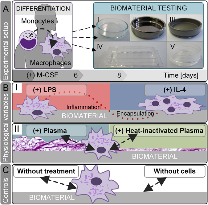
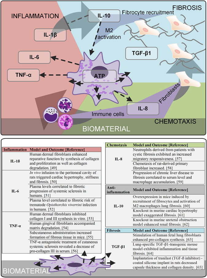
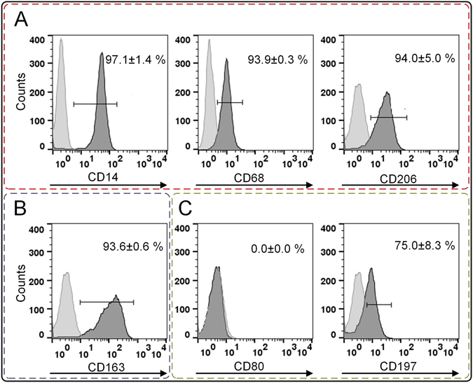
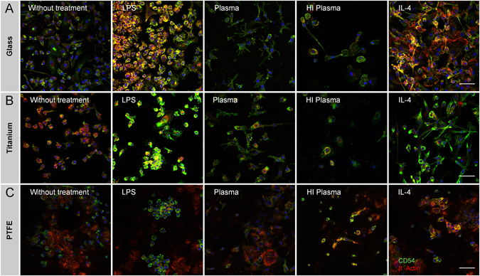
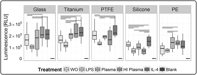
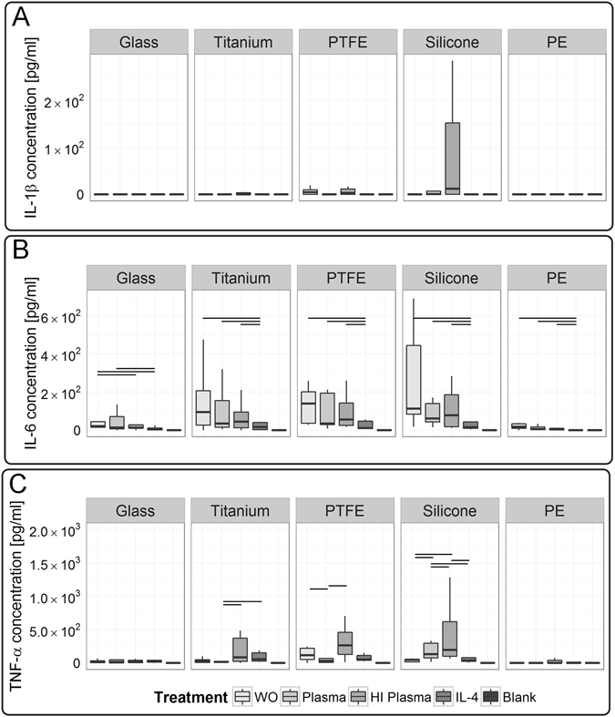
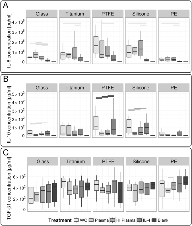
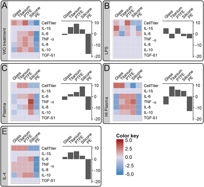
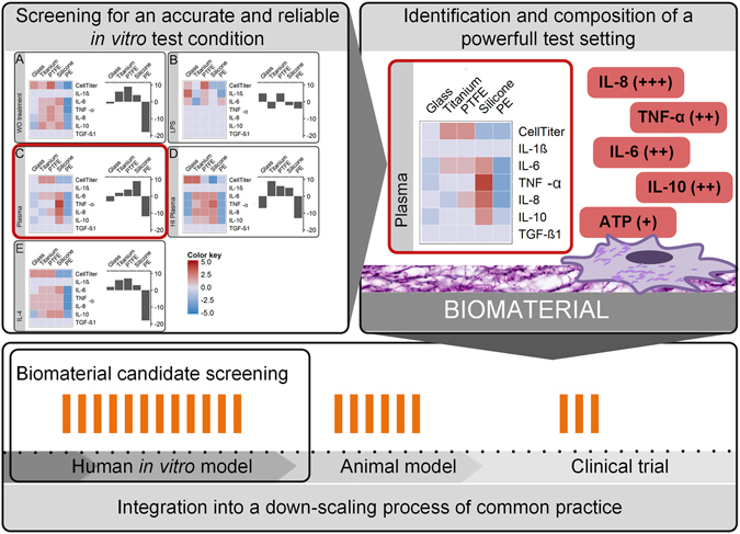
Despite growing effort to advance materials towards a low fibrotic progression, all implants elicit adverse tissue responses. Pre-clinical biomaterial assessment relies on animals testing, which can be complemented by in vitro tests to address the Russell and Burch's 3R aspect of reducing animal burden. However, a poor correlation between in vitro and in vivo biomaterial assessments confirms a need for suitable in vitro biomaterial tests. The aim of the study was to identify a test setting, which is predictive and might be time- and cost-efficient. We demonstrated how sensitive in vitro biomaterial assessment based on human primary macrophages depends on test conditions. Moreover, possible clinical scenarios such as lipopolysaccharide contamination, contact to autologous blood plasma, and presence of IL-4 in an immune niche influence the outcome of a biomaterial ranking. Nevertheless, by using glass, titanium, polytetrafluorethylene, silicone, and polyethylene representing a specific material-induced fibrotic response and by comparison to literature data, we were able to identify a test condition that provides a high correlation to state-of-the-art in vivo studies. Most important, biomaterial ranking obtained under native plasma test conditions showed a high predictive accuracy compared to in vivo assessments, strengthening a biomimetic three-dimensional in vitro test platform.


Conflict of interest statement

The authors declare that they have no competing interests.

Figures

Figure 1
Figure 1
In vitro screening of physiological test conditions for the predictive power to evaluate a biomaterials’ fibrotic potential. (A) In our experimental setup, human blood-derived monocytes were differentiated by supplementation of macrophage colony-stimulating factor (M-CSF) to M2-like macrophages. Subsequently, macrophages were cultured on biomaterials for 48 h: (I) glass, (II) titanium, (III) PTFE, (IV) silicone and (V) PE. (B) On each material, we tested conditions that mimic the physiological in vivo niche on materials’ surface: (I) A common cause of implant failure - LPS contamination - polarizes macrophages’ fate towards pro-inflammation. In contrast, the presence of IL-4 in the immune niche strengthens a pro-survival cellular phenotype – the fusion of macrophages towards foreign body giant cells. (II) A biomimetic approach of protein-material interaction was resembled by applying human autologous blood-derived plasma on biomaterials surface. By calcification of plasma, a primary fibrous three-dimensional niche was formed. In comparison to native blood plasma, the inactivation of heat labile protein, e.g. complement, growth, and coagulation factors was assessed by heat-inactivation (HI) of human plasma. (C) As controls served test conditions without any additions and without cells.
Figure 2
Figure 2
To characterize materials’ fibrotic potential, the influence of the selected test conditions on the net secretion of cytokines was investigated. All selected readout factors represent fibrotic drivers: a strong cellular viability on materials’ surface maintains a chronic responsiveness to the foreign body; a pro-inflammatory component at the initial stage of cell-implant contact induces a fibrotic progression and a chemokine gradient guiding further cells to implant region strengthens both the inflammatory and fibrotic response. Despite the mechanism of surface-induced cytokine secretion and their influence to final fibrotic progression is poorly understood, pre-clinical and clinical studies from adjacent research areas substantiate the selection of readout parameters.
Figure 3
Figure 3
Prior material testing, the differentiation of monocytes to M2-like macrophages was confirmed. (A) Macrophage markers CD14, CD68, and CD206 were expressed. (B) The differentiation towards a M2-like phenotype was shown by CD163 positive profile, whereas (C) CD80 allowed distinguishing between M1 and M2. Here, its absence rejected the M1 differentiation. The marker CD197 was expressed by macrophages differentiated from whole-blood-derived monocytes. Histograms exemplarily represent the differentiation cluster of one donor; whereas mean values ± SD represent data of three blood donors. A high compliance between all three donors was found. The light-grey histograms represent the isotype controls. The following abbreviations are used: SD for standard deviation, CD for cluster of differentiation.
Figure 4
Figure 4
Immune-histological staining of intercellular adhesion molecule CD54 (green) and β-Actin (red) illustrated morphological changes in dependency of test conditions and material on (A) glass, (B) titanium and (C) PTFE. An uneven topography and a long working distance prohibited to capture sharp images on silicone and PE surfaces. The scale bar depicts 50 µm and is valid for all images. The following abbreviations are used: HI for heat-inactivated.
Figure 5
Figure 5
Surface-associated viability of macrophages following test procedure of 48 h was assessed by semi-quantitative ATP measurement, using a luminescence-based assay (CellTiter). Significant differences between the test conditions were found on all tested materials. Data is comprised of ten human macrophage donors (n = 10). Significance level is considered with a p-value ≤ 0.05. The following abbreviations are used: WO for without treatment, HI for heat-inactivated.
Figure 6
Figure 6
Macrophages’ response showed a dependency on test conditions and revealed a material induced secretion of acute pro-inflammatory cytokines. (A) Interestingly, IL-1ß did not resolve differences between test conditions. (B) In contrast, IL-6 showed a strong dependency on test conditions for tests on glass, whereas (C) TNF-α demonstrated a dependency for tests on titanium, PTFE and silicone surfaces. Data of ten donors is shown (n = 10). A p-value ≤ 0.05 is considered as significant. LPS stimulation induced a high cytokine secretion, reasonably leading to provide the obtained data in separate graphs (see Figure S1). The following abbreviations are used: WO for without treatment, HI for heat-inactivated.
Figure 7
Figure 7
Chemokine IL-8, and anti-inflammatory cytokine IL-10 revealed significant differences between the test conditions, whereas pro-fibrotic growth factor TGF-β1 did not resolve differences. (A) Here, we demonstrated a test condition dependency of IL-8 levels for tests on glass. (B) Additionally, IL-10 levels differed significantly between test conditions on glass, PTFE and silicone surfaces. (C) Macrophages’ secretion of TGF-β1 did not exceed the basal level found in the culture medium, thereby no effect of the test conditions were resolved. Those cytokines were analyzed in the supernatant of ten macrophage donors (n = 10). A p-value ≤ 0.05 is considered as significant. LPS-induced secretion of cytokines is provided in separate graphs (see Figure S1). The following abbreviations are used: WO for without treatment, HI for heat-inactivated.
Figure 8
Figure 8
A significance profile of each material in comparison to all other tested materials was translated to a material scoring model. The obtained significance score values were translated into a color map in range of blue for minus values to red for positive values (left graph). Thereby, this scoring model visualized differences between the test conditions and finally allowed the evaluation of each specific readout factor respective to its predictive power. Finally for each material, the material ranking model summarized all readout-dependent scores in a bar plot (right graph). By comparing the literature-based biomaterial ranking to the obtained material rankings, a validation of each test condition for its accuracy to predict a fibrotic progression was performed: (A) without (WO) treatment, (B) LPS stimulation, (C) native plasma and (D) heat-inactivated (HI) plasma treatment as well as (E) IL-4 stimulation.
Figure 9
Figure 9
The integration of human-based in vitro test models into a down-scaling process of common practice is often neglected. One major flaw is a low correlation of in vitro tests to in vivo studies. In this study we propose a procedure to overcome this hurdle. In a large-scale in vitro screening approach, we identified an accurate and suitable condition for biomaterial testing and proved its validity by correlation to a well-described effect-the fibrotic progression - observed in vivo. On basis of this screening approach a well-defined in vitro test setting with a high predictive power was composed of (I) plasma pretreatment of biomaterials’ surface and (II) a set of highly predictive readout factors. We hope that the obtained data justifies a down-scaling of the in vitro test procedure to the identified condition and will be applied for large-scale biomaterial candidate screenings in following studies. Finally, followed by current common practice to study promising candidates in systemic animal models, and carrying the positively-proofed biomaterials in human subjects.

References

    1. Montanaro L, Campoccia D, Arciola CR. Advancements in molecular epidemiology of implant infections and future perspectives. Biomaterials. 2007;28:5155–5168. doi: 10.1016/j.biomaterials.2007.08.003. - DOI - PubMed
    1. Gorbet MB, Sefton MV. Biomaterial-associated thrombosis: roles of coagulation factors, complement, platelets and leukocytes. Biomaterials. 2004;25:5681–5703. doi: 10.1016/j.biomaterials.2004.01.023. - DOI - PubMed
    1. Thevenot P, Hu W, Tang L. Surface chemistry influences implant biocompatibility. Curr Top Med Chem. 2008;8:270–280. doi: 10.2174/156802608783790901. - DOI - PMC - PubMed
    1. Grainger DW. All charged up about implanted biomaterials. Nature biotechnology. 2013;31:507–509. doi: 10.1038/nbt.2600. - DOI - PubMed
    1. Anderson JM, Rodriguez A, Chang DT. Foreign body reaction to biomaterials. Semin Immunol. 2008;20:86–100. doi: 10.1016/j.smim.2007.11.004. - DOI - PMC - PubMed

Publication types