. 2017 May 10;7(1):1682.
doi: 10.1038/s41598-017-01851-9.

Unexpected Efficacy of a Novel Sodium Channel Modulator in Dravet Syndrome

Affiliations

Unexpected Efficacy of a Novel Sodium Channel Modulator in Dravet Syndrome

Lyndsey L Anderson et al. Sci Rep. .

Abstract

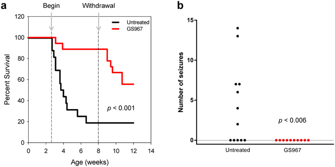
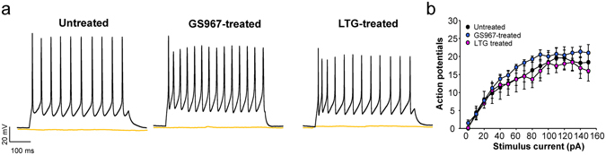
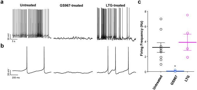
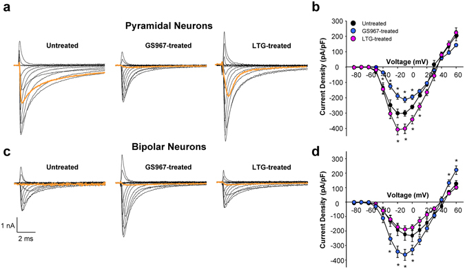
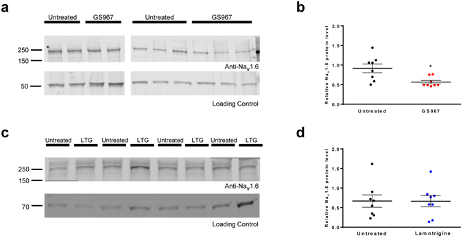
Dravet syndrome, an epileptic encephalopathy affecting children, largely results from heterozygous loss-of-function mutations in the brain voltage-gated sodium channel gene SCN1A. Heterozygous Scn1a knockout (Scn1a +/-) mice recapitulate the severe epilepsy phenotype of Dravet syndrome and are an accepted animal model. Because clinical observations suggest conventional sodium channel blocking antiepileptic drugs may worsen the disease, we predicted the phenotype of Scn1a +/- mice would be exacerbated by GS967, a potent, unconventional sodium channel blocker. Unexpectedly, GS967 significantly improved survival of Scn1a +/- mice and suppressed spontaneous seizures. By contrast, lamotrigine exacerbated the seizure phenotype. Electrophysiological recordings of acutely dissociated neurons revealed that chronic GS967-treatment had no impact on evoked action potential firing frequency of interneurons, but did suppress aberrant spontaneous firing of pyramidal neurons and was associated with significantly lower sodium current density. Lamotrigine had no effects on neuronal excitability of either neuron subtype. Additionally, chronically GS967-treated Scn1a +/- mice exhibited normalized pyramidal neuron sodium current density and reduced hippocampal NaV1.6 protein levels, whereas lamotrigine treatment had no effect on either pyramidal neuron sodium current or hippocampal NaV1.6 levels. Our findings demonstrate unexpected efficacy of a novel sodium channel blocker in Dravet syndrome and suggest a potential mechanism involving a secondary change in NaV1.6.


Conflict of interest statement

Dr. George received a research grant from Gilead Sciences, Inc., the manufacturer of GS967, which partially supported this work. Drs. Anderson, Hawkins, Thompson and Kearney declare no potential conflicts of interest.

Figures

Figure 1
Figure 1
GS967 improved survival and reduced seizure frequency of Scn1a +/− mice. (a) Survival curves comparing untreated and GS967-treated Scn1a +/− mice. Treatment commenced at P18 (first dashed line) and was withdrawn at 8 weeks (second dashed line), with n = 18–37 mice per group. Survival difference between groups was significant (p < 0.001; Mantel-Cox log-rank test). (b) Number of seizures in 48 hours observed for untreated and GS967-treated Scn1a +/− mice (n = 10–12 mice per group) was significantly different (p < 0.006; non-parametric Mann-Whitney test).
Figure 2
Figure 2
GS967 treatment of Scn1a +/− mice does not affect bipolar neuron excitability. (a) Representative action potentials in response to either 0 pA (orange traces) or 50 pA stimulus (black traces) recorded from bipolar neurons acutely isolated from untreated (left), GS967-treated (middle) and lamotrigine (LTG)-treated (right) Scn1a +/− mice. (b) Summary data plotting number of action potentials against stimulus current. Error bars represent SEM, with n = 5–7 per group. There were no significant differences among the groups at any stimulus current level.
Figure 3
Figure 3
GS967 suppresses spontaneous firing of Scn1a +/− pyramidal neurons. (a) Representative spontaneous action potentials recorded from single pyramidal neuron acute isolated from untreated (left), (GS967-treated Scn1a +/− (middle) and LTG-treated (right) Scn1a +/− mice. Membrane potential was clamped at −80 mV, and spontaneous action potentials were recorded from Scn1a +/− neurons. (b) Expansion of the first 1 second shown in panel A for untreated (left), (GS967-treated Scn1a +/− (middle) and LTG-treated (right) Scn1a +/− mice (c) Scatter plot of spontaneous firing frequency. Individual cells are depicted as open circles and average firing frequencies are depicted by bars. Error bars represent standard error of the mean (SEM), with n = 5–9 cells per group (*p < 0.01 for comparison with untreated mice; one-way ANOVA followed by Tukey’s test).
Figure 4
Figure 4
Chronic GS967 treatment of Scn1a +/− mice alters neuronal sodium current density. Representative traces of whole-cell sodium current from (a) pyramidal neurons or (c) bipolar neurons from untreated (left), GS967-treated (middle) and LGT-treated (right) Scn1a +/− mice. Peak sodium current density (normalized to cell capacitance) at tested potentials from (b) pyramidal neurons or (d) bipolar neurons from treated and untreated Scn1a +/− mice. Orange traces represent sodium current elicited by a −40 mV tested pulse, near the action potential threshold potential. Error bars represent SEM, with n = 7–16 per group (*p < 0.05 for comparison with untreated mice; one-way ANOVA followed by Tukey’s test).
Figure 5
Figure 5
GS967 reduces neuronal NaV1.6 expression in Scn1a +/− mice. (a) Western blot analysis of Nav1.6 protein levels in hippocampal membrane preparations from Scn1a +/− mice. Representative blots of five biological replicates are shown for untreated and GS967-treated mice. (b) Scatter plot of densitometric analysis of Western blot data. Data points represent the average densitometry (≥2 technical replicates) for individual untreated or GS967-treated mice. The average densitometry values are depicted by the thick black line. Error bars represent SEM, with n = 2–8 mice per group (*p < 0.016; Student’s t-test). (c) Western blot analysis of NaV1.6 protein levels in hippocampal membrane preparations from Scn1a +/− mice. Representative blot from four biological replicates are shown for untreated and lamotrigine-treated mice. (d) Scatter plot of densitometric analysis of Western blot data. Data points represent the individual densitometry for untreated or lamotrigine-treated (LTG) mice. The average densitometry values are depicted by the thick black line. Error bars represent SEM, with n = 8 per treatment. There is no significant difference between groups. Images were cropped to improve conciseness and full length western blot images are presented in Supplemental Fig. S7.

References

    1. Mefford HC, et al. Genome-wide copy number variation in epilepsy: novel susceptibility loci in idiopathic generalized and focal epilepsies. PLoS. Genet. 2010;20:e1000962. doi: 10.1371/journal.pgen.1000962. - DOI - PMC - PubMed
    1. Martin HC, et al. Clinical whole-genome sequencing in severe early-onset epilepsy reveals new genes and improves molecular diagnosis. Hum. Mol. Genet. 2014;23:3200–3211. doi: 10.1093/hmg/ddu030. - DOI - PMC - PubMed
    1. Carvill GL, et al. Targeted resequencing in epileptic encephalopathies identifies de novo mutations in CHD2 and SYNGAP1. Nature Genet. 2013;45:825–830. doi: 10.1038/ng.2646. - DOI - PMC - PubMed
    1. Allen AS, et al. De novo mutations in epileptic encephalopathies. Nature. 2013;501:217–221. doi: 10.1038/nature12439. - DOI - PMC - PubMed
    1. Dravet, C., Bureau, M., Guerrini, R., Giraud, N. & Toger, J. Epileptic Syndromes in Infancy, Childhood and Adolescence. Rogers, J., Bureau, M., Dravet, C., Dreifuss, F. E. & Wolf, P. (eds.), pp. 75–88 (John Libbey, London, 1992).

Publication types

MeSH terms

Substances