. 2017 May 10;7(1):1704.
doi: 10.1038/s41598-017-01651-1.

Dendritic cells provide a therapeutic target for synthetic small molecule analogues of the parasitic worm product, ES-62

Affiliations

Dendritic cells provide a therapeutic target for synthetic small molecule analogues of the parasitic worm product, ES-62

Felicity E Lumb et al. Sci Rep. .

Abstract

ES-62, a glycoprotein secreted by the parasitic filarial nematode Acanthocheilonema viteae, subverts host immune responses towards anti-inflammatory phenotypes by virtue of covalently attached phosphorylcholine (PC). The PC dictates that ES-62 exhibits protection in murine models of inflammatory disease and hence a library of drug-like PC-based small molecule analogues (SMAs) was synthesised. Four sulfone-containing SMAs termed 11a, 11e, 11i and 12b were found to reduce mouse bone marrow-derived dendritic cell (DC) pathogen-associated molecular pattern (PAMP)-induced pro-inflammatory cytokine production, inhibit NF-κB p65 activation, and suppress LPS-induced up-regulation of CD40 and CD86. Active SMAs also resulted in a DC phenotype that exhibited reduced capacity to prime antigen (Ag)-specific IFN-γ production during co-culture with naïve transgenic TCR DO.11.10 T cells in vitro and reduced their ability, following adoptive transfer, to prime the expansion of Ag-specific T lymphocytes, specifically TH17 cells, in vivo. Consistent with this, mice receiving DCs treated with SMAs exhibited significantly reduced severity of collagen-induced arthritis and this was accompanied by a significant reduction in IL-17+ cells in the draining lymph nodes. Collectively, these studies indicate that drug-like compounds that target DCs can be designed from parasitic worm products and demonstrate the potential for ES-62 SMA-based DC therapy in inflammatory disease.


Conflict of interest statement

The authors declare that they have no competing interests.

Figures

Figure 1
Figure 1
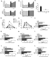
Small molecule analogues (SMAs) of the parasitic worm product ES-62 inhibit PAMP-induced cytokine production. The structures of SMAs 11a (A), 11e (B), 11i (C) and 12b (D) are shown. BmDCs were incubated with SMA (5 μg/ml) for 18 hours before stimulation with LPS, BLP (both 100 ng/ml) or CpG (0.1 μM) and the production of IL-6 (E) TNF-α (F) and IL-12p70 (G) was measured by ELISA. Data are expressed as means (of triplicate determinations) ± SD and analysed using one-way ANOVA with Bonferroni post-test where *p < 0.05; **p < 0.01; ***p < 0.001 compared to relevant PAMP. All data are representative of at least two independent experiments.
Figure 2
Figure 2
ES-62 SMAs target cytokine production at the level of transcription and display differing effects on the activation of MAPKs and NF-κBp65. BmDCs were treated with SMAs 11a, 11e, 11i or 12b (5 μg/ml) for 18 hours, matured with LPS for 4 h, before extracting RNA and analysing the expression levels of IL-12p40 (A), IL-12p35 (B), IL-6 (C) and TNF-α (D) by qRT-PCR. Cytokine expression was normalised to GAPDH and the levels of mRNA in SMA + LPS (from all experiments [n = 3]) compared to LPS (100%) DC calculated and compared using a one sample t test. Results are expressed as the mean ± SEM. BmDCs were treated with SMA 12b for 18 hours and then stimulated with LPS for 10 minutes and the levels of total and phosphorylated p38 (E) and ERK (F) measured by fast-activated cell ELISA (FACE). Phosphorylated p38 or ERK1/2 absorbance were normalised to the corresponding total p38 or ERK1/2 absorbance and the data expressed as means (of triplicate determinations) ± SD. Data were analysed using one way ANOVA with Bonferroni post-test where for statistical analysis, *p < 0.05; **p < 0.01. As above, bmDCs were incubated with SMAs for 18 hours before stimulation with LPS for 30 minutes and the level of p65 activation in duplicate samples measured by TransAM (G). All data are representative of at least two independent experiments.
Figure 3
Figure 3
ES-62 SMAs modulate bmDC priming of T cell responses. BmDCs were incubated with SMAs for 18 hours, stimulated with LPS (100 ng/ml) for 24 hours, and CD40 (A) and CD86 (B) expression by CD11c+ cells measured by flow cytometry in term of % cells (upper panels) and Median Fluorescence Intensity (MFI; lower panels). (C) BmDCs from BALB/c mice pre-incubated with/without SMAs (5 μg/mL), stimulated for 24 hours with LPS and then pulsed with OVA peptide, were co-cultured with naive OVA-specific CD4+ T cells for 3 days before measuring IFN-γ release by ELISA. Inhibitory effects were observed at several peptide concentrations over two independent experiments and the results shown are for 300 μM. Data are presented as means ± SD and were analysed by one way ANOVA with Bonferroni post-test where *p < 0.05; **p < 0.01; ***p < 0.001. For panel (C), “ND” = not detected. BmDCs were incubated with/without SMA 11a or 11i and pulsed with OVA overnight before stimulation ± LPS for 24 hours and injected into BALB/c mice that had received 106 naïve CD4+KJ1.26+ T cells from donor DO11.10 mice 24 h previously. The number (D) and percentage (E) of KJ1.26+ CD4 T cells in the popliteal dLNs of adoptively transferred mice on different days is shown. Data are expressed as the mean percentage of 3 mice per group per day and the error bars (SEM) are omitted for clarity. On days 3 and 5 all treatments except OVA+SMA on day 3 and OVA+11i on day 5 demonstrated a significant increase in the proportion of KJ1.26+CD4+ T cells compared to RPMI DCs alone. The numbers of these T cells on day 5 (F) and their expression of intracellular IL-17 (G,H), IFNγ (I,J) and IL-4 (K,L) on this day were analysed by flow cytometry. Data are expressed as mean ± SEM for 3 mice per group and analysed using Fishers LSD test where *p < 0.05, **p < 0.01 and ***p < 0.001; ****p < 0.0001 compared to RPMI; formula image p < 0.05 compared to OVA DCs and formula image p < 0.05 and formula image p < 0.01 compared to OVA+LPS DCs.
Figure 4
Figure 4
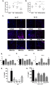
SMA-treated dendritic cells protect against CIA. DBA/1 bmDCs were incubated with SMAs 11a and 12b (2.5 μg/ml of each) and CII overnight, washed, and injected into CIA-mice on day −2, 0 and 21. Disease is shown by mean arthritic score (A) (PBS, n = 3; RPMI-DCs and SMA-DCs, n = 6). Data are expressed as means ± SEM and analysed using an unpaired t test where *p < 0.05 compared to PBS. Joint sections (x10 magnification) from individual mice representative of each treatment group were assessed for histopathology by Trichrome staining (B). CII-specific IgG1 (C) and IgG2a (D) levels in serum samples from naïve (no CII treatment; n = 3), PBS- (n = 3), RPMI DC- (n = 6) and SMA DC- (n = 6) treated CIA mice were determined by ELISA. Results are expressed as mean (of duplicate determinations) for each mouse in the group. The percentage of IL-10+ FOXP3+ CD4+ T cells (E), IL-10+ CD4+ T cells (F) and CD4IL-10+ cells (G) in the DLNs from all paws from CIA-mice treated with PBS, RPMI-DCs or SMA-DCs were analysed by flow cytometry. Results from individual mice in each group are shown; numbers as above. Data were analysed using one-way ANOVA employing Fishers LSD test.
Figure 5
Figure 5
SMA-treated bmDCs inhibit IL-17 and IL-1β responses. The percentage of IL-17-expressing CD4+ cells (A) and IL-17-expressing CD4 cells (B) in the DLNs from paws from naïve or CIA-mice treated with PBS, RPMI-DCs or SMA-DCs was determined by flow cytometry. Results from individual mice in each group (Naïve n = 3, PBS n = 3, RPMI-DCs n = 6 and SMA-DCs n = 6) are shown. Data are compared using one way ANOVA and Fishers LSD test where *p < 0.05 **p < 0.01. (C) Joint sections from individual mice representative of each treatment group were assessed for IL-17 and IL-1β expression by immunofluorescence (magnification 20x; scale bars 20 μm). The strong IL-1β positive staining in the SMA-DCs image (denoted by white arrow) reflects high production of IL-1β by keratinocytes in the skin and is an additional control for antibody specificity. BmDCs were incubated with the indicated SMAs (5 μg/ml) for 4 hours, the RNA was extracted and the expression levels of IL-1β (D), NLRP3 (E) and HMOX-1 (F) measured by qRT-PCR. Cytokine expression was normalised to GAPDH and then expressed as a fold change with respect to the relevant RPMI control. The data shown are collated from 3 independent experiments and presented as means of mean values (triplicate assays in each experiment) ± SEM, n = 3. (G) BmDCs were incubated with the indicated SMAs (5 μg/ml) for 18 hours before stimulation with BLP for 24 hours and the levels of IL-1β measured by ELISA. Data are expressed as means (of triplicate determinations) ± SD. (H) BmDCs were incubated with the indicated SMAs (5 μg/ml) for 18 hours before being primed with LPS (100 ng/ml) for 5 hours then stimulated with 1 mM ATP for 30 minutes and the levels of IL-1β measured by ELISA. Data are expressed as means (of triplicate determinations) ± SD. Data in D-H were analysed using students t-test (or Wilcoxin signed rank test for normalised qRT-PCR data) where *p < 0.05, **p < 0.01, ***p < 0.001, ****p < 0.0001; data in G & H are from single experiments that are representative of at least two independent experiments.

Similar articles

Cited by

References

    1. Kawai T, Akira S. The role of pattern-recognition receptors in innate immunity: update on Toll-like receptors. Nat. Immunol. 2010;11:373–384. doi: 10.1038/ni.1863. - DOI - PubMed
    1. Kumar H, Kawai T, Akira S. Pathogen Recognition by the Innate Immune System. Int. Rev. Immunol. 2011;30:16–34. doi: 10.3109/08830185.2010.529976. - DOI - PubMed
    1. Carvalho L, et al. Review series on helminths, immune modulation and the hygiene hypothesis: Mechanisms underlying helminth modulation of dendritic cell function. Immunology. 2009;126:28–34. doi: 10.1111/j.1365-2567.2008.03008.x. - DOI - PMC - PubMed
    1. MacDonald AS, Straw AD, Bauman B, Pearce EJ. CD8− Dendritic Cell Activation Status Plays an Integral Role in Influencing Th2 Response Development. J. Immunol. 2001;167:1982–1988. doi: 10.4049/jimmunol.167.4.1982. - DOI - PubMed
    1. Falcón C, et al. Excretory-secretory products (ESP) from Fasciola hepatica induce tolerogenic properties in myeloid dendritic cells. Vet. Immunol. Immunopathol. 2010;137:36–46. doi: 10.1016/j.vetimm.2010.04.007. - DOI - PubMed

Publication types

MeSH terms