Review
. 2017:2017:5468023.
doi: 10.1155/2017/5468023. Epub 2017 Apr 8.

Adipokine Contribution to the Pathogenesis of Osteoarthritis

Affiliations
Review

Adipokine Contribution to the Pathogenesis of Osteoarthritis

Daniel Azamar-Llamas et al. Mediators Inflamm. 2017.

Abstract

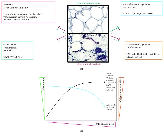
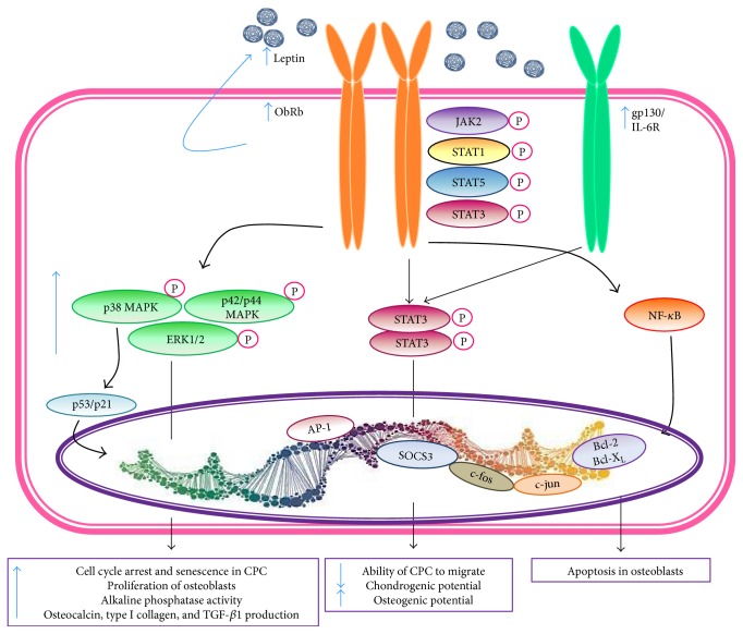
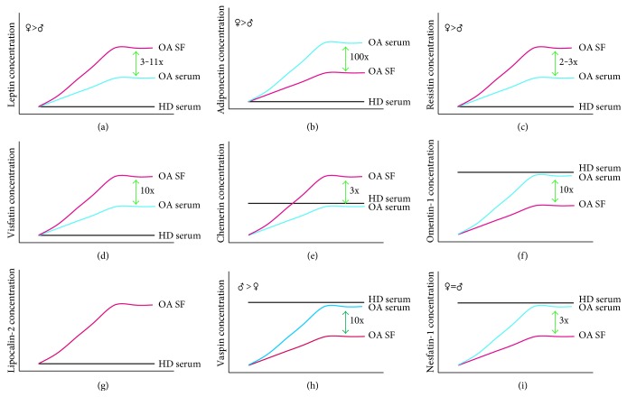
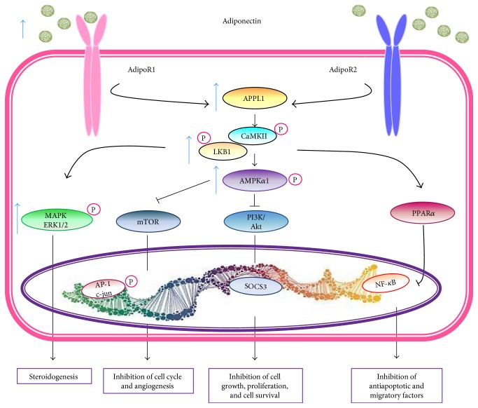
Recent studies have shown that overweight and obesity play an important role in the development of osteoarthritis (OA). However, joint overload is not the only risk factor in this disease. For instance, the presence of OA in non-weight-bearing joints such as the hand suggests that metabolic factors may also contribute to its pathogenesis. Recently, white adipose tissue (WAT) has been recognized not only as an energy reservoir but also as an important secretory organ of adipokines. In this regard, adipokines have been closely associated with obesity and also play an important role in bone and cartilage homeostasis. Furthermore, drugs such as rosuvastatin or rosiglitazone have demonstrated chondroprotective and anti-inflammatory effects in cartilage explants from patients with OA. Thus, it seems that adipokines are important factors linking obesity, adiposity, and inflammation in OA. In this review, we are focused on establishing the physiological mechanisms of adipokines on cartilage homeostasis and evaluating their role in the pathophysiology of OA based on evidence derived from experimental research as well as from clinical-epidemiological studies.


Figures

Figure 1
Figure 1
(a) Soluble mediators synthesized by white adipose tissue. Solid red arrows represent cytokines, growth factors, and hormones produced by obese white adipose tissue. Dotted red lines represent the inhibition of the soluble mediator expression by obese white adipose tissue. Solid green arrows depict endocrine and immune soluble mediators synthesized by lean white adipose tissue. Dotted green lines represent the inhibition of the soluble mediator expression by lean white adipose tissue. (b) Relationship of adipokines with the inflammation and the fat mass index in OA patients. sAdipokine: serum adipokine; BMI: body mass index.
Figure 2
Figure 2
Leptin signal transduction. The Ob receptor b (ObR b) isoform of leptin binds to the JAK-STAT intracellular signaling system. As a consequence of leptin binding to its receptor, JAK2 is activated by the autophosphorylation. STAT1 and STAT5 bind tyrosine residues. STAT3 proteins form dimers and translocate to the nucleus and regulate c-fos, c-jun, SOCS3, and AP1 gene expression. Src homology domains of receptor (SHP2) activate MAPK pathways (p38, p42/44, and ERK1/2). These pathways induce the expression of cytokine and chemokine genes. Moreover, ObRb/leptin also induces the transcription of metalloproteinases and aggrecanases, cartilage degradation proteins, and the signaling pathways of inflammatory cytokines through activation of NF-kB and AP-1 that transcribe the genes of inflammatory proteins (IL-1β, IL-6, TNF-α, and induced nitric oxide synthase among others). Leptin, through interleukin 6 (IL-6)/gp130 pathway activates STAT3, which in the nucleus, transcribes the gene of SOCS3 that suppresses the leptin signaling pathways. Bcl: B cell lymphoma; ERK: extracellular signal-regulated kinase; JAK: c-Jun N-terminal kinase-associated kinase; MAPK: mitogen-activated protein kinase; NF-kB: nuclear factor-kappa B; ObR: Ob receptor; SOCS3: suppressor of cytokine signaling-3; STAT: signal transducer and activator of transcription.
Figure 3
Figure 3
Adipokines in synovial fluid and serum from OA patients and their relative expression compared with healthy individuals. (a) Leptin levels, (b) adiponectin levels, (c) resistin levels, (d) visfatin levels, (e) chemerin levels, (f) omentin-1 levels, (g) lipocalin-2 levels, (h) vaspin levels, and (i) nesfatin-1 levels. Red lines: synovial fluid concentration in patients with OA; blue lines: serum concentration in patients with OA; black lines: serum concentrations in healthy donors; SF: synovial fluid; HD: healthy donors; ♀: female levels; ♂: male levels.
Figure 4
Figure 4
Adiponectin signaling via AdipoR1 and AdipoR2 activation. Adiponectin is decreased in obesity. AdipoRs can lead to stimulation of various signaling pathways. AMPK blocks angiogenesis via mTOR and cell growth and proliferation via PI3K/Akt. Antiapoptotic and migratory proteins induced by p65/p50 of the NF-kB pathway is inhibited by PPAR-α. Adipo R: adiponectin receptors; APPL1: adaptor protein containing pleckstrin homology domain, phosphotyrosine-binding domain, and leucine zipper motif 1; PPAR-α: peroxisome proliferator-activated receptor α; AMPK: 5′-adenosine monophosphate-activated protein kinase; MAPK: mitogen-activated protein kinase; ERK1/2: extracellular signal-regulated kinases 1/2; SOCS3: suppressor of cytokine signaling-3; mTOR: mammalian target of rapamycin; LKB1: liver kinase B1.
Figure 5
Figure 5
Resistin signaling. Resistin is recognized by TLR4 receptor. Two signaling pathways are triggered through the recruitment of the adaptor molecules TIRAP and MyD88. The first through PI3K followed by Akt and NF-kB. The second through MAPK pathway, followed by upregulation of NF-kB.
Figure 6
Figure 6
Visfatin signaling. Visfatin stimulates monocytes to release IL-6. IL-6 signals increase the expression level of STAT3 which upregulates the active enzymatic form of visfatin/PBEF/Nampt. Visfatin/PBEF/Nampt can increase cell survival through Sirt-1 and Sirt-6 stimulating the release of TNF-α inducing a chronic low grade inflammation. In the second pathway, visfatin signals through the cells surface receptor β1 integrin. This binding upregulates and activates p38 MAPK and ERK1/2. The MAPK cascade increases the expression of AP-1 and NF-kB that upregulate SDF-1, leading to increased survival and migration. The third pathway was demonstrated through the activation of unknown receptor increasing the antioxidative enzymes superoxide dismutase (SOD) and catalase (CAT).
Figure 7
Figure 7
Chemerin signaling. Chemerin binds to three different G protein-coupled receptors: CMKLR1 (chemokine-like receptor 1), GPR1 (G protein-coupled receptor 1), and CCRL2 (chemokine (CC motif) receptor-like 2). The latter does not transduce any signal; once activated, CMKLR1 and GPR1 stimulate or inhibit different signaling pathways including MAPK ERK1/2, Akt, and AMPK to regulate different biological processes such as angiogenesis, inflammation, and steroidogenesis.
Figure 8
Figure 8
Omentin signaling. Omentin activates AMPK, which further blocks E-selectin expression and reduces endothelial inflammation. AMPK also activates endothelial nitric oxide (eNOS), also known as nitric oxide synthase 3 (NOS3) or constitutive NOS (cNOS), which has vasodilation effect and blocks JNK signaling. JNK activates inflammation through TNF-α-mediated COX2 effect. Moreover, omentin inhibits NF-κB signaling pathway and thus inhibits inflammation.
Figure 9
Figure 9
Vaspin signaling. Vaspin binds its receptor, glucose-regulated protein 78 (GRP78) GRP78, and activates the expression of Bcl-2 and downregulates that of Bax. Moreover, vaspin stimulates the PI3K signaling pathway with a specific phosphorylation of ERK and AKT. Vaspin has antiapoptotic effects in vascular endothelial cells and human osteoblasts.
Figure 10
Figure 10
Expression of adipokines in large and small joints. Hand: Different studies have demonstrated that adiponectin may have a protective role in knee OA and it may be related to erosive hand OA [163, 164, 166]. Choe et al. showed that serum levels of resistin correlate with radiographic changes, in specific with subchondral erosions but with no pain [173]. Knee: there are multiple studies that show a higher level of leptin in synovial fluid and serum, and this correlates with the damage of the joint. The higher SF levels are thought to be related to the presence of infrapatellar fat (IFP) pad that produces leptin locally [215, 216]. Resistin and visfatin are produced by the IFP, and their levels correlate with joint damage and the levels of IL-6. Resistin also correlates with menisci damage. Hip: even though the hip and knee are under similar stress conditions, the clinical features and the adipocytokine profile are different, with lower levels of leptin and higher adiponectin, resistin, and visfatin levels within the joint. Only the levels of visfatin correlate with hip pain [140]. Shoulder: the leptin and adiponectin levels correlate with joint damage, but appear to have a different profile of adipocytokines in shoulder, with lower SF and serum levels of leptin and adiponectin, but a higher serum/SF ratio for both, especially adiponectin [168].

Similar articles

Cited by

References

    1. Hart D. J., Doyle D. V., Spector T. D. Association between metabolic factors and knee osteoarthritis in women: the Chingford study. The Journal of Rheumatology. 1995;22(6):1118–1123. - PubMed
    1. Aspden R. M., Scheven B. A., Hutchison J. D. Osteoarthritis as a systemic disorder including stromal cell differentiation and lipid metabolism. Lancet. 2001;357(9262):1118–1120. doi: 10.1016/S0140-6736(00)04264-1. - DOI - PubMed
    1. Parimisetty A., Dorsemans A. C., Awada R., Ravanan P., Diotel N., Lefebvre d'Hellencourt C. Secret talk between adipose tissue and central nervous system via secreted factors-an emerging frontier in the neurodegenerative research. Journal of Neuroinflammation. 2016;13(1):p. 67. doi: 10.1186/s12974-016-0530-x. - DOI - PMC - PubMed
    1. Maury E., Ehala-Aleksejev K., Guiot Y., Detry R., Vandenhooft A., Brichard S. M. Adipokines oversecreted by omental adipose tissue in human obesity. American Journal of Physiology. Endocrinology and Metabolism. 2007;293(3):E565–E565. doi: 10.1152/ajpendo.00127.2007. - DOI - PubMed
    1. Gierman L. M., van der Ham F., Koudijs A., et al. Metabolic stress-induced inflammation plays a major role in the development of osteoarthritis in mice. Arthritis and Rheumatism. 2012;64(4):1172–1181. doi: 10.1002/art.33443. - DOI - PubMed

LinkOut - more resources