. 2017 Mar-Apr;22(2):181-192.
doi: 10.1016/j.rpor.2017.02.007. Epub 2017 Apr 14.

Other non-surgical treatments for liver cancer

Affiliations

Other non-surgical treatments for liver cancer

Paul Revel-Mouroz et al. Rep Pract Oncol Radiother. 2017 Mar-Apr.

Abstract

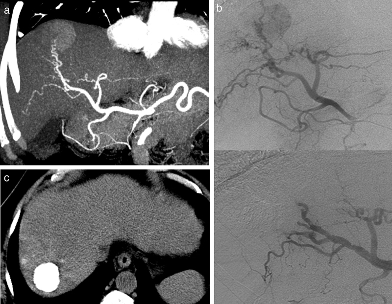
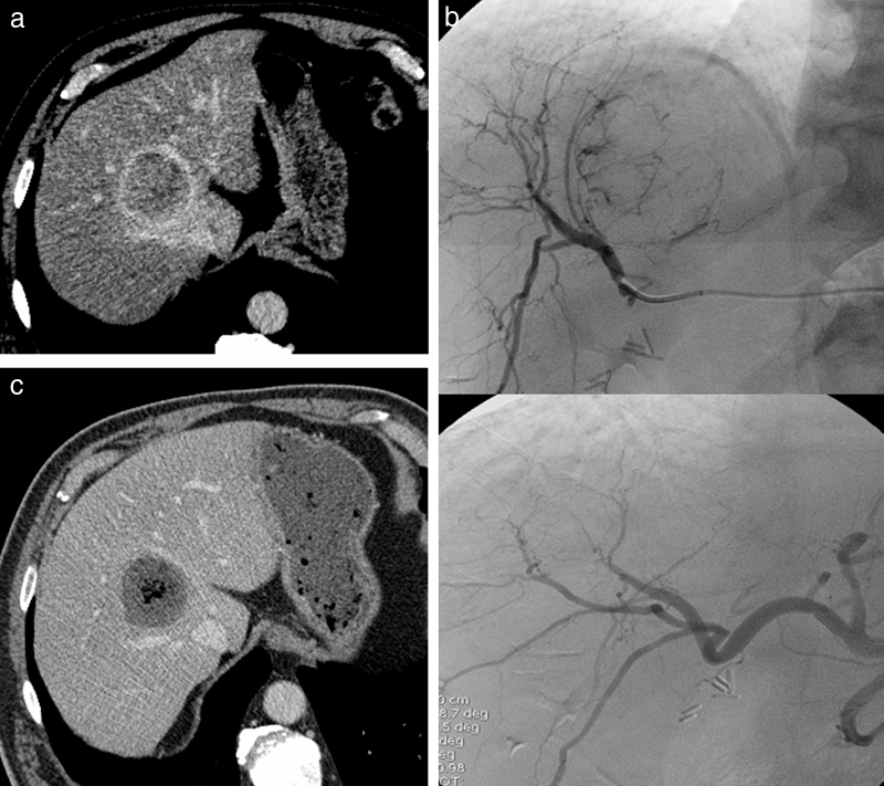
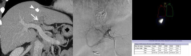
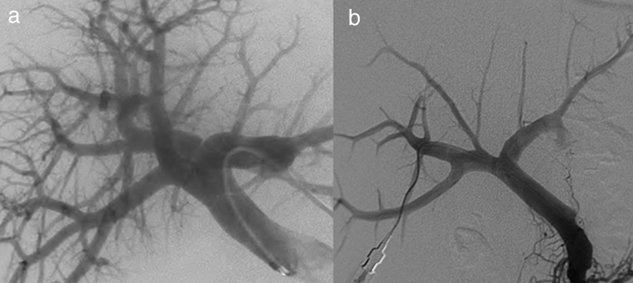
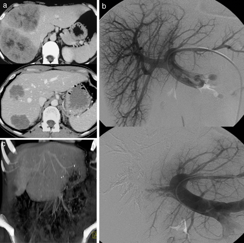
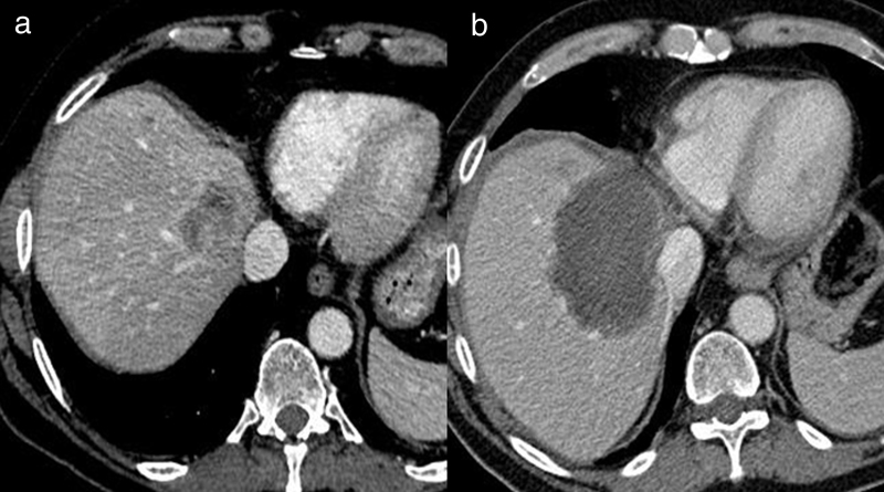
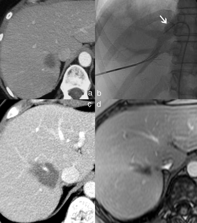
Interventional radiology plays a major role in the modern management of liver cancers, in primary hepatic malignancies or metastases and in palliative or curative situations. Radiological treatments are divided in two categories based on their approach: endovascular treatment and direct transcapsular access. Endovascular treatments include mainly three applications: transarterial chemoembolization (TACE), transarterial radioembolization (TARE) and portal vein embolization (PVE). TACE and TARE share an endovascular arterial approach, consisting of a selective catheterization of the hepatic artery or its branches. Subsequently, either a chemotherapy (TACE) or radioembolic (TARE) agent is injected in the target vessel to act on the tumor. PVE raises the volume of the future liver remnant in extended hepatectomy by embolizing a portal vein territory which results in hepatic regeneration. Direct transcapsular access treatments involve mainly three techniques: radiofrequency thermal ablation (RFA), microwave thermal ablation (MWA) and percutaneous ethanol injection (PEI). RFA and MWA procedures are almost identical, their clinical applications are similar. A probe is deployed directly into the tumor to generate heat and coagulation necrosis. PEI has known implications based on the chemical toxicity of intra-tumoral injection with highly concentrated alcohol by a thin needle.

Keywords: Microwave thermal ablation; Percutaneous ethanol injection; Portal vein embolization; Radiofrequency thermal ablation; Transarterial chemoembolization; Transarterial radioembolization.


Figures

Fig. 1
Fig. 1
Transarterial oily-chemoembolization for hepatocellular carcinoma. (a) Enhanced CT at the arterial phase (same case as in Fig. 3). Hepatocellular carcinoma with arterial enhancement. (b) Corresponding selective angiography of the right branch of the hepatic artery shows tumoral blush (top). Subsequently, a transarterial oily-chemoembolization is performed. The control angiography assessed at the end of the procedure (bottom image) shows the disappearance of the blush. (c) Abdominal non-enhanced CT performed 6 weeks later, shows an homogeneous and intense lipiodol uptake of the tumor.
Fig. 2
Fig. 2
Transarterial chemoembolization for hepatocellular carcinoma with drug-eluting beads. (a) Enhanced CT at the venous phase, showing an HCC with wash-out. (b) Selective catheterization and opacification of the right branch of the hepatic artery (top), showing the branches feeding the tumor. The angiography after DC Bead injection (bottom) shows the disappearance of the tumoral blush. (c) Enhanced CT at the portal venous phase 6 weeks later, showing a complete tumoral necrosis.
Fig. 3
Fig. 3
Transarterial radioembolization for HCC with 131I-Lipiodol®. (a) Enhanced CT at the arterial phase, MPR reconstructions showing a large heterogeneous hepatocellular carcinoma with marked enhancement. There is a large extension of the tumor into the portal vein (arrow). (b) Enhanced CT at the portal arterial phase, performed 6 weeks after a transarterial radioembolization with 131I-Lipiodol®. MPR reconstructions shows an heterogeneous lipiodol uptake, a shrinkage of the tumor and a substantial reduction of the extension of the tumor into the portal vein.
Fig. 4
Fig. 4
Transarterial radioembolization for hepatocellular carcinoma with yttrium-90-labeled microspheres. (a) Enhanced CT at the portal venous phase, performed in order to evaluate the effectiveness of a transarterial chemoembolization for a hepatocellular carcinoma. The primitive tumor shows an incomplete lipiodol uptake (arrowhead) and a portal vein tumor thrombosis (arrow). (b) Selective opacification of the artery feeding the segment IV of the liver, as 1st step of the radioembolization, consisting of the injection of a low dose of 99mTc-labeled macroaggregated albumin. (c) Immediately after the infusion of the 99mTc-labeled macroaggregated albumin, a single-photon emission computed tomography is performed, which also allows to plan the therapeutic dose of 90Y after checking the lack of pulmonary and gastro-intestinal shunting.
Fig. 5
Fig. 5
Portal vein embolization of the right liver. The procedure can be performed either with a contralateral (a) or a homolateral approach (b).
Fig. 6
Fig. 6
Portal vein embolization of the right liver for colorectal cancer metastasis. (a) Enhanced CT at the portal venous phase before (top) and after (bottom) a systemic chemotherapy treatment showing a good response. The patient could be eligible to a curative right hepatectomy provided that the future liver remnant reaches a sufficient volume. (b) Portal vein embolization of the right liver assessed with a contralateral approach. (c) Enhanced CT at the portal venous phase, volume rendering shows a hypertrophied left lobe allowing surgical resection.
Fig. 7
Fig. 7
Overcoming the heat-sink effect in radiofrequency thermal ablation: a peroperative approach. (a) Enhanced CT at the portal venous phase, showing a colic metastasis in the upper aspect of the right liver, whose posterior aspect has close contact with the right hepatic vein. Patient also required a left lobectomy. Peroperative thermal ablation of the right lesion was performed under Pringle maneuver (clamping of the hepatoduodenal ligament) with a 4-cm diameter needle, to overcome the heat-sink effect. (b) Abdominal CT follow-up at two months shows a complete devascularized thermal lesion, larger than expected.
Fig. 8
Fig. 8
Overcoming the heat-sink effect in radiofrequency thermal ablation: a percutaneous approach. (a) Enhanced CT at the portal venous phase showing a breast metastasis whose posterior aspect has close contact with the right hepatic vein. (b) Selective occlusion of the right hepatic vein with a Fogarty balloon during the application of the thermal ablation. (c) Abdominal CT follow-up at three months showing an homogeneous thermal lesion without enhancement. The right hepatic vein remains patent. (d) Liver MRI follow-up at two years, showing the shrinkage of the thermal lesion, without enhancement.

References

    1. Wang P.M., Chung N.N., Hsu W.C., Chang F.L., Jang C.J., Scorsetti M. Stereotactic body radiation therapy in hepatocellular carcinoma: optimal treatment strategies based on liver segmentation and functional hepatic reserve. Rep Pract Oncol Radiother. 2015;20(November–December (6)):417–424. - PMC - PubMed
    1. Comito T., Clerici E., Tozzi A., D’agostino G. Liver metastases and SBRT: a new paradigm? Rep Pract Oncol Radiother. 2015;20(November–December (6)):464–471. - PMC - PubMed
    1. Mondazzi L., Bottelli R., Brambilla G. Transarterial oily chemoembolization for the treatment of hepatocellular carcinoma: a multivariate analysis of prognostic factors. Hepatology. 1994;19(May (5)):1115–1123. - PubMed
    1. Dumortier J., Chapuis F., Borson O. Unresectable hepatocellular carcinoma: survival and prognostic factors after lipiodol chemoembolisation in 89 patients. Dig Liver Dis. 2006;38(February (2)):125–133. - PubMed
    1. Tam K.Y., Leung K.C., Wang Y.X. Chemoembolization agents for cancer treatment. Eur J Pharm Sci. 2011;44(September (1–2)):1–10. - PubMed