. 2017 Apr 26:8:502.
doi: 10.3389/fimmu.2017.00502. eCollection 2017.

ISA-2011B, a Phosphatidylinositol 4-Phosphate 5-Kinase α Inhibitor, Impairs CD28-Dependent Costimulatory and Pro-inflammatory Signals in Human T Lymphocytes

Affiliations

ISA-2011B, a Phosphatidylinositol 4-Phosphate 5-Kinase α Inhibitor, Impairs CD28-Dependent Costimulatory and Pro-inflammatory Signals in Human T Lymphocytes

Martina Kunkl et al. Front Immunol. .

Abstract

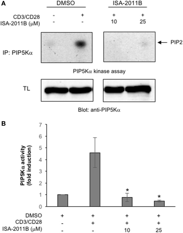
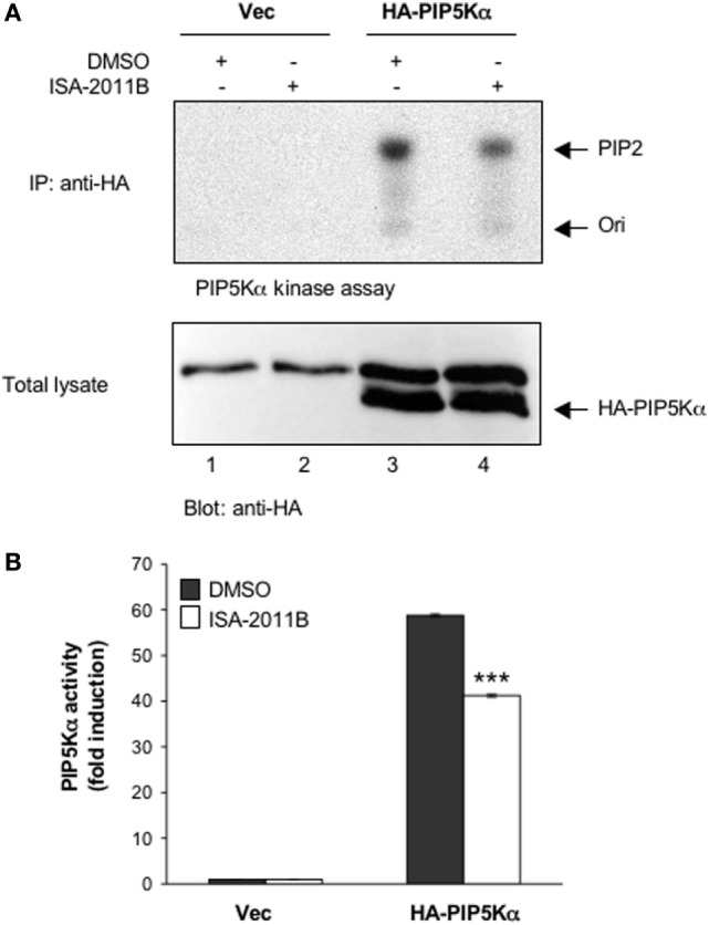
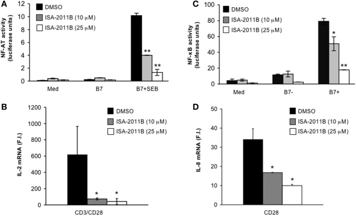
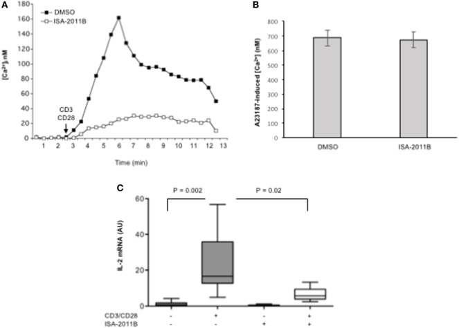
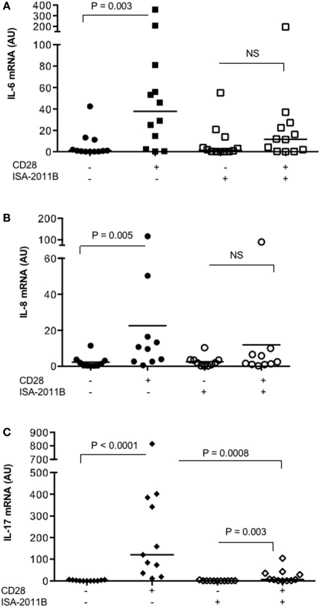
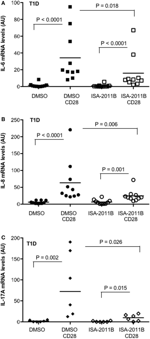
Phosphatidylinositol 4,5-biphosphate (PIP2) is a membrane phospholipid that controls the activity of several proteins regulating cytoskeleton reorganization, cytokine gene expression, T cell survival, proliferation, and differentiation. Phosphatidylinositol 4-phosphate 5-kinases (PIP5Ks) are the main enzymes involved in PIP2 biosynthesis by phosphorylating phosphatidylinositol 4-monophosphate (PI4P) at the D5 position of the inositol ring. In human T lymphocytes, we recently found that CD28 costimulatory molecule is pivotal for PIP2 turnover by recruiting and activating PIP5Kα. We also found that PIP5Kα is the main regulator of both CD28 costimulatory signals integrating those delivered by TCR as well as CD28 autonomous signals regulating the expression of pro-inflammatory genes. Given emerging studies linking alterations of PIP2 metabolism to immune-based diseases, PIP5Kα may represent a promising target to modulate immunity and inflammation. Herewith, we characterized a recently discovered inhibitor of PIP5Kα, ISA-2011B, for its inhibitory effects on T lymphocyte functions. We found that the inhibition of PIP5Kα lipid-kinase activity by ISA-2011B significantly impaired CD28 costimulatory signals necessary for TCR-mediated Ca2+ influx, NF-AT transcriptional activity, and IL-2 gene expression as well as CD28 autonomous signals regulating the activation of NF-κB and the transcription of pro-inflammatory cytokine and chemokine genes. Moreover, our data on the inhibitory effects of ISA-2011B on CD28-mediated upregulation of inflammatory cytokines related to Th17 cell phenotype in type 1 diabetes patients suggest ISA-2011B as a promising anti-inflammatory drug.

Keywords: CD28 co-stimulation; PIP5K; T lymphocytes; T1D; pro-inflammatory cytokines.


Figures

Figure 1
Figure 1
ISA-2011B inhibits phosphatidylinositol 4-phosphate 5-kinase (PIP5K)α lipid-kinase activity in primary T cells. (A) Primary T cells from healthy donors (HD) were treated for 6 h with DMSO or the indicated concentrations of ISA-2011B and then unstimulated (ctr) or stimulated for 5 min with saturating concentration of anti-CD3 (5 µg/ml) plus anti-CD28 Abs crosslinked with goat anti-mouse (20 µg/ml). PIP5K kinase assays were performed on anti-PIP5Kα immunoprecipitations and the reaction products were subjected to thin-layer chromatography followed by autoradiography (upper panel). An equal amount of cell lysate was analyzed for PIP5Kα content by Western blotting (lower panel). (B) Fold inductions were quantified by densitometric analysis and normalized to PIP5Kα levels. Bars show the mean ± SD of two experiments. Asterisks (*) indicate P < 0.05 calculated by Student’s t-test.
Figure 2
Figure 2
ISA-2011B inhibits exogenously expressed PIP5Kα lipid-kinase activity. (A) Jurkat cells were transfected and cultured for 24 h with 20 µg control empty vector (Vec) or 20 µg HA-PIP5Kα and then treated for further 6 h with DMSO or 25 µM ISA-2011B. PIP5Kα kinase assays performed on anti-HA IP (upper panel). HA-PIP5Kα content was analyzed by western blotting (lower panel). (B) Fold inductions were quantified by densitometric analysis and normalized to HA-PIP5Kα levels. Data are expressed as fold induction over the basal level of cells transfected with empty vector and treated with DMSO. Bars show the mean ± SD of two experiments. Asterisks (***) indicate P < 0.001 calculated by Student’s t-test, compared with cells transfected with HA-PIP5Kα and treated with DMSO.
Figure 3
Figure 3
ISA-2011B impairs CD28 costimulatory functions in Jurkat T cells. (A) NF-AT luciferase activity of Jurkat cells treated for 6 h with DMSO or the indicated concentration of ISA-2011B and stimulated in the absence (med) or presence of 5-3.1/B7 (B7) cells pre-pulsed or not with 1 µg/ml SEB. The results are expressed as the mean of luciferase units ± SD after normalization to GFP values. The data are representative of three independent experiments. (B,D) Real-time PCR was used to measure IL-2 mRNA (B) and IL-8 mRNA levels (D) in Jurkat cells treated with DMSO or the indicated concentration of ISA-2011B and stimulated for 6 h with control IgG or anti-CD28 Abs (D) or anti-CD3 plus anti-CD28 Abs (B). Data are expressed as fold inductions (F.I.) over the basal level of cells stimulated with control IgG and treated with DMSO. Bars show the mean ± SD of three independent experiments. (C) NF-κB luciferase activity of Jurkat cells treated for 6 h with DMSO or the indicated concentration of ISA-2011B and stimulated in the absence (med) or presence of B7-negative (%-3.1) or B7-positive (5-3.1/B7) cells. The results are expressed as the mean of luciferase units ± SD after normalization to GFP values. The data are representative of three independent experiments. Asterisks (*) and (**) indicate P < 0.05 and P < 0.01, respectively, calculated by Student’s t-test.
Figure 4
Figure 4
ISA-2011B impairs TCR- and CD28-induced increase of [Ca2+]i and IL-2 gene expression in primary T cells. (A,B) Primary T cells from HD were treated for 6 h with DMSO or ISA-2011B (10 µM) and after loading T cells with Fluo-3-AM, Ca2+ levels ([Ca2+]i) were measured by cytofluorimetric analysis after stimulation with crosslinked anti-CD3 plus anti-CD28 Abs (A) or A23871 (2 µg/ml) for 10 min. Data are representative of three independent experiments. (C) Real-time PCR was used to measure IL-2 mRNA levels in primary T cells from healthy donors (n = 5) treated with DMSO or ISA-2011B (10 µg/ml) and stimulated for 6 h with crosslinked anti-CD3 plus anti-CD28 Abs. Values, normalized on GAPDH, are expressed as arbitrary units. Median values: DMSO = 0.55; DMSO CD3 plus CD28 = 16.59; ISA-2011B = 0.25: ISA-2011B CD3 plus CD28 = 5.68. Lines represent median values and P values are indicated where significant (Mann–Whitney).
Figure 5
Figure 5
ISA-2011B impairs CD28 autonomous signals inducing the expression of pro-inflammatory cytokine/chemokine. Primary T cells from healthy donors subjects (n = 12) were treated with DMSO or ISA-2011B (10 µM) and stimulated for 6 h (A,B) or 24 h (C) with control isotype-matched mAb or crosslinked anti-CD28.2 Abs. IL-6 (A), IL-8 (B), and IL-17A (C) mRNA levels were measured by real-time PCR and values, normalized on GAPDH, expressed as arbitrary units. Median values: IL-6, DMSO = 0.89, DMSO CD28 = 37.66, ISA-2011B = 1.33, ISA-2011B CD28 = 11.66; IL-8, DMSO = 0.9, DMSO CD28 = 10.19, ISA-2011B = 1.9, ISA-2011B CD28 = 2.0; IL-17A, DMSO = 0.76, DMSO + CD28 = 120.4, ISA-2011B = 0.69, ISA-2011B CD28 = 7.0. Lines represent median values and P values are indicated where significant (Mann–Whitney). NS = not significant.
Figure 6
Figure 6
PIP5Kα inhibition by ISA-2011B impairs CD28-mediated upregulation of pro-inflammatory cytokines in primary T cells from type 1 diabetes (T1D) patients. (A) Primary T cells from T1D patients subjects were treated with DMSO or ISA-2011B (10 µg/ml) and stimulated for 6 h (A,B) or 24 h (C) with control isotype-matched mAb or crosslinked anti-CD28.2 Abs (T1D CD28). IL-6 (A), IL-8 (B), and IL-17A (C) mRNA levels were measured by real-time PCR and values, normalized on GAPDH, expressed as arbitrary units. Median values: IL-6 (n = 10), DMSO = 0.77, DMSO CD28 = 18.99, ISA-2011B = 0.26, ISA-2011B CD28 = 8.27; IL-8 (n = 10), DMSO = 4, DMSO CD28 = 37.74, ISA-2011B = 4.75, ISA-2011B CD28 = 20.3; IL-17A (n = 6), DMSO = 1.18, DMSO CD28 = 72.42, ISA-2011B = 0.27, ISA-2011B CD28 = 10.32. Lines represent mean values and P values are indicated where significant (Mann–Whitney).

Similar articles

Cited by

References

    1. Van Den Bout I, Divecha N. PIP5K-driven PtdIns(4,5)P2 synthesis: regulation and cellular functions. J Cell Sci (2009) 122(Pt 21):3837–50.10.1242/jcs.056127 - DOI - PubMed
    1. Kallikourdis M, Trovato AE, Roselli G, Muscolini M, Porciello N, Tuosto L, et al. Phosphatidylinositol 4-phosphate 5-kinase β controls recruitment of lipid rafts into the immunological synapse. J Immunol (2016) 196(4):1955–63.10.4049/jimmunol - DOI - PubMed
    1. Muscolini M, Camperio C, Capuano C, Caristi S, Piccolella E, Galandrini R, et al. Phosphatidylinositol 4-phosphate 5-kinase alpha activation critically contributes to CD28-dependent signaling responses. J Immunol (2013) 190(10):5279–86.10.4049/jimmunol.1203157 - DOI - PubMed
    1. Muscolini M, Camperio C, Porciello N, Caristi S, Capuano C, Viola A, et al. Phosphatidylinositol 4-phosphate 5-kinase alpha and Vav1 mutual cooperation in CD28-mediated actin remodeling and signaling functions. J Immunol (2015) 194(3):1323–33.10.4049/jimmunol.1401643 - DOI - PubMed
    1. Porciello N, Kunkl M, Viola A, Tuosto L. Phosphatidylinositol 4-phosphate 5-kinases in the regulation of T cell activation. Front Immunol (2016) 7:186.10.3389/fimmu.2016.00186 - DOI - PMC - PubMed