

Diagnosis of abnormally invasive posterior placentation: the role of MR imaging
- PMID: 28491174
- PMCID: PMC5417735
- DOI: 10.1016/j.radcr.2017.01.014
Diagnosis of abnormally invasive posterior placentation: the role of MR imaging
Abstract
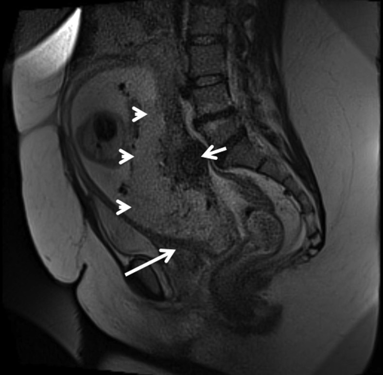
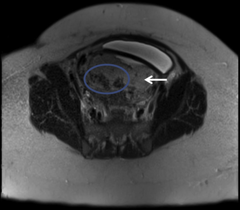
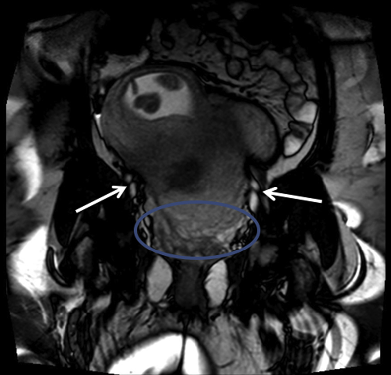
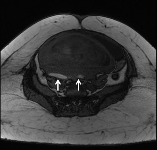
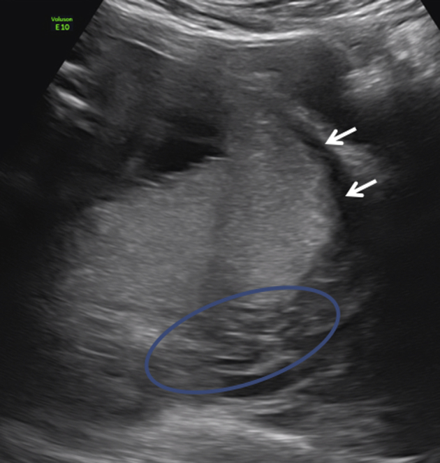
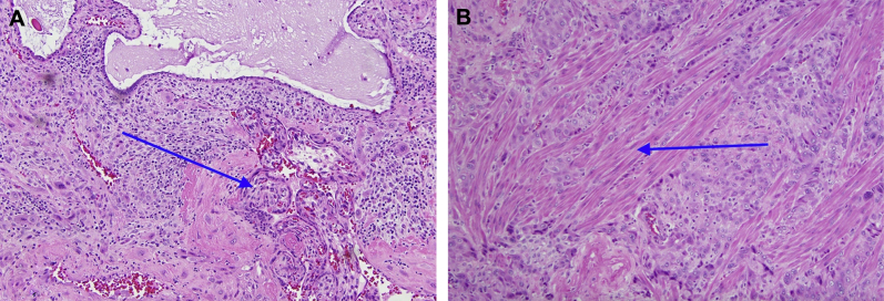
Abnormally invasive placentation is becoming more common with a recent increase in cesarean sections and maternal age, among other risk factors. Ultrasonography is the first line-imaging, but it can be difficult to diagnose when limiting factors are present. Failure to recognize this serious placental abnormality precludes us from making the appropriate plan for the delivery and consequently can lead to fatal results. In this report, we present a case in which magnetic resonance imaging was used to diagnose posterior placenta increta missed by multiple sonographic examinations in a patient with previous myomectomies, and we also include a review of the literature on this topic. It is our conclusion that magnetic resonance imaging is superior to sonography to diagnose abnormally invasive placentation in cases of posterior placenta previa and high pretesting probability.
Keywords: Abnormal invasive placentation; Magnetic resonance imaging; Posterior placenta previa.
Figures
Similar articles
-
Abnormal placentation.Semin Perinatol. 2019 Feb;43(1):51-59. doi: 10.1053/j.semperi.2018.11.009. Epub 2018 Nov 15. Semin Perinatol. 2019. PMID: 30578147 Review.
-
Maternal outcome after conservative management of abnormally invasive placenta.Taiwan J Obstet Gynecol. 2017 Jun;56(3):353-357. doi: 10.1016/j.tjog.2017.04.016. Taiwan J Obstet Gynecol. 2017. PMID: 28600047
-
Placenta percreta is associated with more frequent severe maternal morbidity than placenta accreta.Am J Obstet Gynecol. 2018 Aug;219(2):193.e1-193.e9. doi: 10.1016/j.ajog.2018.04.049. Epub 2018 May 5. Am J Obstet Gynecol. 2018. PMID: 29733839
-
The gravid uterus: MR imaging and reporting of abnormal placentation.Abdom Radiol (NY). 2016 Dec;41(12):2411-2423. doi: 10.1007/s00261-016-0752-5. Abdom Radiol (NY). 2016. PMID: 27472936 Review.
-
Management of anesthesia for cesarean section in parturients with placenta previa with/without placenta accreta: a retrospective study.Ginekol Pol. 2012 Feb;83(2):99-103. Ginekol Pol. 2012. PMID: 22568353
Cited by
-
Identification of suspicious invasive placentation based on clinical MRI data using textural features and automated machine learning.Eur Radiol. 2019 Nov;29(11):6152-6162. doi: 10.1007/s00330-019-06372-9. Epub 2019 Aug 23. Eur Radiol. 2019. PMID: 31444599
-
Management of Myomectomy Scar Pregnancy: A Scoping Review.Medicina (Kaunas). 2025 Apr 29;61(5):817. doi: 10.3390/medicina61050817. Medicina (Kaunas). 2025. PMID: 40428776 Free PMC article.
References
-
- O'Brien J.M., Barton J.R., Donaldson E.S. The management of placenta percreta: conservative and operative strategies. Am J Obstet Gynecol. 1996;175(6):1632–1638. - PubMed
-
- Aitken K., Allen L., Pantazi S., Kingdom J., Keating S., Pollard L. MRI significantly improves disease staging to direct surgical planning for abnormal invasive placentation: a single centre experience. J Obstet Gynaecol Can. 2016;38(3):246–251.e1. - PubMed
-
- Balcacer P., Pahade J., Spektor M., Staib L., Copel J.A., McCarthy S. Magnetic resonance imaging and sonography in the diagnosis of placental invasion. J Ultrasound Med. 2016;35(7):1445–1456. - PubMed
-
- D'Antonio F., Palacios-Jaraquemada J., Lim P.S., Forlani F., Lanzone A., Timor-Tritsch I. Counseling in fetal medicine: evidence-based answers to clinical questions on morbidly adherent placenta. Ultrasound Obstet Gynecol. 2016;47(3):290–301. - PubMed
-
- Shetty M.K., Dryden D.K. Morbidly adherent placenta: ultrasound assessment and supplemental role of magnetic resonance imaging. Semin Ultrasound CT MR. 2015;36(4):324–331. - PubMed
Publication types
LinkOut - more resources
Full Text Sources
Other Literature Sources
