

MRI characteristics of primary fallopian tube choriocarcinoma: a case report
- PMID: 28491175
- PMCID: PMC5417728
- DOI: 10.1016/j.radcr.2017.01.015
MRI characteristics of primary fallopian tube choriocarcinoma: a case report
Abstract
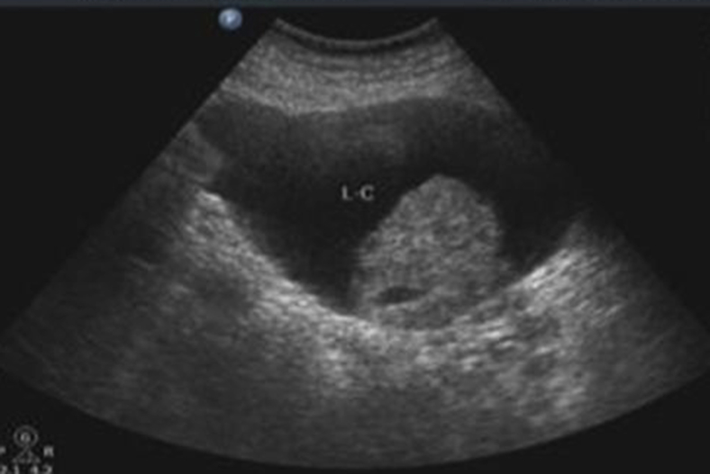
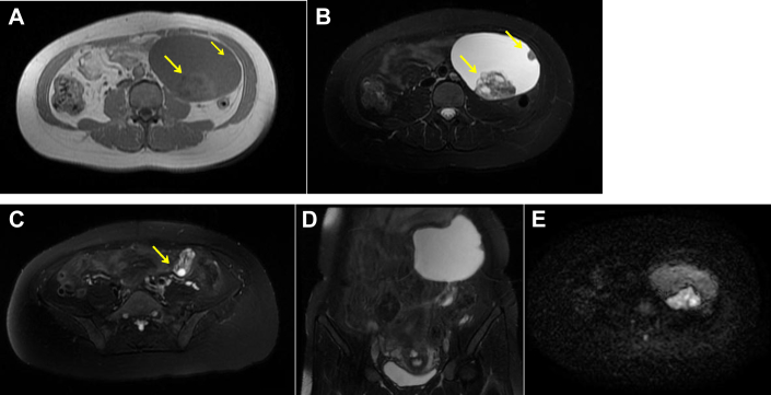
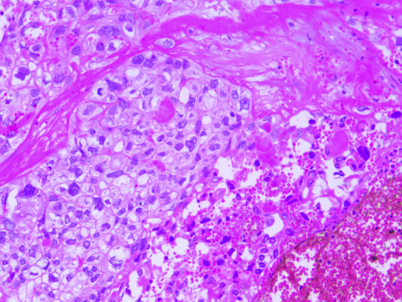
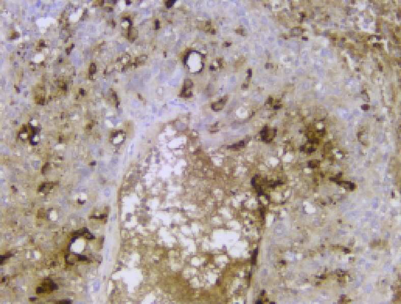
Tubal choriocarcinoma is uncommon, and its magnetic resonance imaging characteristics have not yet been reported. In this report, a 39-year-old woman presented with irregular painless vaginal bleeding and a palpable left lower abdominal lump for 2 months following 6 weeks' amenorrhea and positive urine pregnancy test. Her serum β-human chorionic gonadotropin value was significantly increased. Ultrasound revealed a left adnexal mass, which showed no blood flow signal on Color doppler flow imaging. A further MR examination showed a well-defined cystic-solid mass with cystic component accounting for a large proportion in the left lower abdomen. The solid part with mixed signals resembled a honeycomb. Finally, the left tubal choriocarcinoma was confirmed by pathology. When the solid parts of cystic-solid mass appeared as "honeycomb appearance" and the ovaries were normal by magnetic resonance imaging, together with typical symptoms and significantly elevated β-human chorionic gonadotropin values, radiologists should feel more confident in suspecting tubal choriocarcinoma and reporting it on their differential.
Keywords: Choriocarcinoma; Fallopian tube; MRI.
Figures
Similar articles
-
Choriocarcinoma in tubal pregnancy: A case report.Clin Case Rep. 2023 Sep 27;11(10):e7977. doi: 10.1002/ccr3.7977. eCollection 2023 Oct. Clin Case Rep. 2023. PMID: 37780932 Free PMC article.
-
Primary choriocarcinoma of the fallopian tube: a case report and literature review.Eur J Gynaecol Oncol. 2014;35(5):604-7. Eur J Gynaecol Oncol. 2014. PMID: 25423716 Review.
-
Extrauterine Choriocarcinoma in the Fallopian Tube Following Infertility Treatment: Implications for the Management of Early-Detected Ectopic Pregnancies.J Minim Invasive Gynecol. 2017 Jul-Aug;24(5):855-858. doi: 10.1016/j.jmig.2017.03.006. Epub 2017 Mar 14. J Minim Invasive Gynecol. 2017. PMID: 28315411
-
Primary Tubal Choriocarcinoma Presented as Ruptured Ectopic Pregnancy.J Clin Diagn Res. 2015 Sep;9(9):QD17-8. doi: 10.7860/JCDR/2015/15828.6534. Epub 2015 Sep 1. J Clin Diagn Res. 2015. PMID: 26500968 Free PMC article.
-
Gestational choriocarcinoma of the fallopian tube.Diagn Gynecol Obstet. 1981 Fall;3(3):213-31. Diagn Gynecol Obstet. 1981. PMID: 7035113 Review.
Cited by
-
Gestational Tubal Choriocarcinoma Presenting as a Pregnancy of Unknown Location following Ovarian Induction.Case Rep Obstet Gynecol. 2018 May 3;2018:4705192. doi: 10.1155/2018/4705192. eCollection 2018. Case Rep Obstet Gynecol. 2018. PMID: 29854512 Free PMC article.
-
A rare case of pure non-gestational ovarian choriocarcinoma: Diagnostic mimicry and management strategies.Oncoscience. 2025 Jul 28;12:70-78. doi: 10.18632/oncoscience.622. eCollection 2025. Oncoscience. 2025. PMID: 40735309 Free PMC article.
-
Choriocarcinoma in tubal pregnancy: A case report.Clin Case Rep. 2023 Sep 27;11(10):e7977. doi: 10.1002/ccr3.7977. eCollection 2023 Oct. Clin Case Rep. 2023. PMID: 37780932 Free PMC article.
-
Rare non-serous fallopian tube cancers: institutional experience and literature review.Wien Med Wochenschr. 2024 Jun;174(9-10):199-207. doi: 10.1007/s10354-023-01027-3. Epub 2023 Nov 28. Wien Med Wochenschr. 2024. PMID: 38015299 Review. English.
References
-
- Rettenmaier M.A., Khan H.J., Epstein H.D., Nguyen D., Abaid L.N., Goldstein B.H. Gestational choriocarcinoma in the fallopian tube. J Obstet Gynaecol. 2013;33(8):912–914. - PubMed
-
- Ubayasiri K., Hancock B., Duncan T. A case of primary choriocarcinoma of the fallopian tube. J Obstet Gynaecol. 2010;30(8):881–883. - PubMed
-
- Davies J., Butler R., Chadha Y., Singh M. Primary tubal choriocarcinoma. J Clin Pathol. 2010;63(12):1130–1132. - PubMed
Publication types
LinkOut - more resources
Full Text Sources
Other Literature Sources
