

Pyogenic brain abscess with atypical features resembling glioblastoma in advanced MRI imaging
- PMID: 28491190
- PMCID: PMC5417631
- DOI: 10.1016/j.radcr.2016.12.007
Pyogenic brain abscess with atypical features resembling glioblastoma in advanced MRI imaging
Abstract
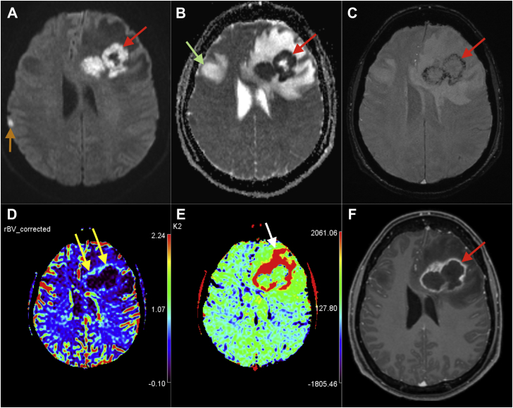
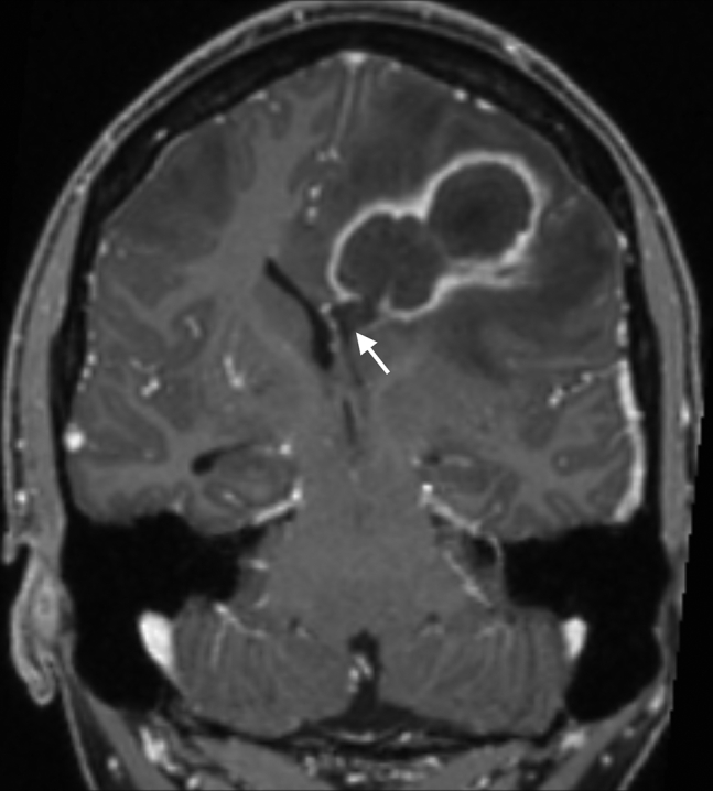
Differentiation between infectious and neoplastic brain processes is crucial for treatment planning. Advanced magnetic resonance imaging techniques, such as diffusion, perfusion, susceptibility weighted imaging, and magnetic resonance spectroscopy, enhance the imaging differences between these two pathologies. However, despite the utilization of these advanced techniques, the pathologic process may be confound by atypical findings. Here, we report a case of an autistic patient with multiple brain lesions with diffusion weighted imaging, susceptibility weighted imaging, and perfusion patterns resembling features of a multicentric glioblastoma, which were confirmed surgically, neuropathologically, and bacteriologically as brain abscesses. We discuss the differentiation of these different entities in the light of advanced magnetic resonance imaging techniques.
Keywords: Diffusion; Infectious; Neoplastic; Perfusion; Susceptibility.
Figures
Similar articles
-
Atypical pyogenic brain abscess evaluation by diffusion-weighted imaging: diagnosis with multimodality MR imaging.Jpn J Radiol. 2015 Oct;33(10):668-71. doi: 10.1007/s11604-015-0472-8. Epub 2015 Aug 15. Jpn J Radiol. 2015. PMID: 26275792
-
Pyogenic brain abscess, ventriculitis and diffuse meningitis with fatal outcome in an adult: Radiologic-pathologic correlation☆,.Radiol Case Rep. 2018 May 18;13(5):1063-1068. doi: 10.1016/j.radcr.2018.04.019. eCollection 2018 Oct. Radiol Case Rep. 2018. PMID: 30228844 Free PMC article.
-
Brain abscess and cystic brain tumor: discrimination with dynamic susceptibility contrast perfusion-weighted MRI.J Comput Assist Tomogr. 2005 Sep-Oct;29(5):663-7. doi: 10.1097/01.rct.0000168868.50256.55. J Comput Assist Tomogr. 2005. PMID: 16163039
-
Magnetic resonance features of pyogenic brain abscesses and differential diagnosis using morphological and functional imaging studies: a pictorial essay.J Neuroradiol. 2014 Jul;41(3):153-67. doi: 10.1016/j.neurad.2014.05.004. Epub 2014 Jun 21. J Neuroradiol. 2014. PMID: 24957685 Review.
-
Imaging of skull base pathologies: Role of advanced magnetic resonance imaging techniques.Neuroradiol J. 2015 Aug;28(4):426-37. doi: 10.1177/1971400915609341. Epub 2015 Oct 1. Neuroradiol J. 2015. PMID: 26427895 Free PMC article. Review.
Cited by
-
Inflammatory lesions and brain tumors: is it possible to differentiate them based on texture features in magnetic resonance imaging?J Venom Anim Toxins Incl Trop Dis. 2020 Sep 4;26:e20200011. doi: 10.1590/1678-9199-JVATITD-2020-0011. J Venom Anim Toxins Incl Trop Dis. 2020. PMID: 32952531 Free PMC article.
-
Abnormal magnetic resonance imaging signal in the burr hole and the identification of the intracranial infection of a deep brain stimulation device: illustrative case.J Neurosurg Case Lessons. 2023 Aug 21;6(8):CASE23370. doi: 10.3171/CASE23370. Print 2023 Aug 21. J Neurosurg Case Lessons. 2023. PMID: 37728295 Free PMC article.
-
Challenges, limitations, and pitfalls of PET and advanced MRI in patients with brain tumors: A report of the PET/RANO group.Neuro Oncol. 2024 Jul 5;26(7):1181-1194. doi: 10.1093/neuonc/noae049. Neuro Oncol. 2024. PMID: 38466087 Free PMC article. Review.
References
-
- Schwartz K.M., Erickson B.J., Lucchinetti C. Pattern of T2 hypointensity associated with ring-enhancing brain lesions can help to differentiate pathology. Neuroradiology. 2006;48:143–149. - PubMed
-
- Smirniotopoulos J.G., Murphy F.M., Rushing E.J., Rees J.H., Schroeder J.W. Patterns of contrast enhancement in the brain and meninges. Radiographics. 2007;27:525–551. - PubMed
-
- Schaefer P.W., Grant P.E., Gonzalez R.G. Diffusion-weighted MR imaging of the brain. Radialogy. 2000;217:331–345. - PubMed
-
- Ebisu T., Tanaka C., Umeda M., Kitamura M., Naruse S., Higuchi T. Discrimination of brain abscess from necrotic or cystic tumors by diffusion-weighted echo planar imaging. Magn Reson Imaging. 1996;14:1113–1116. - PubMed
Publication types
LinkOut - more resources
Full Text Sources
Other Literature Sources
