

Traumatic rupture of a giant congenital splenic cyst presenting as peritonitis
- PMID: 28491197
- PMCID: PMC5417624
- DOI: 10.1016/j.radcr.2017.01.001
Traumatic rupture of a giant congenital splenic cyst presenting as peritonitis
Abstract
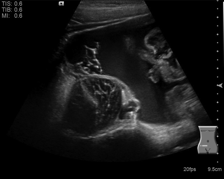
Splenic cysts are uncommon, with large cysts and complications being rare. We describe a 6-year-old patient who initially presented 1 day after falling onto her abdomen at the playground with worsening abdominal pain and distention. An ultrasound of the abdomen demonstrated free abdominal fluid in all four quadrants. A subsequent contrast-enhanced computed tomography scan of the abdomen and pelvis was performed which showed a large splenic cyst with open communication to the peritoneal cavity. A congenital primary cyst was confirmed on pathology after partial splenectomy was performed. Although the majority of splenic cysts are asymptomatic, rupture can lead to acute peritoneal signs and mimic other significant causes of abdominal pain such as viscous injury or acute appendicitis.
Keywords: Child; Cyst; Laparoscopy; Peritonitis; Rupture; Spleen; Splenectomy.
Figures


Similar articles
-
Treatment of splenic cyst by laparoscopic partial splenectomy: case report.Cir Cir. 2010 Jan-Feb;78(1):83-5. Cir Cir. 2010. PMID: 20226133 English, Spanish.
-
Laparoscopic marsupialization of a giant posttraumatic splenic cyst.JSLS. 2004 Oct-Dec;8(4):384-8. JSLS. 2004. PMID: 15554287 Free PMC article.
-
Giant splenic cyst: A case series of rare and challenging cases from the last 22 years.Int J Surg Case Rep. 2023 May;106:108263. doi: 10.1016/j.ijscr.2023.108263. Epub 2023 Apr 26. Int J Surg Case Rep. 2023. PMID: 37116278 Free PMC article.
-
[Laparoscopic splenectomy for epithelial cyst of the spleen].Rev Med Chir Soc Med Nat Iasi. 2005 Jul-Sep;109(3):548-55. Rev Med Chir Soc Med Nat Iasi. 2005. PMID: 16607748 Review. Romanian.
-
Spontaneous giant splenic hydatid cyst rupture causing fatal anaphylactic shock: a case report and brief literature review.Turk J Gastroenterol. 2014 Feb;25(1):88-91. doi: 10.5152/tjg.2014.3521. Turk J Gastroenterol. 2014. PMID: 24918138 Review.
Cited by
-
Carbohydrate antigen 19-9-producing splenic cyst: a case report.Radiol Case Rep. 2021 Oct 28;17(1):19-22. doi: 10.1016/j.radcr.2021.09.063. eCollection 2022 Jan. Radiol Case Rep. 2021. PMID: 34760035 Free PMC article.
-
Emergent laparoscopic dome resection and omental suturing to the splenic parenchymal edge for a spontaneously ruptured non-parasitic large splenic cyst in a pediatric patient: a case report.Surg Case Rep. 2019 Dec 18;5(1):201. doi: 10.1186/s40792-019-0750-2. Surg Case Rep. 2019. PMID: 31853667 Free PMC article.
-
Watchful waiting for large primary nonparasitic splenic cysts.Can J Surg. 2023 Jul 27;66(4):E390-E395. doi: 10.1503/cjs.010322. Print 2023 Jul-Aug. Can J Surg. 2023. PMID: 37500107 Free PMC article.
-
A case of ruptured splenic cyst with elevated serum levels of CEA treated by laparoscopic unroofing.Clin J Gastroenterol. 2019 Dec;12(6):642-649. doi: 10.1007/s12328-019-00980-0. Epub 2019 Apr 10. Clin J Gastroenterol. 2019. PMID: 30972710
-
Spontaneous rupture of a non-parasitic splenic cyst.BMJ Case Rep. 2019 Oct 30;12(10):e231473. doi: 10.1136/bcr-2019-231473. BMJ Case Rep. 2019. PMID: 31666253 Free PMC article.
References
Publication types
LinkOut - more resources
Full Text Sources
Other Literature Sources
