Fibrosarcoma of the urinary bladder in a cat
- PMID: 28491352
- PMCID: PMC5362888
- DOI: 10.1177/2055116915585019
Fibrosarcoma of the urinary bladder in a cat
Abstract
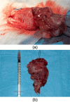
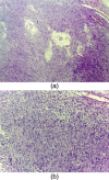
Case summary: A 5-year-old female spayed domestic shorthair cat was presented with haematuria, pollakiuria and stranguria of 2 months' duration, and a firm non-painful mass in the urinary bladder was palpated. Abdominal radiographs showed thickening and irregular cranial margins of the urinary bladder wall. Abdominal ultrasound showed a vascularised mass of mixed echogenicity almost entirely occupying the urinary bladder lumen. During explorative laparotomy, the mass appeared pedunculated and was totally excised. Histopathology was characterised by infiltration of the mucosal, submucosal and muscular layers by proliferated atypical mesenchymal cells; immunochemistry confirmed the diagnosis of fibrosarcoma. The cat was discharged with normal urination 5 days after surgery. The owner declined any imaging follow-up but reported the cat to be free of any clinical signs at 16 months after surgery.
Relevance and novel information: To the best of our knowledge, this is the first case of primary fibrosarcoma of the urinary bladder in the cat. Fibrosarcoma should be included in the differential diagnosis of urinary bladder neoplasia.
Conflict of interest statement
Conflict of interest: The authors do not have any potential conflicts of interest to declare.
Figures


Similar articles
-
Ultrasonographic features of a urinary bladder leiomyoma in a cat.JFMS Open Rep. 2023 May 22;9(1):20551169231165247. doi: 10.1177/20551169231165247. eCollection 2023 Jan-Jun. JFMS Open Rep. 2023. PMID: 37249943 Free PMC article.
-
Primary fibrosarcoma of the urinary bladder in a cat: follow-up after incomplete surgical excision.JFMS Open Rep. 2017 Jun 19;3(1):2055116917714881. doi: 10.1177/2055116917714881. eCollection 2017 Jan-Jun. JFMS Open Rep. 2017. PMID: 28680699 Free PMC article.
-
Extranodal B-cell lymphoma in the urinary bladder with cytological evidence of concurrent involvement of the gall bladder in a cat.J Small Anim Pract. 2010 May;51(5):280-7. doi: 10.1111/j.1748-5827.2010.00911.x. J Small Anim Pract. 2010. PMID: 20536697
-
[Multiple synchronous primary malignant tumors of fibrosarcoma and squamous cell carcinoma in the urinary bladder: a case report].Nihon Hinyokika Gakkai Zasshi. 2002 Jul;93(5):642-7. doi: 10.5980/jpnjurol1989.93.642. Nihon Hinyokika Gakkai Zasshi. 2002. PMID: 12174642 Review. Japanese.
-
[Inflammatory pseudotumor of the urinary bladder. Study of 4 cases and review of the literature].Pathologica. 1995 Dec;87(6):653-8. Pathologica. 1995. PMID: 8927426 Review. Italian.
Cited by
-
Ultrasonographic features of a urinary bladder leiomyoma in a cat.JFMS Open Rep. 2023 May 22;9(1):20551169231165247. doi: 10.1177/20551169231165247. eCollection 2023 Jan-Jun. JFMS Open Rep. 2023. PMID: 37249943 Free PMC article.
-
Long-Term Survival of a Cat with Primary Leiomyosarcoma of the Urinary Bladder.Vet Sci. 2019 Jun 27;6(3):60. doi: 10.3390/vetsci6030060. Vet Sci. 2019. PMID: 31252551 Free PMC article.
-
Primary fibrosarcoma of the urinary bladder in a cat: follow-up after incomplete surgical excision.JFMS Open Rep. 2017 Jun 19;3(1):2055116917714881. doi: 10.1177/2055116917714881. eCollection 2017 Jan-Jun. JFMS Open Rep. 2017. PMID: 28680699 Free PMC article.
-
Feline abdominal ultrasonography: What's normal? What's abnormal? Renal pelvis, ureters and urinary bladder.J Feline Med Surg. 2020 Sep;22(9):847-865. doi: 10.1177/1098612X20941786. J Feline Med Surg. 2020. PMID: 32845227 Free PMC article. Review.
References
-
- Osborne CA, Low DG, Perman V, et al. Neoplasms of the canine and feline urinary bladder: incidence, etiologic factors, occurrence and pathologic features. Am J Vet Res 1968; 29: 2041–2055. - PubMed
-
- Mutsaers AJ, Widmer WR, Knapp DW. Canine transitional cell carcinoma. J Vet Intern Med 2003; 17: 136–144. - PubMed
-
- Wimberly HC, Lewis RM. Transitional cell carcinoma in the domestic cat. Vet Pathol 1979; 16: 223–228. - PubMed
-
- Schwarz PD, Greene RW, Patnaik AK. Urinary bladder tumors in the cat: a review of 27 cases. J Am Anim Hosp Assoc 1985; 21: 237–245.
-
- Wilson HM, Chun R, Larson VS, et al. Clinical signs, treatments, and outcome in cats with transitional cell carcinoma of the urinary bladder: 20 cases (1990–2004). J Am Vet Med Assoc 2007; 231: 101–106. - PubMed
Publication types
LinkOut - more resources
Full Text Sources
Other Literature Sources
Miscellaneous
