

Selective fatty replacement of the exocrine pancreas in a domestic shorthair cat: histopathological findings with long-term outcome
- PMID: 28491371
- PMCID: PMC5362021
- DOI: 10.1177/2055116915593967
Selective fatty replacement of the exocrine pancreas in a domestic shorthair cat: histopathological findings with long-term outcome
Abstract
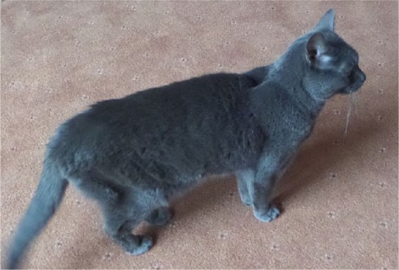
The clinical, histopathological findings and eventual outcome of a cat with marked and selective fatty replacement of the exocrine pancreas are described in this case report. A 9-year-old female neutered domestic shorthair cat presenting with polyphagia, weight loss and intermittent vomiting was diagnosed on histopathology with severe exocrine pancreatic atrophy, with relative sparing of the endocrine pancreas and replacement of the acinar cells by mature adipose tissue. This case report discusses the histological findings in this case and the eventual outcome, as well as the potential underlying causes of this histological change.
Conflict of interest statement
Conflict of interest: The authors do not have any potential conflicts of interest to declare.
Figures


Similar articles
-
Hyperinsulinaemic, hypoglycaemic syndrome due to acquired nesidioblastosis in a cat.JFMS Open Rep. 2016 Jul 7;2(2):2055116916657846. doi: 10.1177/2055116916657846. eCollection 2016 Jul-Dec. JFMS Open Rep. 2016. PMID: 28491431 Free PMC article.
-
Pancreatic leiomyosarcoma in a domestic shorthair cat.JFMS Open Rep. 2022 Jun 7;8(1):20551169221098328. doi: 10.1177/20551169221098328. eCollection 2022 Jan-Jun. JFMS Open Rep. 2022. PMID: 35693479 Free PMC article.
-
Hyalinizing pancreatic adenocarcinoma in a cat.JFMS Open Rep. 2025 Apr 16;11(1):20551169251325333. doi: 10.1177/20551169251325333. eCollection 2025 Jan-Jun. JFMS Open Rep. 2025. PMID: 40291437 Free PMC article.
-
Structure and function of the exocrine pancreas in patients with type 1 diabetes.Rev Endocr Metab Disord. 2019 Jun;20(2):129-149. doi: 10.1007/s11154-019-09501-3. Rev Endocr Metab Disord. 2019. PMID: 31077020 Review.
-
The Pancreas: Causes for Malabsorption.Viszeralmedizin. 2014 Jun;30(3):190-7. doi: 10.1159/000363778. Viszeralmedizin. 2014. PMID: 26288593 Free PMC article. Review.
References
-
- Rothuizen J, Bunch SE, Charles JA, et al. WSAVA standards for clinical and histological diagnosis of caninr and feline liver disease. Philadelphia, PA: Saunders Elsevier, 2006.
-
- Day MJ, Bilzer T, Mansell J, et al. Histopathological standards for the diagnosis of gastrointestinal inflammation in endoscopic biopsy samples from the dog and cat: a report from the World Small Animal Veterinary Association Gastrointestinal Standardization Group. J Comp Pathol 2008; 138: S1–S43. - PubMed
-
- De Cock HEV, Forman MA, Farver TB, et al. Prevalence and histopathologic characteristics of pancreatitis in cats. Vet Pathol 2007; 44: 39–49. - PubMed
Publication types
LinkOut - more resources
Full Text Sources
Other Literature Sources
Miscellaneous