

Benign cementoblastoma (true cementoma) in a cat
- PMID: 28491408
- PMCID: PMC5362872
- DOI: 10.1177/2055116915626847
Benign cementoblastoma (true cementoma) in a cat
Abstract
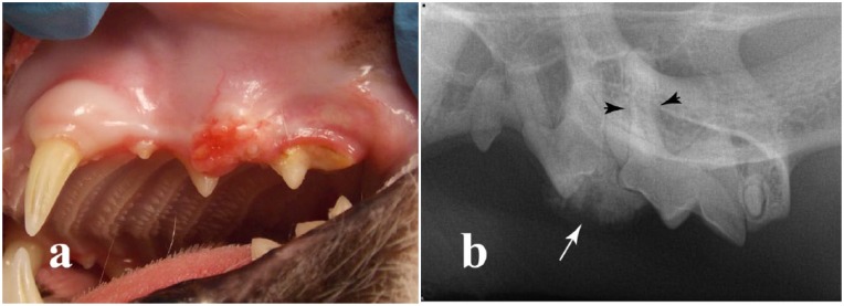
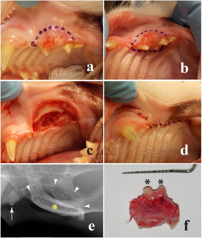
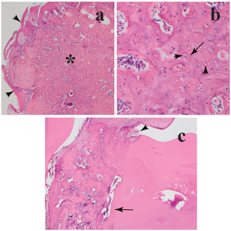
Case summary: A 10-year-old castrated male domestic shorthair cat was presented for assessment of a gingival mass surrounding the left maxillary third and fourth premolar teeth. The mass was surgically removed by means of a marginal rim excision, and the tissue was submitted for histological assessment. It was identified as a benign cementoblastoma (true cementoma). There was proliferation of mineralized eosinophilic material with multiple irregularly placed lacunae and reversal lines, reminiscent of cementum. The cat recovered uneventfully from the anesthesia, and there was no evidence of tumor recurrence 6 months after surgery.
Relevance and novel information: Cementoblastomas (true cementomas) in domestic animals are rare, with just a few reports in ruminants, monogastric herbivores and rodents. Cementoblastoma is considered a benign tumor that arises from the tooth root. The slow, expansive and constant growth that characterizes these masses may be accompanied by signs of oral discomfort and dysphagia. This case report is intended to increase knowledge regarding this tumor in cats and also highlights the importance of complete excision of the neoplasm. To our knowledge, there are no previous reports in the literature of cementoblastoma in the cat.
Conflict of interest statement
Conflict of interest: The authors declared no potential conflicts of interest with respect to the research, authorship, and/or publication of this article.
Figures
Similar articles
-
Benign cementoblastoma of the anterior mandible: an unusual case report.J Korean Assoc Oral Maxillofac Surg. 2016 Aug;42(4):231-5. doi: 10.5125/jkaoms.2016.42.4.231. Epub 2016 Aug 24. J Korean Assoc Oral Maxillofac Surg. 2016. PMID: 27595092 Free PMC article.
-
Cementoblastoma of a primary molar: A rare pediatric occurrence.J Oral Maxillofac Pathol. 2020 Sep-Dec;24(3):548-553. doi: 10.4103/jomfp.JOMFP_307_19. Epub 2021 Jan 9. J Oral Maxillofac Pathol. 2020. PMID: 33967495 Free PMC article.
-
[Maxillary cementoblastoma. A case report].Rev Med Brux. 2009 May-Jun;30(3):185-8. Rev Med Brux. 2009. PMID: 19642490 French.
-
Benign cementoblastoma involving multiple maxillary teeth: report of a case with a review of the literature.Oral Surg Oral Med Oral Pathol Oral Radiol Endod. 2004 Jan;97(1):53-8. doi: 10.1016/j.tripleo.2003.08.012. Oral Surg Oral Med Oral Pathol Oral Radiol Endod. 2004. PMID: 14716256 Review.
-
Cemento-ossifying fibroma and benign cementoblastoma.Semin Diagn Pathol. 1999 Nov;16(4):302-7. Semin Diagn Pathol. 1999. PMID: 10587273 Review.
Cited by
-
Caudal and middle segmental mandibulectomies for the treatment of unilateral temporomandibular joint ankylosis in cats.JFMS Open Rep. 2022 Mar 31;8(1):20551169221086438. doi: 10.1177/20551169221086438. eCollection 2022 Jan-Jun. JFMS Open Rep. 2022. PMID: 35386208 Free PMC article.
-
Comparison of unilateral rostral, middle and caudal segmental mandibulectomies as an alternative treatment for unilateral temporomandibular joint ankylosis in cats: an ex vivo study.J Feline Med Surg. 2021 Aug;23(8):783-793. doi: 10.1177/1098612X20977134. Epub 2020 Dec 8. J Feline Med Surg. 2021. PMID: 33289444 Free PMC article.
References
-
- Head KW, Else RW, Dubielzig RR. Tumors of the alimentary tract. In: Meuten DJ. (ed). Tumors in domestic animals. 4th ed. Ames, IA: Iowa State Press, 2002, pp 407–408.
-
- Regezi JA, Sciubba JJ, Jordan RCK. Odontogenic tumors. In: Oral pathology clinical pathology correlations. 5th ed. St Louis, MO: Saunders Elsevier, 2008, pp 261–281.
-
- Brannon RB, Fowler CB, Carpenter WM, et al. Cementoblastoma: an innocuous neoplasm? A clinicopathologic study of 44 cases and review of the literature with special emphasis on recurrence. Oral Surg Oral Med Oral Pathol Oral Radiol Endod 2002; 93: 311–320. - PubMed
-
- Neves FS, Falcão AF, Dos Santos JN, et al. Benign cementoblastoma: case report and review of the literature. Minerva Stomatol 2009; 58: 55–59. - PubMed
Publication types
LinkOut - more resources
Full Text Sources
Other Literature Sources
Miscellaneous