

A case of acute acquired obstructive hydrocephalus in a cat with suspected ischaemic cerebellar infarct
- PMID: 28491457
- PMCID: PMC5405888
- DOI: 10.1177/2055116917704089
A case of acute acquired obstructive hydrocephalus in a cat with suspected ischaemic cerebellar infarct
Abstract
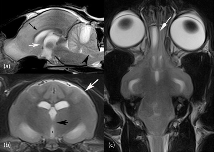
Case summary: A case of acquired acute obstructive hydrocephalus that developed as a complication of an ischaemic infarct in the vascular territory of the rostral cerebellar artery is described in an adult domestic shorthair cat. The clinical findings, diagnostic investigations, treatment and prognosis are reported. MRI findings are described in detail.
Relevance and novel information: This is the first report of obstructive hydrocephalus as a complication of an ischaemic infarct in the region of the rostral cerebellar artery in a cat. MRI findings are described in detail with regard to the recognition of the early signs of obstructive hydrocephalus. A brief review of the literature is included, as this complication has been frequently reported in humans.
Conflict of interest statement
Conflict of interest: The authors declared no potential conflicts of interest with respect to the research, authorship, and/or publication of this article.
Figures


Similar articles
-
Neurological signs in 23 dogs with suspected rostral cerebellar ischaemic stroke.Acta Vet Scand. 2016 Jun 7;58(1):40. doi: 10.1186/s13028-016-0219-2. Acta Vet Scand. 2016. PMID: 27267355 Free PMC article.
-
Acute hydrocephalus in cerebellar infarct and hemorrhage.Neurology. 1979 Mar;29(3):409-13. doi: 10.1212/wnl.29.3.409. Neurology. 1979. PMID: 571991
-
Delayed Obstructive Hydrocephalus After Cardiac Surgery With Cardiopulmonary Bypass in a Patient With Cerebellar Infarction: A Case Report.A A Pract. 2021 Mar 30;15(4):e01439. doi: 10.1213/XAA.0000000000001439. A A Pract. 2021. PMID: 33783405
-
Cerebellar ataxia and obstructive hydrocephalus, rare neurologic presentations in patients with systemic lupus erythematosus.Rheumatol Int. 2017 Nov;37(11):1917-1930. doi: 10.1007/s00296-017-3773-7. Epub 2017 Jul 13. Rheumatol Int. 2017. PMID: 28707035 Review.
-
Fourth ventricular entrapment caused by rostrocaudal herniation following shunt malfunction.Pediatr Neurosurg. 1993 Jul-Aug;19(4):209-14. doi: 10.1159/000120733. Pediatr Neurosurg. 1993. PMID: 8329307 Review.
References
-
- Garosi LS. Cerebrovascular disease in dogs and cats. Vet Clin North Am Small Anim Pract 2010; 40: 65–79. - PubMed
-
- Mcconnell JF, Garosi LS, Platt SR, et al. Magnetic resonance imaging findings of presumed cerebellar cerebrovascular accident in twelve dogs. Vet Radiol Ultrasound 2005; 46: 1–10. - PubMed
-
- Garosi LS, McConnell JF, Platt SR, et al. Clinical and topographic magnetic resonance characteristics of suspected brain infarction in 40 dogs. J Vet Intern Med 2006; 20: 311–321. - PubMed
-
- Tidwell AS, Robertson ID. Magnetic resonance imaging of normal and abnormal brain perfusion. Vet Radiol Ultrasound 2011; 52 Suppl 1: S62–S71. - PubMed
Publication types
LinkOut - more resources
Full Text Sources
Other Literature Sources
Miscellaneous