

A Novel Three-Dimensional Vector Analysis of Axial Globe Position in Thyroid Eye Disease
- PMID: 28491471
- PMCID: PMC5401755
- DOI: 10.1155/2017/7253898
A Novel Three-Dimensional Vector Analysis of Axial Globe Position in Thyroid Eye Disease
Abstract
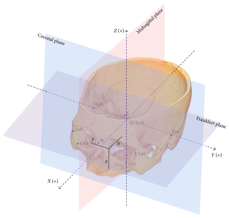
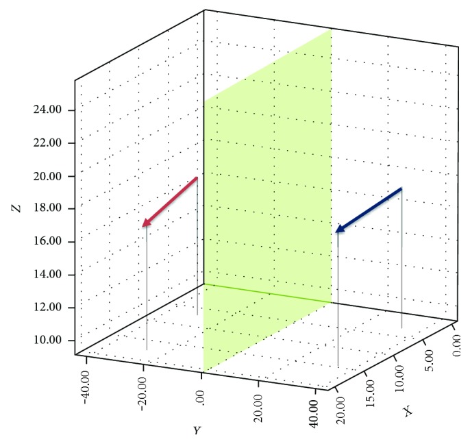
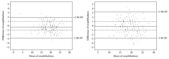
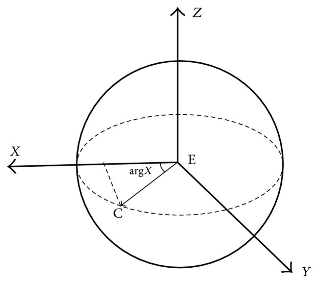
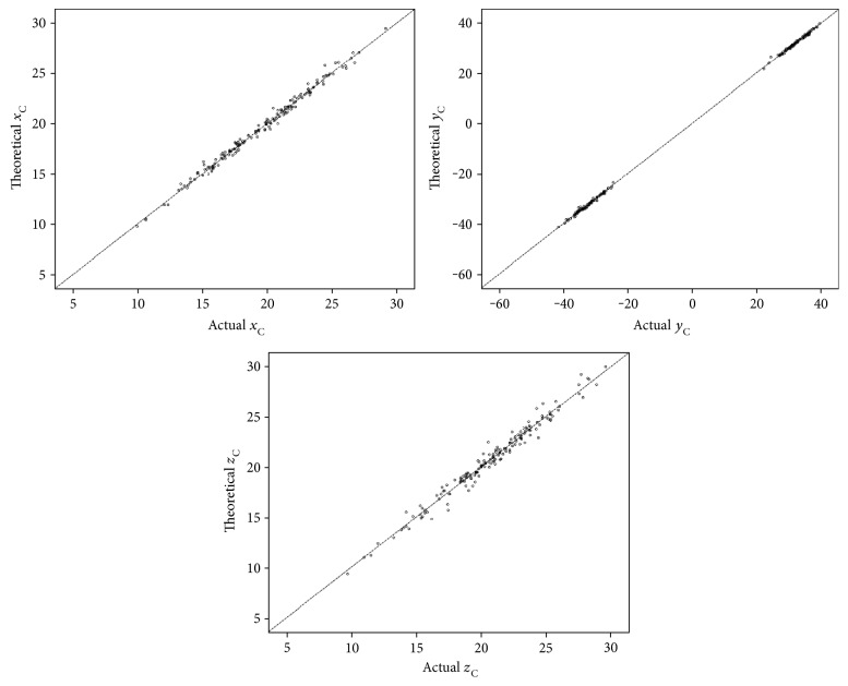
Purpose. To define a three-dimensional (3D) vector method to describe the axial globe position in thyroid eye disease (TED). Methods. CT data from 59 patients with TED were collected and 3D images were reconstructed. A reference coordinate system was established, and the coordinates of the corneal apex and the eyeball center were calculated to obtain the globe vector [Formula: see text]. The measurement reliability was evaluated. The parameters of [Formula: see text] were analyzed and compared with the results of two-dimensional (2D) CT measurement, Hertel exophthalmometry, and strabismus tests. Results. The reliability of [Formula: see text] measurement was excellent. The difference between [Formula: see text] and 2D CT measurement was significant (p = 0.003), and [Formula: see text] was more consistent with Hertel exophthalmometry than with 2D CT measurement (p < 0.001). There was no significant difference between [Formula: see text] and Hirschberg test, and a strong correlation was found between [Formula: see text] and synoptophore test. When one eye had a larger deviation angle than its fellow, its corneal apex shifted in the corresponding direction, but the shift of the eyeball center was not significant. The parameters of [Formula: see text] were almost perfectly consistent with the geometrical equation. Conclusions. The establishment of a 3D globe vector is feasible and reliable, and it could provide more information in the axial globe position.
Figures
Similar articles
-
Computed Tomography Measurements as a Standard of Exophthalmos? Two-Dimensional Versus Three-Dimensional Techniques.Curr Eye Res. 2018 May;43(5):647-653. doi: 10.1080/02713683.2018.1431285. Epub 2018 Jan 29. Curr Eye Res. 2018. PMID: 29377729
-
Reliability and correlation analysis of computed methods to convert conventional 2D radiological hindfoot measurements to a 3D setting using weightbearing CT.Int J Comput Assist Radiol Surg. 2018 Dec;13(12):1999-2008. doi: 10.1007/s11548-018-1727-5. Epub 2018 Mar 9. Int J Comput Assist Radiol Surg. 2018. PMID: 29524088
-
Relative en- and exophthalmometry in zygomatic fractures comparing optical non-contact, non-ionizing 3D imaging to the Hertel instrument and computed tomography.J Craniomaxillofac Surg. 2003 Dec;31(6):362-8. doi: 10.1016/j.jcms.2003.07.001. J Craniomaxillofac Surg. 2003. PMID: 14637065
-
Atomic scale displacements detected by optical image cross-correlation analysis and 3D printed marker arrays.Sci Rep. 2021 Jan 27;11(1):2304. doi: 10.1038/s41598-021-81712-8. Sci Rep. 2021. PMID: 33504911 Free PMC article.
-
Brane-world singularities and asymptotics of five-dimensional bulk fluids.Philos Trans A Math Phys Eng Sci. 2022 Aug 22;380(2230):20210180. doi: 10.1098/rsta.2021.0180. Epub 2022 Jul 4. Philos Trans A Math Phys Eng Sci. 2022. PMID: 35785973 Review.
Cited by
-
Eyeball descending identification using MRI-based spatial coordinates in thyroid-associated orbitopathy patients with unilateral upper eyelid retraction.Quant Imaging Med Surg. 2025 Feb 1;15(2):1287-1296. doi: 10.21037/qims-24-1659. Epub 2025 Jan 9. Quant Imaging Med Surg. 2025. PMID: 39995720 Free PMC article.
-
Automatic measurement of exophthalmos based orbital CT images using deep learning.Front Cell Dev Biol. 2023 Feb 24;11:1135959. doi: 10.3389/fcell.2023.1135959. eCollection 2023. Front Cell Dev Biol. 2023. PMID: 36910161 Free PMC article.
References
-
- Bingham C. M., Sivak-Callcott J. A., Gurka M. J., Nguyen J., Hogg J. P., Feldon S. E. Axial globe position measurement: a prospective multicenter study by the International Thyroid Eye Disease Society. Ophthalmic Plastic and Reconstructive Surgery. 2015;32(2):106–112. doi: 10.1097/IOP.0000000000000437. - DOI - PMC - PubMed
-
- Segni M., Bartley G. B., Garrity J. A., Bergstralh E. J., Gorman C. A. Comparability of proptosis measurements by different techniques. American Journal of Ophthalmology. 2002;133(6):813–818. - PubMed
LinkOut - more resources
Full Text Sources
Other Literature Sources
Miscellaneous
