Review
. 2017 Apr 20:15:95-105.
doi: 10.1016/j.nicl.2017.04.017. eCollection 2017.

Towards in vivo focal cortical dysplasia phenotyping using quantitative MRI

Affiliations
Review

Towards in vivo focal cortical dysplasia phenotyping using quantitative MRI

Sophie Adler et al. Neuroimage Clin. .

Abstract

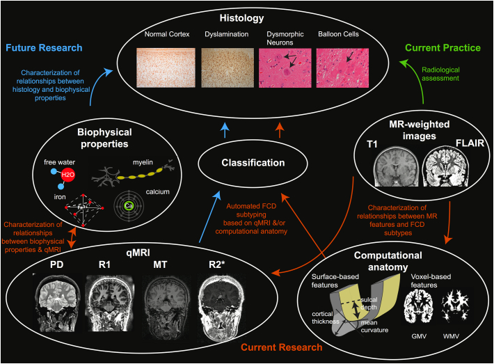
Focal cortical dysplasias (FCDs) are a range of malformations of cortical development each with specific histopathological features. Conventional radiological assessment of standard structural MRI is useful for the localization of lesions but is unable to accurately predict the histopathological features. Quantitative MRI offers the possibility to probe tissue biophysical properties in vivo and may bridge the gap between radiological assessment and ex-vivo histology. This review will cover histological, genetic and radiological features of FCD following the ILAE classification and will explain how quantitative voxel- and surface-based techniques can characterise these features. We will provide an overview of the quantitative MRI measures available, their link with biophysical properties and finally the potential application of quantitative MRI to the problem of FCD subtyping. Future research linking quantitative MRI to FCD histological properties should improve clinical protocols, allow better characterisation of lesions in vivo and tailored surgical planning to the individual.

Keywords: Biophysical tissue properties; Epilepsy surgery; Focal cortical dysplasia; Histology; MRI; Malformation of cortical development; Quantitative MRI; Quantitative mapping; Radiology; qMRI.


Figures

Fig. 1
Fig. 1
Overview of relationship between histological features, biophysical tissue properties, MR images, computational anatomy measures and quantitative neuroimaging. Green arrow indicates current link between MR-weighted images and ex vivo histology. Orange arrows indicate areas of current research using computational anatomy based on MR-weighted images for FCD subtyping. Blue arrows indicate future directions to characterise biophysical tissue properties of FCDs using qMRI. qMRI maps are sensitive to biophysical tissue properties, such as myelin, iron, calcium and free water content. The qMRI profiles of different FCD subtypes are currently unknown but may offer a technique to probe histology in vivo. (For interpretation of the references to color in this figure legend, the reader is referred to the online version of this chapter.)
Fig. 2
Fig. 2
Computational and quantitative MRI features used by Hong et al., Morphology features include cortical thickness and sulcal depth. Intensity features include normalized T1 and FLAIR signal intensity sampled at multiple intra- and sub-cortical surfaces, as well as horizontal and vertical intensity gradients. Diffusion tensor based features include fractional anisotropy (FA) and mean diffusivity (MD). Resting-state fMRI features include amplitude of local functional fluctuation (ALFF) and regional homogeneity (ReHo).

References

    1. Adler S., Wagstyl K., Gunny R., Ronan L., Carmichael D., Cross J.H., Fletcher P.C., Baldeweg T. Novel surface features for automated detection of focal cortical dysplasias in paediatric epilepsy. Neuroimage. Clin. 2016:18–27. - PMC - PubMed
    1. Ahmed B., Brodley C.E., Blackmon K.E., Kuzniecky R., Barash G., Carlson C., Quinn B.T., Doyle W., French J., Devinsky O., Thesen T. Cortical feature analysis and machine learning improves detection of “MRI-negative” focal cortical dysplasia. Epilepsy Behav. 2015;48:21–28. - PMC - PubMed
    1. Alexander A.L., Hurley S.A., Samsonov A.A., Adluru N., Hosseinbor A.P., Mossahebi P., Tromp do P.M., Zakszewski E., Field A.S. Characterization of cerebral white matter properties using quantitative magnetic resonance imaging stains. Brain Connect. 2012;1:423–446. - PMC - PubMed
    1. Alhusaini S., Whelan C.D., Doherty C.P., Delanty N., Fitzsimons M., Cavalleri G.L. Temporal cortex morphology in mesial temporal lobe epilepsy patients and their asymptomatic siblings. Cereb. Cortex. 2016;26:1234–1241. - PubMed
    1. Barkovich A.J., Kuzniecky R.I., Bollen A.W., Grant P.E. Focal transmantle dysplasia: a specific malformation of cortical development. Neurology. 1997;49:1148–1152. - PubMed

MeSH terms