

Case Reports
doi: 10.1016/j.hrcr.2015.07.003.
eCollection 2015 Nov.
Left atrial access via an unroofed coronary sinus to eliminate fast/slow atypical AVNRT: A case report
Affiliations
- PMID: 28491606
- PMCID: PMC5419727
- DOI: 10.1016/j.hrcr.2015.07.003
Item in Clipboard
Case Reports
Left atrial access via an unroofed coronary sinus to eliminate fast/slow atypical AVNRT: A case report
HeartRhythm Case Rep.
.
No abstract available
Keywords: ABL, ablation catheter; AV, atrioventricular; AVNRT; AVNRT, atrioventricular nodal reentrant tachycardia; Ablation; Atypical AVNRT; CS, coronary sinus; EPS, electrophysiologic study; HIS, His-bundle recording catheter; HRA, high right atrial recording catheter; LA, left atrium; LAO, left anterior oblique; Mapping; PVC, premature ventricular contractions; RA, right atrium; RAO, right anterior oblique; RF, radiofrequency; RV, right ventricle; RVA, right ventricular apex; SVC, superior vena cava; SVT, supraventricular tachycardia; Unroofed coronary sinus.
Figures

Cardiac computed tomography angiogram. A: Multiplanar reconstruction at the level of the atrial septal defect demonstrating completely unroofed coronary sinus, with the cardiac vein (red arrows) draining noncontrasted blood directly into the left atrium. Blue dotted lines represent the coronary sinus ostium and the area of the absent coronary sinus (CS) roof. B: A 3-dimernsional reconstruction demonstrating the cardiac vein (white arrow) entering into the coronary sinus. Notice that there is no definite separation between the mid CS and left atrium, confirming CS unroofing. LA = left atrium; RA = right atrium.

A: A 12-lead electrocardiogram of the clinical arrhythmia. Notice a long RP tachycardia with superior P-wave axis. Note the irregularity of the fourth and fifth beats that appear more premature. Those beats were spontaneous atrial ectopic beats occurring frequently at baseline, during the electrophysiologic study and during arrhythmia. B: Intracardiac electrogram recorded during the electrophysiologic study. From top to bottom: 12 lead electrocardiogram leads I, II, III, V1, and V2; high right atrial recording catheter (HRA), His-bundle recording catheter (HIS), coronary sinus recording catheter (CS) from CS proximal (9,10) to distal (1,2), and right ventricular apex (RVA). The tachycardia cycle length is 368 milliseconds with a long VA time (210 milliseconds).
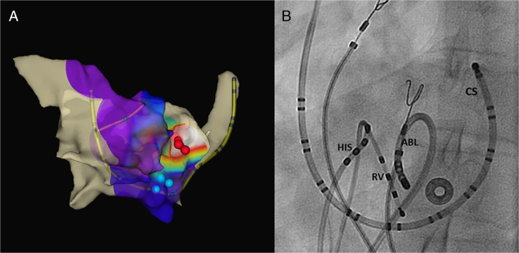
A: Right anterior oblique (RAO) projection of electroanatomic map of the right and left atria with atrial activation sequence during arrhythmia. Blue dots represent the unsuccessful initial sites of radiofrequency (RF) ablation in the low right atrial septum at the typical anatomic location of the slow AV node pathway. Notice the earliest area of atrial activation (coded in white) corresponds to the left atrial septum level. Red dots represent RF ablation at the successful site. B: Left anterior oblique (LAO) fluoroscopic view of the successful ablation site. The ablation catheter (ABL) enters in the left atrium through the proximal unroofed coronary sinus. The other catheters are quadripolar His (HIS) and right ventricular (RV) catheters and the duodecapolar catheter located in the coronary sinus (CS).
References
-
- Chiang C.E., Chen S.A., Yang C.R., Cheng C.C., Wu T.R., Tsai D.S., Chiou C.W., Chen C.Y., Wang S.P., Chiang B.N. Major coronary sinus abnormalities: identification of occurrence and significance in radiofrequency ablation of supraventricular tachycardia. Am Heart J. 1994;127:1279–1289. - PubMed
-
- Corno A. Unroofed coronary sinus. In: Corno A., editor. Vol. 1. Steinkopff; Darmstadt, Germany: 2003. pp. 21–23. (Congenital Heart Defects: Decision Making for Surgery).
-
- Kouchoukos N.T., Blackstone E.H., Doty D.B., Hanley F.L., Karp R.B. Kirklin/Barratt-Boyes Cardiac Surgery. 3rd ed. Churchill Livingstone; Philadelphia, PA: 2003. Unroofed coronary sinus syndromes; pp. 790–799.
Publication types
LinkOut - more resources
Full Text Sources
Other Literature Sources
Miscellaneous
