

Case Reports
doi: 10.1016/j.hrcr.2015.08.012.
eCollection 2016 Jan.
Bradycardia-dependent rise in the atrial capture threshold early after cardiac pacemaker implantation in patients with sick sinus syndrome
Affiliations
- PMID: 28491626
- PMCID: PMC5412638
- DOI: 10.1016/j.hrcr.2015.08.012
Item in Clipboard
Case Reports
Bradycardia-dependent rise in the atrial capture threshold early after cardiac pacemaker implantation in patients with sick sinus syndrome
HeartRhythm Case Rep.
.
No abstract available
Keywords: Bradycardia; ECG, electrocardiography; EPS, electrophysiological study; Pacemaker; Phase 4 block; SSS, sick sinus syndrome; Sick sinus syndrome; Threshold; bpm, beats per minute.
Figures

Programmed atrial stimulation was performed on the fifth day after implantation. The basic stimuli (S1) cycle length was 600 milliseconds, and one atrial extrastimulus (S2) was delivered 1714 milliseconds to 250 milliseconds after S1. When the S1-S2 interval was 1714 milliseconds (35 bpm), S2 was captured with reproducibility. When the S1-S2 interval ranged from 1500 milliseconds (40 bpm) to 750 milliseconds (80 bpm), S2 did not capture the atrium. When the S1-S2 interval ranged from 706 milliseconds (85 bpm) to 260 milliseconds, S2 captured the atrium. When the S1-S2 interval was ≤ 250 milliseconds, S2 did not capture the atrium (atrial effective refractory period = 260 milliseconds). The “noncapture range” lay between 1500 milliseconds and 750 milliseconds.

Programmed atrial stimulation was performed 1 month after implantation. The S2 stimulus failed to capture the atrium only when the S1-S2 interval was 1000 milliseconds. The “noncapture range” was clearly reduced.
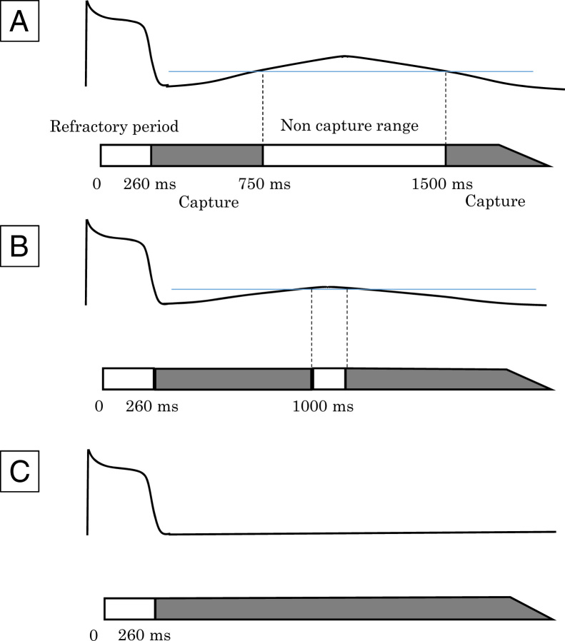
Our assumption as to the membrane potentials of atrial myocardium around the pacing lead according to the results from the electrophysiological study of Case 1. A: The greater diastolic (phase 4) depolarization caused by acute histologic changes creates a wide noncapture range on the fifth day after implantation. B: As diastolic depolarization gradually improves, the noncapture range is reduced at 1 month after implantation. C: Three months after implantation, the noncapture range has disappeared.
Similar articles
-
Sinus and paced P wave duration and dispersion as predictors of atrial fibrillation after pacemaker implantation in patients with isolated sick sinus syndrome.Pacing Clin Electrophysiol. 2004 May;27(5):606-14. doi: 10.1111/j.1540-8159.2004.00494.x. Pacing Clin Electrophysiol. 2004. PMID: 15125716
-
Overdrive suppression in diagnosis of sick sinus syndrome.J Electrocardiol. 1975 Jul;8(3):209-16. doi: 10.1016/s0022-0736(75)80047-1. J Electrocardiol. 1975. PMID: 1159346
-
Underrecognized entity of the transient rise in the atrial capture threshold early after dual-chamber pacemaker implantation.Pacing Clin Electrophysiol. 2017 Dec;40(12):1396-1404. doi: 10.1111/pace.13235. Epub 2017 Dec 5. Pacing Clin Electrophysiol. 2017. PMID: 29139149
-
[Atrioventricular block/sick sinus syndrome].Nihon Rinsho. 2002 Jul;60(7):1401-7. Nihon Rinsho. 2002. PMID: 12136621 Review. Japanese.
-
[Sinus node syndrome].Z Kardiol. 1975 Aug;64(8):697-721. Z Kardiol. 1975. PMID: 1099830 Review. German.
Cited by
-
Rate-dependent change in capture threshold following implantation of a leadless pacemaker.HeartRhythm Case Rep. 2021 Dec 9;8(3):183-186. doi: 10.1016/j.hrcr.2021.12.004. eCollection 2022 Mar. HeartRhythm Case Rep. 2021. PMID: 35492834 Free PMC article. No abstract available.
-
Mechanistic implication of decreased plasma atrial natriuretic peptide level for transient rise in the atrial capture threshold early after ICD or CRT-D implantation.J Interv Card Electrophysiol. 2018 Oct;53(1):131-140. doi: 10.1007/s10840-018-0409-0. Epub 2018 Jul 17. J Interv Card Electrophysiol. 2018. PMID: 30019272
References
-
- Hellestrand K.J., Nathan A.W., Bexton R.S., Camm A.J. Electrophysiologic effects of flecainide acetate on sinus node function, anomalous atrioventricular connections, and pacemaker thresholds. Am J Cardiol. 1984;53:30b–38b. - PubMed
-
- Scoblionko D.P., Rolett E.L. Short-term threshold behavior of human ventricular pacing electrode: Noninvasive monitoring with a multiprogrammable pacing system. Pacing Clin Electrophysiol. 1981;4:631–637. - PubMed
-
- Katsumoto K., Niibori T., Watanabe Y. Rate-dependent threshold changes during atrial pacing: Clinical and experimental studies. Pacing Clin Electrophysiol. 1990;13:1009–1019. - PubMed
-
- Fujiki A., Tani M., Mizumaki K., Yoshida S., Sasayama S. Rate-dependent accessory pathway conduction due to phase 3 and phase 4 block. Antegrade and retrograde conduction properties. J Electrocardiol. 1992;25:25–31. - PubMed
Publication types
LinkOut - more resources
Full Text Sources
Other Literature Sources
