

Review
doi: 10.1016/j.ijwd.2015.11.001.
eCollection 2016 Mar.
Amyloidosis: A story of how inframammary erosions eclipsed inconspicuous periorbital ecchymoses
Affiliations
- PMID: 28491996
- PMCID: PMC5412114
- DOI: 10.1016/j.ijwd.2015.11.001
Item in Clipboard
Review
Amyloidosis: A story of how inframammary erosions eclipsed inconspicuous periorbital ecchymoses
Int J Womens Dermatol.
.
Abstract
Systemic amyloidosis is a rare disease that can be rapidly progressive due to widespread organ involvement. There are well-described renal, cardiac, pulmonary, neurological, and dermatologic findings. Here, we outline one patient's experience with the condition from presentation to making the diagnosis. She presented with pathognomonic dermatologic findings including pinch purpura and ecchymoses found in the skin folds.
Keywords: Congo red; amyloid; amyloidosis; apple-green birefringence; inframammary erosions; periorbital ecchymoses; pinch purpura; plasma-cell dyscrasia; serum protein electrophoresis.
Figures

Ecchymoses and erosions in the inframammary region. Note how the skin at the inframammary crease is intact whereas skin where friction is likely to be applied is most affected.

Waxy ecchymosis on the medial portion of the left upper eyelid. With the appropriate history, this may represent a pinch purpura classically described as a waxy, indurated, plaque after minor trauma.
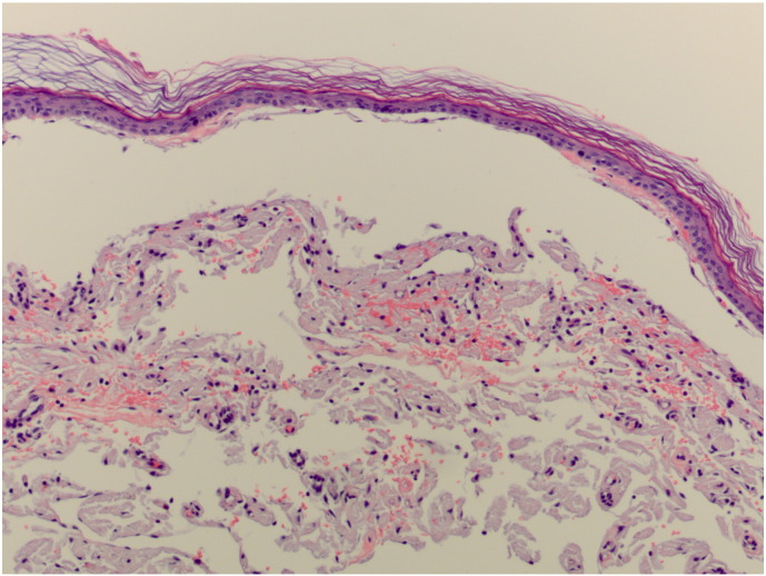
Hematoxylin and eosin slide from a punch biopsy showing amorphous, eosinophilic material in the dermis. There appears to be increased pigmentation in the basal layer of the epidermis.

Close up of the previous slide showing amorphous deposition of eosinophilic material. Classically, these depositions are found within the papillary dermis.

Congo-red stain highlighting the deposition of amyloid protein.

Congo-red stained slide under polarization demonstrating apple-green birefringence of the misfolded amyloid protein.
References
-
- Campbell M., Rosenthal A., Kundranda M., Pickert A., Dicaudo D., Dogan A. The blue man: a novel cutaneous manifestation of systemic amyloidosis. Amyloid. 2011;18:156–159. - PubMed
-
- Chandran N.S., Goh B.K., Lee S.S., Goh C.L. Case of primary localized cutaneous amyloidosis with protean clinical manifestations: lichen, poikiloderma-like, dyschromic and bullous variants. J Dermatol. 2011;38:1066–1071. - PubMed
-
- Colucci G., Alberio L., Demarmels Biasiutti F., Lämmle B. Bilateral periorbital ecchymoses. An often missed sign of amyloid purpura. Hamostaseologie. 2014;34:249–252. - PubMed
-
- Falk R.H., Comenzo R.L., Skinner M. The systemic amyloidoses. N Engl J Med. 1997;337:898–909. - PubMed
Publication types
LinkOut - more resources
Full Text Sources
Other Literature Sources
