Clinical Trial
. 2017 May 11:8:15086.
doi: 10.1038/ncomms15086.

Circulating tumour DNA sequence analysis as an alternative to multiple myeloma bone marrow aspirates

Affiliations
Clinical Trial

Circulating tumour DNA sequence analysis as an alternative to multiple myeloma bone marrow aspirates

Olena Kis et al. Nat Commun. .

Abstract

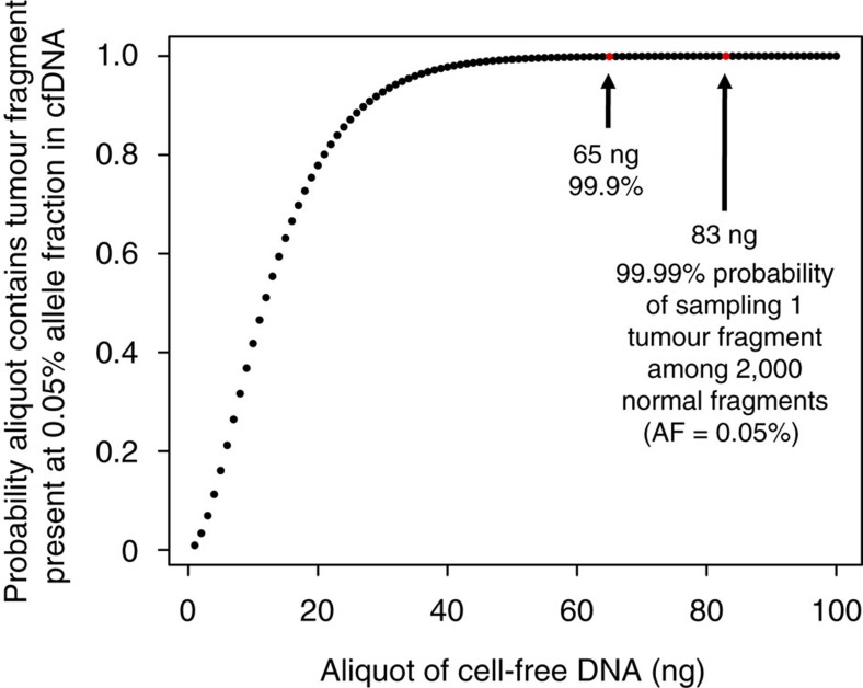
The requirement for bone-marrow aspirates for genomic profiling of multiple myeloma poses an obstacle to enrolment and retention of patients in clinical trials. We evaluated whether circulating cell-free DNA (cfDNA) analysis is comparable to molecular profiling of myeloma using bone-marrow tumour cells. We report here a hybrid-capture-based Liquid Biopsy Sequencing (LB-Seq) method used to sequence all protein-coding exons of KRAS, NRAS, BRAF, EGFR and PIK3CA in 64 cfDNA specimens from 53 myeloma patients to >20,000 × median coverage. This method includes a variant filtering algorithm that enables detection of tumour-derived fragments present in cfDNA at allele frequencies as low as 0.25% (median 3.2%, range 0.25-46%). Using LB-Seq analysis of 48 cfDNA specimens with matched bone-marrow data, we detect 49/51 likely somatic mutations, with subclonal hierarchies reflecting tumour profiling (96% concordance), and four additional mutations likely missed by bone-marrow testing (>98% specificity). Overall, LB-Seq is a high fidelity adjunct to genetic profiling of bone-marrow in multiple myeloma.


Conflict of interest statement

S.T. received research support from GlaxoSmithKline, Amgen, Oncoethix and Astellas and provided consultant services to Novartis. T.J.P. received research support from Boehringer Ingelheim. The remaining authors declare no competing financial interests.

Figures

Figure 1
Figure 1. Cell-free DNA concentrations in blood plasma of MM patients.
(a) Comparison of cfDNA yields in blood plasma samples from patients with MM and other cancer types (Supplementary Table 2). Effect of MM stage at diagnosis (b), disease status at blood draw, classified as newly diagnosed or early (1–3 prior lines of therapy) or late relapsed (>3 prior lines of therapy) (c), and lactase dehydrogenase concentrations in plasma at blood draw (d) on the cfDNA concentrations detectable in blood plasma of MM patients. For each group, the exact sample size is indicated in the figure, underneath group name. All data points represent unique measurements and do not include any technical or biological replicates. The distributions of cfDNA concentrations in each group are shown as box plots, where the central rectangle spans the first to the third quartile (interquartile range or IQR). A segment inside the rectangle shows the median, and ‘whiskers' above and below the box show the value 1.5 × IQR above or below the third or the first quartile, respectively. Wilcoxon signed-rank test or Kruskal–Wallis rank sum test was used for comparison of two or multiple groups, respectively, with P value of 0.05 considered statistically significant. Additional clinical measurements and cfDNA yields are provided in Supplementary Data 1.
Figure 2
Figure 2. Relationship between the amounts of cfDNA used for sequencing and the probability of capturing tumour-derived fragment.
A potential risk for allele drop-out exists when the aliquot of DNA used for these assays is insufficiently large to sample tumour fragments at low concentration within all circulating DNA. The binomial sampling model shown depicts the relationship between the potential risk of allele dropout and the amount of cfDNA used for preparation of DNA sequencing libraries and downstream sequencing. Under a binomial sampling model assuming a haploid genome mass of 3.5 pg, we calculated that to have a 99.99% chance of capturing a tumour fragment at 0.05% concentration, we need to input at least 83 ng of cfDNA into our assay.
Figure 3
Figure 3. Comparison of mutations identified by cfDNA sequence analysis and genetic profiling of BM-derived tumour DNA.
Sample IDs followed by (2) or (3) represent serial samples obtained on a separate clinical visit. EGFR protein changes are annotated using ENST00000275493 transcript. *Serial plasma samples from patients included in the training cohort that were excluded from validation of the Z-score threshold but were included in the overall calculations of sensitivity (concordance between LB-Seq and BM tumour DNA profiling data) and specificity of mutation calling across all samples analysed in this study (Supplementary Fig. 10); Single amino acid substitution resulting from a two-base genomic substitution. For BM samples with data from more than one source (specified by black diamond in BM analysis column), all sequencing results are presented in Supplementary Table 1.
Figure 4
Figure 4. Distribution of tumour LOD scores in cfDNA sequencing data.
For each sample, all candidate mutation calls generated by muTect version 1.1.4 were divided into subgroups based on the type of mutation or the filtering step at which they were removed, as indicated in the legend. The stripcharts showing the tumour LOD scores for each subgroup of mutations were overlaid with the boxplot demonstrating the distribution of tumour LOD scores for all mutation calls kept by muTect within each sample, prior to downstream filtering. The central rectangle spans the first to the third quartile (IQR), and segment inside the rectangle shows the median tumour LOD score for each sample. Sample-specific thresholds for calling likely somatic mutations (−) were determined as the tumour LOD score corresponding to the modified Z-score of 20 (that is, 20 MADs above the median). Unfiltered annotated data for all samples are available as Supplementary Data 2. Custom Rscripts (in R version 3.2.2) for filtering and plotting LOD score distribution data are available at www.github.com/pughlab/lb-seq.
Figure 5
Figure 5. Comparison of mutant allele frequencies in cfDNA and BM-derived tumour DNA.
For 11 subjects with multiple mutations in one or more genes, a scatter plot of cfDNA and BM-derived DNA allele fractions was generated for each mutation in each subject. Linear regression was then assessed for patients with three or more mutations to determine the strength of correlation (R2 value) (a). Comparison of relative tumour and cfDNA AFs and clonal hierarchies determined from cfDNA and BM in two patients, MYL-023 and 058, with three or more mutations in multiple genes (b) and two patients, MYL-020 and 028, with multiple mutations in the same gene (c). AF correlation plot for all cfDNA samples with matching BM samples (n=48) and bar graphs for other samples with multiple mutations excluded from b,c of this figure are presented as Supplementary Figures 14–16.

References

    1. Gertz M. A. et al.. Clinical implications of t(11;14)(q13;q32), t(4;14)(p16.3;q32), and -17p13 in myeloma patients treated with high-dose therapy. Blood 106, 2837–2840 (2005). - PMC - PubMed
    1. Kumar S. K. et al.. Improved survival in multiple myeloma and the impact of novel therapies. Blood 111, 2516–2520 (2008). - PMC - PubMed
    1. Chapman M. A. et al.. Initial genome sequencing and analysis of multiple myeloma. Nature 471, 467–472 (2011). - PMC - PubMed
    1. Lohr J. G. et al.. Widespread genetic heterogeneity in multiple myeloma: implications for targeted therapy. Cancer Cell 25, 91–101 (2014). - PMC - PubMed
    1. Chang-Yew Leow C., Gerondakis S. & Spencer A. MEK inhibitors as a chemotherapeutic intervention in multiple myeloma. Blood Cancer J. 3, e105 (2013). - PMC - PubMed

Publication types

MeSH terms