. 2017 May 11:8:15236.
doi: 10.1038/ncomms15236.

PPKs mediate direct signal transfer from phytochrome photoreceptors to transcription factor PIF3

Affiliations

PPKs mediate direct signal transfer from phytochrome photoreceptors to transcription factor PIF3

Weimin Ni et al. Nat Commun. .

Abstract

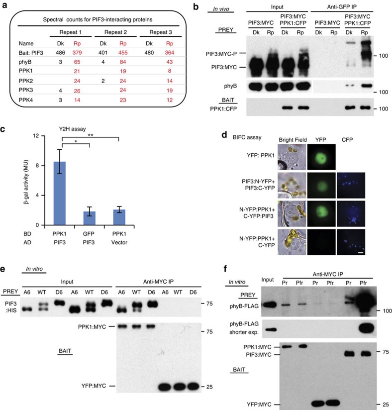
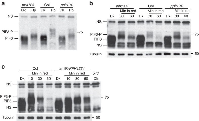
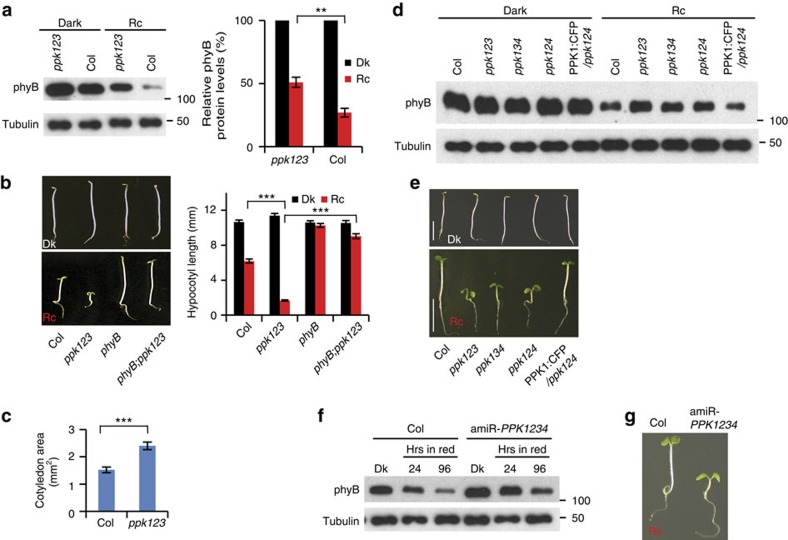
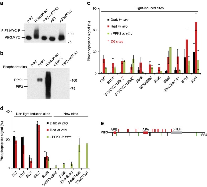
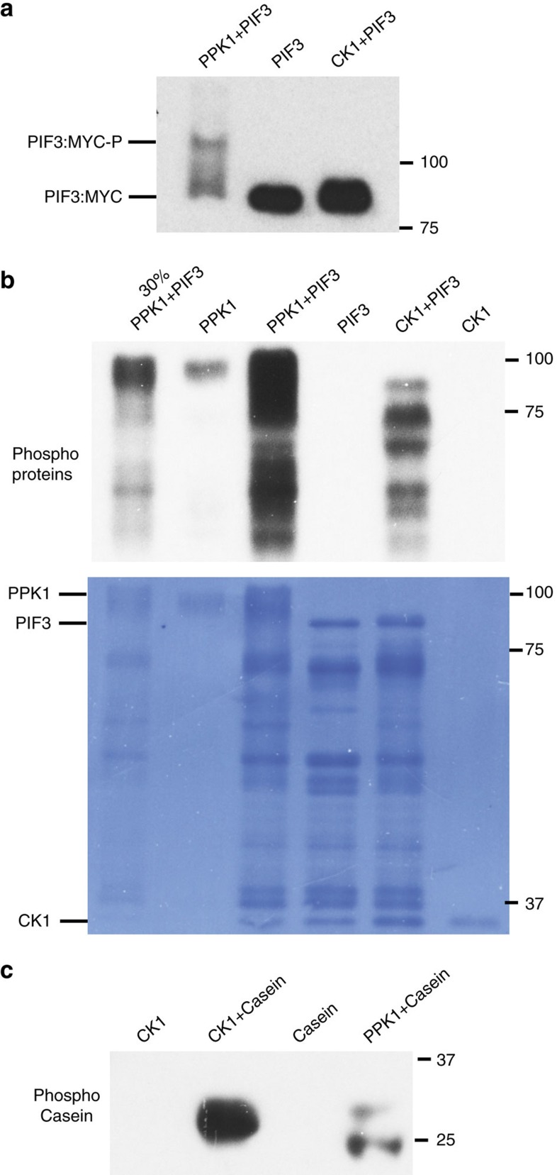
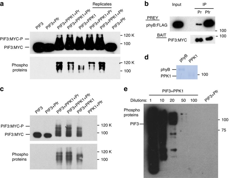
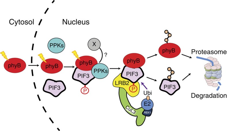
Upon light-induced nuclear translocation, phytochrome (phy) sensory photoreceptors interact with, and induce rapid phosphorylation and consequent ubiquitin-mediated degradation of, transcription factors, called PIFs, thereby regulating target gene expression and plant development. Nevertheless, the biochemical mechanism of phy-induced PIF phosphorylation has remained ill-defined. Here we identify a family of nuclear protein kinases, designated Photoregulatory Protein Kinases (PPK1-4; formerly called MUT9-Like Kinases (MLKs)), that interact with PIF3 and phyB in a light-induced manner in vivo. Genetic analyses demonstrate that the PPKs are collectively necessary for the normal light-induced phosphorylation and degradation of PIF3. PPK1 directly phosphorylates PIF3 in vitro, with a phosphosite pattern that strongly mimics the light-induced pattern in vivo. These data establish that the PPKs are directly involved in catalysing the photoactivated-phy-induced phosphorylation of PIF3 in vivo, and thereby are critical components of a transcriptionally centred signalling hub that pleiotropically regulates plant growth and development in response to multiple signalling pathways.


Conflict of interest statement

The authors declare no competing financial interests.

Figures

Figure 1
Figure 1. In vivo light promotes the interaction of PPKs with PIF3 and phyB.
(a) PIF3-interacting proteins detected by co-immunoprecipitation (co-IP) from cell extracts and subsequent mass spectrometric analysis. Spectral counts from three biological replicates of dark (Dk)-grown and red-light-pulse (Rp)-treated seedlings, respectively. (b) In vivo light-induced interaction of PPK1 with PIF3 and phyB detected by co-IP and subsequent immunoblot analysis. Protein extracts from Dk- or Rp-treated seedlings of the indicated genotypes were immunoprecipitated with anti-GFP antibodies, to pull down CFP-tagged PPK1 as bait, and the immunoblot was probed with either anti-MYC antibody (top, Prey), anti-phyB antibody (middle, Prey) or anti-GFP antibody (bottom, Bait) to detect PIF3:MYC, phyB and PPK1:CFP, respectively. (c) PIF3 and PPK1 interact in yeast-2-hybrid (Y2H) assays. LexA-DNA-binding-domain-fused PPK1 or GFP were used as bait, and B42 activation-domain-fused PIF3 or empty vector were used as prey in a standard Y2H configuration. Error bars represent standard error (s.e.) from three biological replicates. *P<0.05, **P<0.01 (Student's t-test). (d) PIF3 and PPK1 interact in transient-transfection, Bimolecular Fluorescence Complementation (BiFC) assays. Light-grown Nicotiana benthamiana leaves were transfected with the constructs indicated and then exposed to 10 min FR light and incubated in darkness for 72 h before microscopic analysis. Constructs used: YFP:PPK1, yellow fluorescent protein (YFP) fused to PPK1 protein; N-YFP and C-YFP, N- and C-terminal domains of split mVenus210 fluorescent protein, respectively, fused (or not) to PIF3 or PPK1 proteins. All split-Venus constructs also carried the mTq2 Golgi-localized marker as a positive control for transfection. Imaging configuration: Bright field, YFP emission filter; CFP (Cyan Fluorescent Protein) emission filter. Quantification of nuclei displaying split-Venus fluorescence shown in Supplementary Fig. 2a. Scale bar, 5μm. (e) Interaction of in vitro-synthesized recombinant PPK1 and PIF3 proteins detected by co-IP assays as described in b, except that the PPK1 bait was tagged with MYC (PPK1:MYC), and immunoprecipitated with anti-MYC antibodies, and the PIF3 prey was tagged with HIS (PIF3:HIS). YFP:MYC bait was used as a negative control. The immunoblot was probed with either anti-HIS antibody (top, Prey) or anti-MYC antibody (bottom, Bait). WT, wild-type PIF3 sequence; A6, PIF3 variant with phospho-dead mutations; D6, PIF3 variant with phosphomimic mutations. (f) In vitro-synthesized, recombinant PPK1 and phyB interact as detected by co-IP assays as described in b, except that the PPK1 bait was tagged with MYC (PPK1:MYC) and immunoprecipitated with anti-MYC antibodies, and the phyB prey was tagged with FLAG (phyB:FLAG). PIF3:MYC was used as a positive-control bait for light-induced phyB activation and YFP:MYC as a negative control. The immunoblot was probed with either anti-FLAG antibody (top and middle, Prey), or anti-MYC antibody (bottom, Bait). Pfr and Pr, samples were irradiated with R light only, or R followed by FR, respectively, before immunoprecipitation.
Figure 2
Figure 2. PPKs are collectively necessary for light-induced PIF3 phosphorylation and degradation in vivo.
Both light-induced phosphorylation and degradation of endogenous PIF3 are reduced in higher order ppk mutants. (a) Rapid mobility shift (phosphorylation) and (b) subsequent degradation in ppk1ppk2ppk3 (ppk123) and ppk1ppk2ppk4 (ppk124) triple mutants compared to wild-type (Col) in response to light. (c) Phosphorylation and degradation in artificial microRNA PPK1PPK2PPK3PPK4 (amiR-PPK1234) quadruple-knockdown mutant. Dark-grown (Dk) seedlings were irradiated with red light for 10 min (a, Rp), or the period indicated (b,c) before protein extraction and immunoblot analysis using anti-PIF3 antibodies. PIF3-P: phosphorylated PIF3; NS: nonspecific bands.
Figure 3
Figure 3. PPKs function collectively in promoting light-induced phyB degradation.
(a) Light-induced degradation of phyB is reduced in the ppk123 mutant. Seedlings were grown either in the dark (Dk) or continuous red right (Rc) for 4 days before protein extraction and immunoblot analysis using either anti-phyB antibody, or anti-tubulin as a loading control (left panel). Right panel shows quantification of the phyB levels relative to the tubulin protein levels from three biological replicates, expressed as a percentage of the dark-seedling levels for each genotype. Error bars represent s.e. **P<0.01 (Student's t-test). (b) A phyB null mutation suppresses the hypersensitive, short-hypocotyl phenotype of the ppk123 mutant in the light (phyB:ppk123). Average hypocotyl lengths of 4-day Dk- or Rc-grown seedlings are shown at right. Error bars represent s.e. from three biological replicates. ***P<0.001 (Student's t-test). (c) The light-hypersensitive phenotype of the ppk123 triple mutant includes enhanced cotyledon expansion compared to WT. Error bars represent s.e. from three biological replicates. ***P<0.001 (Student's t-test). (d) PPK1, PPK2, PPK3 and PPK4 are collectively necessary for normal PIF3 promoted phyB degradation in the light. Light-induced degradation of phyB is reduced in the ppk123, ppk134 and ppk124 triple-mutant backgrounds, and this reduced phyB degradation in the ppk124 triple-mutant can be rescued by transgenic expression of PPK1:CFP (PPK1:CFP/ppk124). Seedlings of the indicated genotypes were grown for 4 days in the dark or continuous red light (Rc) before protein extraction and western blot analysis using an anti-phyB antibody, or anti-tubulin as a loading control. (e) PPKs are collectively necessary for normal hypocotyl responsiveness to light. Seedlings of the indicated genotypes were grown as in d. Scale bars, 5 mm. (f) Light-induced degradation of phyB is reduced in the amiR-PPK1234 line. Seedling growth and western blot were done as in d. (g) The amiR-PPK1234 line is hypersensitive to red light. Seedlings were grown for 4 days in red light.
Figure 4
Figure 4. PPK1 phosphorylates PIF3 at light-inducible phosphosites in vitro.
(a) PPK1 induces a strong mobility shift in PIF3 in an in vitro kinase assay. PPK1 and MYC-tagged PIF3 variants, affinity-purified after expression in E. coli, were combined under protein-kinase-assay conditions and examined for an induced, potentially phosphorylation-related, mobility-shift in PIF3 by immunoblot blot analysis using anti-MYC antibody. mPPK1: kinase-dead mutant of PPK1; A20: phospho-dead mutant of PIF3 mutated in the 20 phosphoresidues induced by light in vivo. (b) PPK1 phosphorylates PIF3 in vitro. Phosphoproteins from the indicated in vitro protein-kinase-assay combinations were detected by western blot using pIMAGO-biotin. (c,d) Mass-spectrometric analysis of in vitro, PPK1-catalysed phosphosites in PIF3 compared to those sites established as rapidly induced (c), or unaffected (d), by red light (Rp) in vivo. d (left), constitutively phosphorylated, non-light-induced in vivo, (right) not detectably phosphorylated in dark or light in vivo, PPK1-induced in vitro. Phosphopeptide Signal (%) corresponds to the percent of the residues at each site that are phosphorylated in Dk-grown or Rp-treated seedlings, or in PPK1-treated PIF3 in vitro. D6, strongly light-induced sites in vivo. Data are the means of biological repeats±s.e. (e) Schematic depiction of PIF3 phosphosites. Red bars represent phosphosites that are both light-induced in vivo and catalysed by PPK1 in vitro. Black bars represent sites that are constitutively phosphorylated in vivo, but not detectably phosphorylated by PPK1 in vitro, except S323. Green bars represent newly identified phosphosites catalysed by PPK1 in vitro. APB, active phyB-binding domain; APA, active phyA-binding domain; bHLH, basic helix-loop-helix domain.
Figure 5
Figure 5. Specificity of in vitro phosphorylation of PIF3 by PPK1.
(a) Unlike PPK1, CK1 does not induce a strong phosphorylation-related mobility-shift in PIF3 in vitro. PIF3 from the in vitro kinase assays as indicated was detected by immunoblot using a MYC antibody. CK1: rat Casein Kinase 1d. (b) PIF3 phosphorylation by CK1 is qualitatively different and quantitatively less than that by PPK1. Phosphoproteins from the indicated in vitro kinase assays were detected by immunoblot using pIMAGO-biotin. Signals from the kinase-only tracks indicate auto-phosphorylation activity of PPK1. A dilution (30%) of the proteins in the third lane was loaded in the first lane. Bottom panel shows the coomassie-stained membrane. Lower bands are presumptive PIF3 degradation products. (c) Casein phosphorylation by CK1 is greater than, and qualitatively different to, that by PPK1. Phospho-Casein from the indicated in vitro kinase assays was detected by immunoblot using pIMAGO-biotin.
Figure 6
Figure 6. phyB stimulates PPK1-catalysed PIF3-phosphorylation in a non-light-dependent manner in vitro but does not detectably autonomously phosphorylate PIF3.
(a) Mobility-shift (upper panel) and pIMAGO (lower panel) assays detect differential PPK1-catalysed phosphorylation of PIF3 in vitro. In vitro protein kinase reactions containing the protein combinations indicated were performed, followed by detection of the PIF3:MYC fusion protein by immunoblot, using anti-MYC antibodies (upper panel), and detection of phosphoproteins using pIMAGO-biotin on immunoblots (lower panel). Pr or Pfr indicate inclusion of the inactive or active conformer of phyB, respectively. Replicates indicate biological replicates of the kinase assays. (b) The functional activity of phyB-Pfr was verified using an established in vitro pull-down assay with PIF3 as bait. The immunoblot was probed with an MYC (for PIF3 bait) or FLAG (for phyB prey) antibody. (c) Detection of PPK1-catalysed PIF3, and potential phyB, phosphorylation in vitro using mobility-shift (upper panel) and pIMAGO (lower panel) assays as in a. (d) Comassie staining of phyB and PPK1 proteins used in the kinase assays in a. (e) PPK1 kinase activity is at least 50-fold greater than any potential intrinsic phyB kinase activity. Phosphoproteins from the in vitro kinase reactions indicated were detected by immunoblot using pIMAGO-biotin. Numbers refer to the fold-dilutions of the PIF3+PPK1 in vitro kinase reaction shown in a compared to the undiluted PIF3+Pfr reaction.
Figure 7
Figure 7. Model depicting proposed mechanism of light-induced PIF3 phosphorylation.
Photoactivated phyB translocates rapidly into the nucleus and forms a trimolecular complex with PIF3 and one or more PPKs, which catalyse multisite transphosphorylation of PIF3. The LRB Cullin 3 E3 ligases then recognize phosphorylated PIF3, triggering concurrent ubiquitination and degradation of both PIF3 and phyB. X: potential additional factor contributing to Pfr-induced PIF3 phosphorylation in vivo, but missing from in vitro kinase assays performed here.

References

    1. Rockwell N. C., Su Y. S. & Lagarias J. C. Phytochrome structure and signaling mechanisms. Annu. Rev. Plant Biol. 57, 837–858 (2006). - PMC - PubMed
    1. Leivar P. & Quail P. H. PIFs: pivotal components in a cellular signaling hub. Trends Plant Sci. 16, 19–28 (2011). - PMC - PubMed
    1. Ni M., Tepperman J. M. & Quail P. H. Binding of phytochrome B to its nuclear signalling partner PIF3 is reversibly induced by light. Nature 400, 781–784 (1999). - PubMed
    1. Ni W. et al.. Multisite light-induced phosphorylation of the transcription factor PIF3 is necessary for both its rapid degradation and concomitant negative feedback modulation of photoreceptor phyB levels in Arabidopsis. Plant Cell 25, 2679–2698 (2013). - PMC - PubMed
    1. Shen H. et al.. Light-induced phosphorylation and degradation of the negative regulator PHYTOCHROME-INTERACTING FACTOR1 from Arabidopsis depend upon its direct physical interactions with photoactivated phytochromes. Plant Cell 20, 1586–1602 (2008). - PMC - PubMed

Publication types

MeSH terms