Review
. 2017 May 11:3:17026.
doi: 10.1038/nrdp.2017.26.

Pemphigus

Affiliations
Review

Pemphigus

Michael Kasperkiewicz et al. Nat Rev Dis Primers. .

Abstract

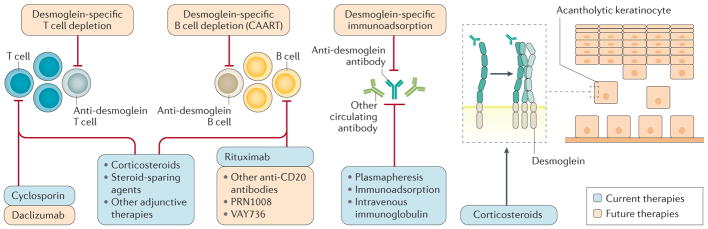
Pemphigus is a group of IgG-mediated autoimmune diseases of stratified squamous epithelia, such as the skin and oral mucosa, in which acantholysis (the loss of cell adhesion) causes blisters and erosions. Pemphigus has three major subtypes: pemphigus vulgaris, pemphigus foliaceus and paraneoplastic pemphigus. IgG autoantibodies are characteristically raised against desmoglein 1 and desmoglein 3, which are cell-cell adhesion molecules found in desmosomes. The sites of blister formation can be physiologically explained by the anti-desmoglein autoantibody profile and tissue-specific expression pattern of desmoglein isoforms. The pathophysiological roles of T cells and B cells have been characterized in mouse models of pemphigus and patients, revealing insights into the mechanisms of autoimmunity. Diagnosis is based on clinical manifestations and confirmed with histological and immunochemical testing. The current first-line treatment is systemic corticosteroids and adjuvant therapies, including immunosuppressive agents, intravenous immunoglobulin and plasmapheresis. Rituximab, a monoclonal antibody against CD20+ B cells, is a promising therapeutic option that may soon become first-line therapy. Pemphigus is one of the best-characterized human autoimmune diseases and provides an ideal paradigm for both basic and clinical research, especially towards the development of antigen-specific immune suppression treatments for autoimmune diseases.


Conflict of interest statement

Competing interests

M.A. receives speaker honoraria and grants from Nihon Pharmaceutical, research support from Medical & Biological Laboratories, Health Sciences Research Grants for Research on Rare and Intractable Diseases from Ministry of Health, Labour, and Welfare, and grants from Japan Society for the Promotion of Science and Agency for Medical Research and Development. H.T. receives grants from Japan Society for the Promotion of Science. A.S.P. is a consultant for Syntimmune and TG Therapeutics and receives grants or research support from the US NIH (R01-AR057001, R56-AR064220 and R01-068288), the Dermatology Foundation and Sanofi. She is co-inventor on a patent related to chimeric immunoreceptor therapy of pemphigus. The content is solely the responsibility of the authors and does not necessarily represent the official views of the US NIH. C.T.E. receives grants or research support from Deutsche Forschungsgemeinschaft (EL711/1-1) and is co-inventor on a patent related to chimeric immunoreceptor therapy of pemphigus. D.Z. is an advisory board member for Roche Pharma, and a consultant for Euroimmun, Almirall, UCB, Fresenius and arGEN-X. He received speakers honoraria and/or travel/accommodations/meeting compensation from Biotest, Fresenius, Miltenyi, Roche Pharma, Biogen Idec, AbbVie, UCB, Janssen and grants or research support from Euroimmun, Miltenyi, Fresenius, Biotest, Dompe, Almirall, Biogen and Roche. He holds the patent Euroimmun: DE 10 2006 059 574 A1. M.K. and J.Y. declare no competing interests.

Figures

Figure 1
Figure 1. Clinical features of pemphigus
Pemphigus vulgaris can present with erosions on the buccal mucosa (part a) and cutaneous flaccid blisters and haemorrhagic erosions (part b). Scaly, crusted erosions in pemphigus foliaceus (part c). Intractable stomatitis in paraneoplastic pemphigus, with erosions and ulcerations that characteristically extend onto the vermilion (the edge between the lip and the adjacent skin; partd).
Figure 2
Figure 2. Pathogenesis of pemphigus
a | The distribution and expression levels of desmoglein 1 (DSG1; blue coloured box in the first column) and DSG3 (yellow coloured box in the first column) vary in the skin and mucous membranes. The compensation theory states that the development of blisters in the skin or mucosa or both depends on the DSG type being targeted by the autoantibodies. In the mucosal-dominant type of pemphigus vulgaris, anti-DSG3 IgG antibodies induce erosions in the oral mucosa, where DSG3 is the most abundant DSG type, but fail to induce cutaneous blisters, as there is compensation from DSG1. Similarly, the anti-DSG1 IgG antibodies in pemphigus foliaceus induce superficial blisters in the skin but not in the oral mucosa. In the mucocutaneous type of pemphigus vulgaris, both anti-DSG3 and anti-DSG1 IgG antibodies are present, resulting in extensive blisters and erosions of the skin and mucous membranes. In paraneoplastic pemphigus, in addition to acantholysis (loss of cell adhesion) induced by humoral autoimmune reactions, a cellular autoimmune response to the epidermis is also observed, which results in keratinocyte apoptosis with T cell infiltration within the epidermis, obscuring of the boundary of epidermis and dermis, and a band-like dense T cell infiltration in the upper dermis indicating interface dermatitis. b | Haematoxylin and eosin staining demonstrates a suprabasilar and subcorneal blister with acantholysis in skin biopsies from patients with pemphigus vulgaris and pemphigus foliaceus, respectively, and interface dermatitis in addition to blister formation in a sample from a patient with paraneoplastic pemphigus.
Figure 3
Figure 3. Mechanisms of acantholysis
Anti-desmoglein autoantibodies can cause acantholysis through steric hindrance of desmoglein-mediated trans- (between molecules on opposing cells) and cis- (between molecules on the same cells) adhesion, inhibition of desmosome assembly and promotion of desmosome disassembly (by clustering and/or endocytosis of desmogleins) and stimulation of signalling pathways that can synergize with these mechanisms to modulate keratinocyte cell adhesion. In pemphigus vulgaris, IgG-induced desmoglein 3 endocytosis is regulated by the p38 mitogen-activated protein kinase (MAPK) pathway and its downstream effector MAPK-activated protein kinase 2 (MK2). Inhibition of either p38 or MK2 prevents pemphigus vulgaris IgG-induced acantholysis and monoclonal antibody-induced spontaneous blistering in passive transfer mouse models,. p38 similarly regulates pemphigus foliaceus IgG-induced acantholysis. The trans-adhesive and cis-adhesive interactions can be homophilic (that is, between desmogleins) or heterophilic (between desmoglein and desmocollin). Dashed arrows indicate that the signal transduction mechanism can affect both steric hindrance and desmoglein depletion. DAG, diacylglycerol; DP, desmoplakin; HSP27, heat shock protein 27; PG, plakoglobin; PIP2, phosphatidylinositol 4,5-bisphosphate; PKC, protein kinase C; PKP, plakophilin; PLC, phospholipase C.
Figure 4
Figure 4. B cell and T cell response in pemphigus
Dendritic cells presenting desmoglein (DSG) antigens can activate CD4+ and CD8+ T cells. These autoreactive T cells can be identified even in healthy individuals and mainly produce IL-10 and interferon-γ (IFNγ). IFNγ has a potential to suppress DSG-specific T helper 2 (TH2) cell development. IL-10 is a key mediator of the peripheral tolerance mechanism that suppresses the activity of pathogenic TH2 cells. However, IL-10 has complex effects, as it can promote immunoglobulin class switching to IgG4, the predominant subclass of anti-DSG antibodies, and has opposite effects in different phases of disease pathogenesis. IL-4-producing TH cells can be isolated from patients with pemphigus (but not healthy individuals) and presumably drive anti-DSG antibody production from B cells. Acantholysis seen in pemphigus vulgaris and pemphigus foliaceus is induced by a humoral autoimmune response. DSG-reactive (or reactive to other epidermal autoantigens) CD4+ and CD8+ T cells with cytotoxic activity can rarely be generated and mediate a cellular autoimmune response that causes interface dermatitis, which can be observed in paraneoplastic pemphigus. TCR, T cell receptor; Tr1, T regulatory type 1.
Figure 5
Figure 5. Mouse models for pemphigus
a | Passive transfer model. IgG fractions prepared from patients’ sera, or other engineered anti-desmoglein 3 (DSG3) antibodies, are injected intraperitoneally or subcutaneously into neonatal mice. The model develops blisters that show the typical histology observed in patients, with IgG deposition on keratinocyte cell surfaces and an acantholytic blister (asterisk).b | Active disease model. DSG3-deficient (Dsg3−/−) mice do not establish tolerance against DSG3 because DSG3 is never exposed to immune cells. After adoptive transfer of lymphocytes from Dsg3−/− mice, immunodeficient mice, such as Rag2−/− mice, stably produce anti-DSG3 IgG antibodies and show the typical gross and histological phenotype of the mucosal-dominant type of pemphigus vulgaris. This model mice also develop telogen hair loss (a feature that is not observed in patients to the same extent) because, in mice, intercellular adhesion of follicular epidermis is mainly mediated by DSG3 during the telogen (rest) phase of hair growth,. Another active disease model is generated by adoptive transfer of DSG3-specific T cells prepared from transgenic mice expressing DSG3-specific T cell receptor (TCR) to immunodeficient mice. Transferred T cells directly infiltrate into the skin and attack DSG3-expressing keratinocytes, causing experimental autoimmune dermatitis (EAD). This model is useful to analyse T cell- mediated cellular autoimmune mechanisms in T cell-mediated skin inflammation. c | Humanized model. In this model, MHC class II (MHC II)-deficient mice are engineered to express transgenic HLA-DRB1*0402. After immunization with human recombinant DSG3, the mice produce anti-human DSG3 antibodies that induce blister formation in human skin specimens. Scale bar indicates 50 μm. Histology image in part c courtesy of M. Hertl and R. Eming, Philipps University of Marburg, Germany.
Figure 6
Figure 6. Immunopathological characteristics of pemphigus vulgaris, pemphigus foliaceus and paraneoplastic pemphigus
Direct immunofluorescence microscopy of skin biopsies of perilesions from patients with pemphigus vulgaris (parta) and pemphigus foliaceus (part b) reveals an intercellular staining of IgG antibodies. Indirect immunofluorescence microscopy of a monkey oesophagus exposed to the serum of a patient with pemphigus vulgaris shows an epithelial intercellular surface staining of IgG (part c). Indirect immunofluorescence microscopy of a rat bladder exposed to the serum of a patient with paraneoplastic pemphigus presents both epithelial intercellular and cytoplasmic staining (part d).
Figure 7
Figure 7. Current and future therapeutic strategies for pemphigus
Current and future therapeutic strategies for pemphigus are outlined based on their mechanisms of action. Whereas conventional treatments such as systemic steroid and immunosuppressive agents affect broad ranges of cells and tissues, antigen-specific treatments targeted against desmoglein-reactive cells or IgG antibodies provide more-tailored and potentially safer strategies. One of these approaches uses T cells that are engineered to express a chimeric immunoreceptor consisting of the desmoglein 3 extracellular domain fused to the T cell receptor cytoplasmic signalling and co-stimulatory domains. These desmoglein 3 chimeric autoantibody receptor T cells (CAARTs) specifically bind to and kill anti-desmoglein 3 B cells, leading to disease remission in a pemphigus mouse model. Owing to the potential for generation of long-term memory CAARTs, this technology offers the potential for long-term disease remission without the immunosuppressive effects from global B cell depletion.

Comment in

References

    1. Amagai M. In: Dermatology. Bolognia JL, Jorizzo JL, Schaffer JV, editors. Vol. 1. Mosby; 2012. pp. 461–474. Ch. 29.
    1. Amagai M, Klaus-Kovtun V, Stanley JR. Autoantibodies against a novel epithelial cadherin in pemphigus vulgaris, a disease of cell adhesion. Cell. 1991;67:869–877. This study reports the cDNA cloning of a pemphigus vulgaris antigen, which turned out to be a desmosomal cadherin called desmoglein 3. - PubMed
    1. Koch PJ, et al. Identification of desmoglein, a constitutive desmosomal glycoprotein, as a member of the cadherin family of cell adhesion molecules. Eur J Cell Biol. 1990;53:1–12. - PubMed
    1. Stanley JR, Amagai M. Pemphigus, bullous impetigo, and the staphylococcal scalded-skin syndrome. N Engl J Med. 2006;355:1800–1810. - PubMed
    1. Anhalt GJ, et al. Paraneoplastic pemphigus. An autoimmune mucocutaneous disease associated with neoplasia. N Engl J Med. 1990;323:1729–1735. - PubMed