. 2017 May 11:8:15295.
doi: 10.1038/ncomms15295.

Tau association with synaptic vesicles causes presynaptic dysfunction

Affiliations

Tau association with synaptic vesicles causes presynaptic dysfunction

Lujia Zhou et al. Nat Commun. .

Abstract

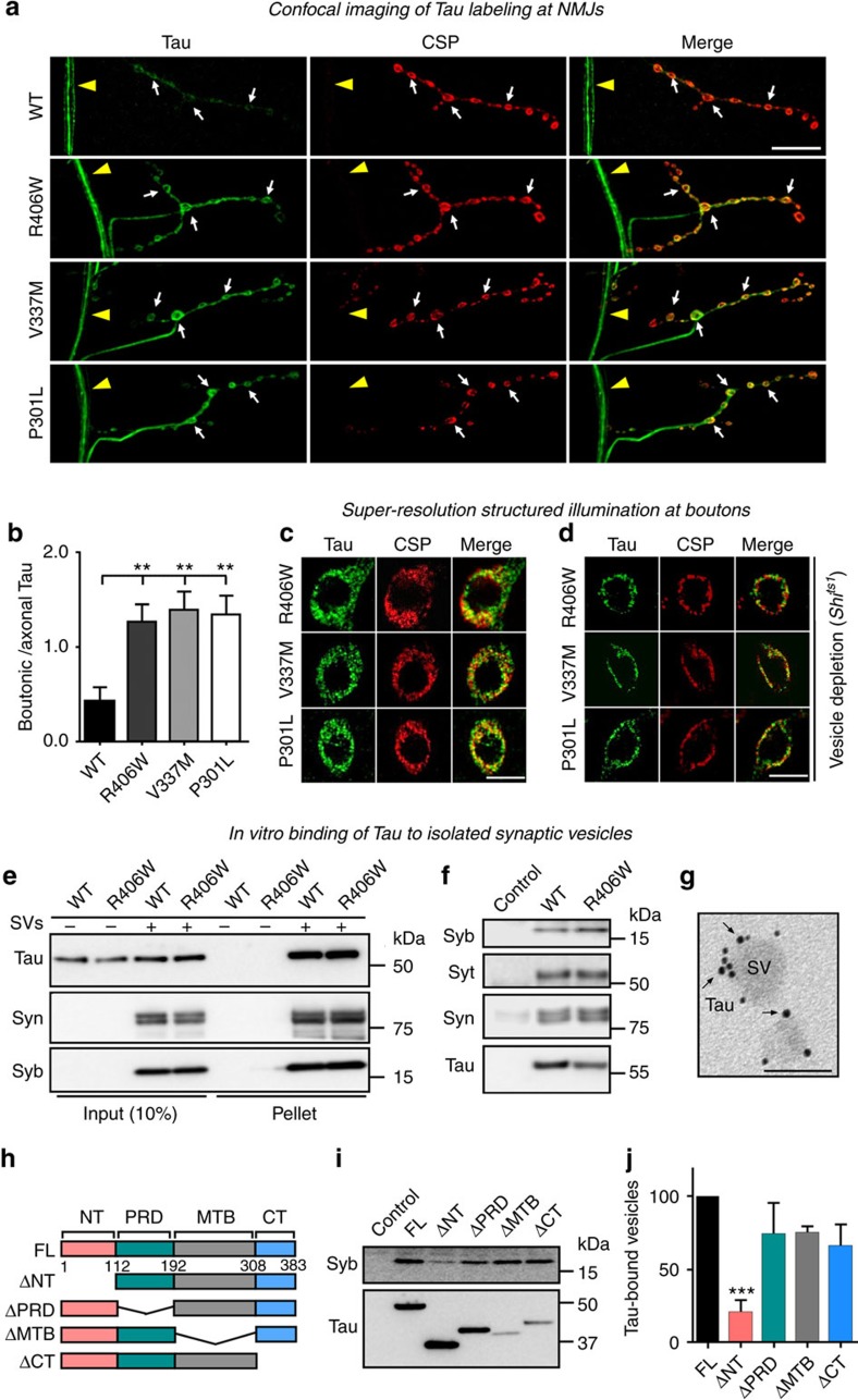
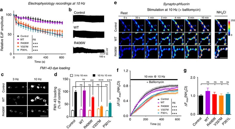
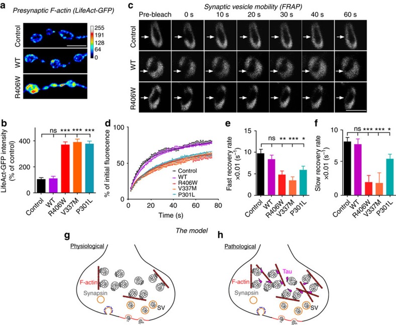
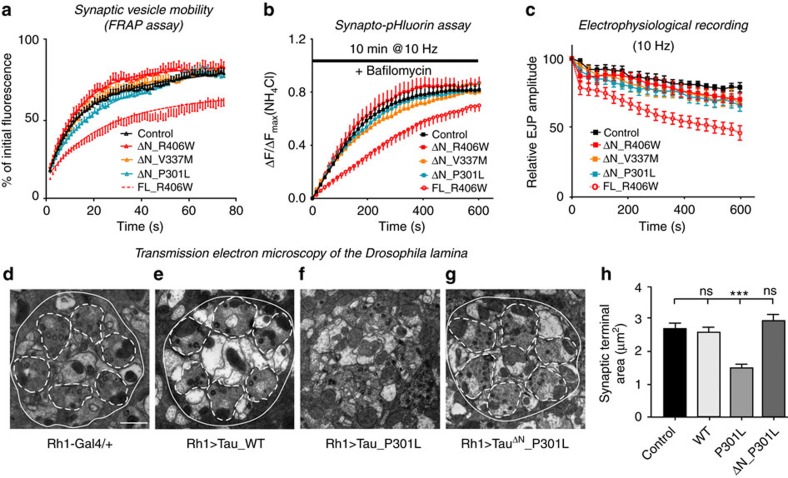
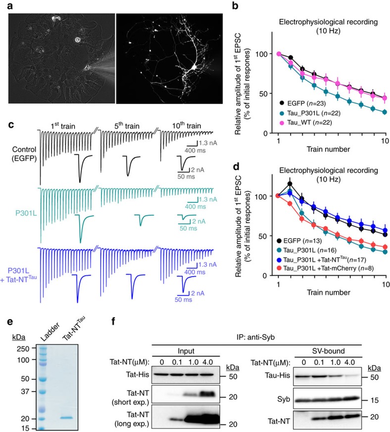
Tau is implicated in more than 20 neurodegenerative diseases, including Alzheimer's disease. Under pathological conditions, Tau dissociates from axonal microtubules and missorts to pre- and postsynaptic terminals. Patients suffer from early synaptic dysfunction prior to Tau aggregate formation, but the underlying mechanism is unclear. Here we show that pathogenic Tau binds to synaptic vesicles via its N-terminal domain and interferes with presynaptic functions, including synaptic vesicle mobility and release rate, lowering neurotransmission in fly and rat neurons. Pathological Tau mutants lacking the vesicle binding domain still localize to the presynaptic compartment but do not impair synaptic function in fly neurons. Moreover, an exogenously applied membrane-permeable peptide that competes for Tau-vesicle binding suppresses Tau-induced synaptic toxicity in rat neurons. Our work uncovers a presynaptic role of Tau that may be part of the early pathology in various Tauopathies and could be exploited therapeutically.


Conflict of interest statement

The authors declare no competing financial interests.

Figures

Figure 1
Figure 1. Tau localizes to presynaptic terminals and binds to synaptic vesicles via its N-terminal domain.
(a) Tau and CSP immunolabeling at neuromuscular junctions (NMJs) of Drosophila larvae expressing WT or FTDP-17 pathogenic mutant Tau (R406W, V337M or P301L) under the D42-Gal4 motor neuron driver. Axons (arrowheads) and synaptic boutons (arrows) are indicated. Scale bar, 20 μm. (b) Quantification of fluorescence intensity of Tau within synaptic boutons (SBs) as ratio to the intensity of axonal Tau. One-way ANOVA, **P=0.0030, 0.0019, 0.0041 (R406W, V337M, P301L) n=10 (R406W, V337M, P301L) or 12 (WT) NMJs from 5 to 6 animals. Data present mean±s.e.m. (c,d) Super-resolution structured illumination microscopy analysis of Tau and CSP immunolabeling within SBs under non-treated condition (c) or after depletion of synaptic vesicles in Shits1 mutant background by KCl stimulation at the non-permissive temperature (d). Scale bar, 5 μm. (e,f) Immunoblots of Tau (anti-His tag) and synaptic vesicle (SV) proteins Synaptobrevin (Syb), Synaptotagmin (Syt) and Synapsin (Syn) from sedimentation assay (e) and co-immunoprecipitation (co-IP) using anti-His antibodies (f) assessing recombinant human Tau binding to purified synaptic vesicles. (g) Electron microscopy imaging of recombinant Tau (probed by Ni-NTA-Nanogold) bound to ultrapure synaptic vesicles in vitro. Scale bar, 50 nm. (hj) Mapping of the vesicle-binding domain of Tau in vitro by co-IP assay. Truncations of the N-terminal (NT), proline-rich (PRD), microtubule-binding (MTB) or C-terminal (CT) domains of Tau were generated as indicated in the schematic (h). Immunoblots of recombinant Tau domain-truncations and Syb (SV marker) from co-IP using anti-His antibodies (i) and quantification of relative Syb intensity (j). One-way ANOVA, ***P=0.002, n=3 independent experiments.
Figure 2
Figure 2. Pathogenic mutant Tau reduces synaptic transmission and vesicle cycling/release during sustained high-frequency stimulation.
Drosophila larvae used in these assays express UAS-Tau (WT, R406W, V337M or P301L) under the D42-Gal4 motor neuron driver. (a,b) Electrophysiological recordings of synaptic transmission during 10 Hz stimulation for 10 min. Plot of evoked junction potential (EJP) amplitudes (a) and representative traces (b). Two-way ANOVA, ***P<0.0001, n=7 (Control, WT) or 9 (R406W, V337M, P301L) NMJs (animals). (c,d) FM1-43 dye loading with stimulation at 3 Hz (recycling vesicle pool) or 10 Hz (reserve vesicle pool) for 10 min. Representative images of FM1-43 dye loading (c) and plot of FM1-43 dye loading intensity (d). One-way ANOVA, **P=0.0028 (R406W), ***P=0.0001(V337M, P301L), n=14 (WT, R406W, V337M, P301L) or 20 (Control) NMJs (animals). Scale bar, 10 μm. (eg) Synapto-pHluorin (SpH) responses to stimulation at 10 Hz with the presence of bafilomycin. Representative images of SpH responses (e) and plot of fluorescence change ΔF at ratio to maximal ΔF (NH4Cl dequenching) (f). Two-way ANOVA, ***P<0.0001, n=7 (WT, R406W, V337M, P301L) or 11 (Control) NMJs (animals). Plot of maximal ΔF (NH4Cl dequenching) calibrated to control levels (g). One-way ANOVA, ns, not significant. Scale bar, 20 μm. Data present mean±s.e.m.
Figure 3
Figure 3. Pathogenic mutant Tau increases F-actin levels and reduces synaptic vesicle mobility at presynaptic terminals.
Drosophila larvae used in these assays express UAS-Tau (WT, R406W, V337M or P301L) under the D42-Gal4 motor neuron driver. (a,b) Immunolabeling of LifeAct-GFP probing for F-actin within synaptic boutons. Representative images of immunolabeling (a) and quantification of LifeAct-GFP intensity (b). One-way ANOVA, ***P<0.0001, n=12 (WT, R406W, V337M, P301L) or 14 (Control) NMJs (two NMJs per animal). Scale bar, 5 μm. (cf) FRAP measurement of vesicle mobility within synaptic boutons. Representative images acquired immediately before photobleaching (pre-bleach) and immediately after bleaching at 0–60 s post-bleaching time points (c). Plot of fluorescence recovery (% of initial fluorescence) over time and fit with double-exponential curve (d). Plot of fast recovery rates calculated from fluorescence recovery curve (e) One-way ANOVA, **P=0.0011 (R406W), ***P=0.0001 (V337M), *P=0.0158 (P301L), n=24, 21, 22, 20, 25 (Control, WT, R406W, V337M, P301L) boutons (3–5 boutons per animal). Plot of slow recovery rates (f). One-way ANOVA, *P=0.0001 (R406W, V337M), *P=0.0250 (P301L), n=24, 21, 22, 20, 25 (Control, WT, R406W, V337M, P301L) boutons (3–5 boutons per animal). Scale bar, 5 μm. Data present mean±s.e.m. (g,h) Proposed model of Tau clustering synaptic vesicles to F-actin to restrict reserve pool vesicle mobilization and release.
Figure 4
Figure 4. Interfering with Tau N-terminal-dependent vesicle-binding reverts Tau-induced presynaptic deficits in fly neurons.
Drosophila larvae used in (ac) express UAS-TauΔN (R406W, V337M or P301L) under the D42-Gal4 motor neuron driver. (a) FRAP measurements of vesicle mobility within synaptic boutons. Fluoresence recovery (% of initial fluorescence) was plotted over time and fit with double-exponential curves. n=22 (R406W), 20 (ΔN_R406W, ΔN_V337M), 24 (ΔN_P301L) or 25 (Control) boutons (3–5 boutons per animal). (b) Synapto-pHluorin responses to stimulation at 10 Hz with the presence of bafilomycin. Fluorescence change ΔF at ratio to maximal ΔF (NH4Cl dequenching) was plotted over time during the stimulation. Two-way ANOVA, n=7 (R406W, ΔN_R406W, ΔN_V337M, ΔN_P301L),9 (Control) NMJs (animals). (c) Electrophysiological recordings of EJP amplitudes during stimulation at 10 Hz. Two-way ANOVA, n=7 (R406W, ΔN_R406W), 8 (ΔN_V337M, ΔN_P301L), or 9 (Control) NMJs (animals). (dh) Transmission electron microscopy (TEM) of lamina sections of 9-day-old flies expressing UAS-Tau (WT, P301L or ΔN_P301L) under the late-onset retinal driver Rhodopsin1-Gal4 (Rh1). In control Rh1-Gal4/+ animals (d), six photoreceptor synaptic terminals (outlined in dashed white lines) are converged in a ‘cartridge' (outlined in white line). Disrupted synaptic terminal organization and morphology are detected in Rh1>Tau_P301L (f) flies, whereas no obvious abnormalities at the synaptic terminals of Rh1>Tau_WT (e) and Rh1>TauΔN_P301L (g) flies. Scale bar, 1 μm. (h) Quantification of the synaptic terminal area based on the electromicrographs. One-way ANOVA, ***P=0.0001. n=5 animals per genotype. Data present mean±s.e.m.
Figure 5
Figure 5. Interfering with Tau N-terminal-dependent vesicle-binding rescues Tau-induced presynaptic deficits in cultured rat hippocampal neurons.
Autaptic rat hippocampal neuronal cultures were transduced with AAV viral vectors expressing GFP or Tau variants. (a) Autaptic neuronal culture transduced with AAV-EGFP is visualized by bright-field illumination (left) and GFP fluorescence (right). (bd) Electrophysiological recordings using patch clamp in response to 10 consecutive high frequency stimulation trains (10 Hz for 10 s with 30 s interval). The representative traces are shown in (c) and the relative first EPSCs were plotted to train numbers (b,d). In (d), TauP301L expressing neurons were acutely treated with 5 μM Tat-NTTau or control Tat-mCherry peptides before patch clamp recordings. The experiments were independently repeated at least three times and the number of neurons recorded is indicated in the graph. Data present mean±s.e.m. (e) Colloidal coomassie staining of purified Tat-NTTau peptide; NTTau corresponds to the N-terminal domain (aa 1–113) of Tau. (f) Tat-NTTau peptide competes with full-length Tau for vesicle binding in vitro. Intact synaptic vesicles (SVs) were immobilized to Dynabeads using anti-Synaptobrevin 2 (Syb) antibodies. Following immobilization, SVs were incubated with recombinant full-length Tau (40 nM) with or without the presence of 0.1, 1.0 or 4.0 μM purified Tat-NTTau peptide. Immunoblots were probed for full-length Tau, Tat-NTTau and the synaptic vesicle marker Syb. Note that Tat-NTTau reduced the amount of full-length Tau bound to SVs in a dose-dependent manner.

References

    1. Morris M., Maeda S., Vossel K. & Mucke L. The many faces of tau. Neuron 70, 410–426 (2011). - PMC - PubMed
    1. Ballatore C., Lee V. M. & Trojanowski J. Q. Tau-mediated neurodegeneration in Alzheimer's disease and related disorders. Nat. Rev. Neurosci. 8, 663–672 (2007). - PubMed
    1. Wang Y. & Mandelkow E. Tau in physiology and pathology. Nat. Rev. Neurosci. 17, 22–35 (2016). - PubMed
    1. Kwok J. B. et al.. Tau haplotypes regulate transcription and are associated with Parkinson's disease. Ann. Neurol. 55, 329–334 (2004). - PubMed
    1. Refenes N. et al.. Role of the H1 haplotype of microtubule-associated protein tau (MAPT) gene in Greek patients with Parkinson's disease. BMC Neurol. 9, 26 (2009). - PMC - PubMed

Publication types