. 2017 May 11;8(5):e2773.
doi: 10.1038/cddis.2017.183.

Non-junctional Cx32 mediates anti-apoptotic and pro-tumor effects via epidermal growth factor receptor in human cervical cancer cells

Affiliations

Non-junctional Cx32 mediates anti-apoptotic and pro-tumor effects via epidermal growth factor receptor in human cervical cancer cells

Yifan Zhao et al. Cell Death Dis. .

Abstract

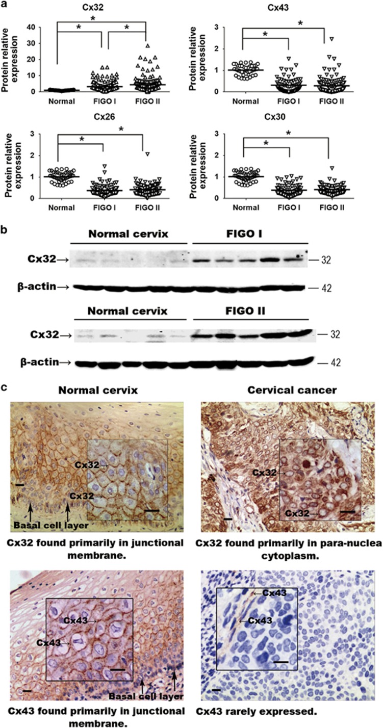
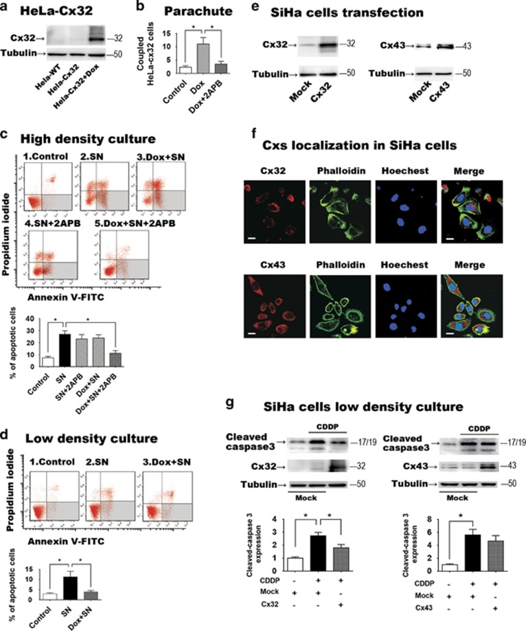
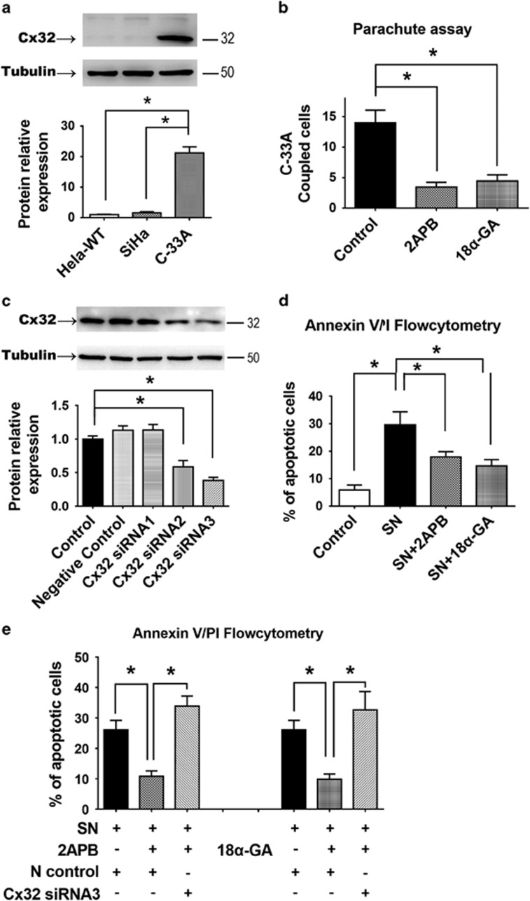
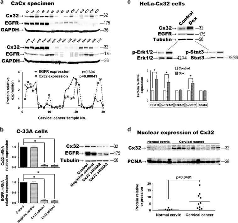
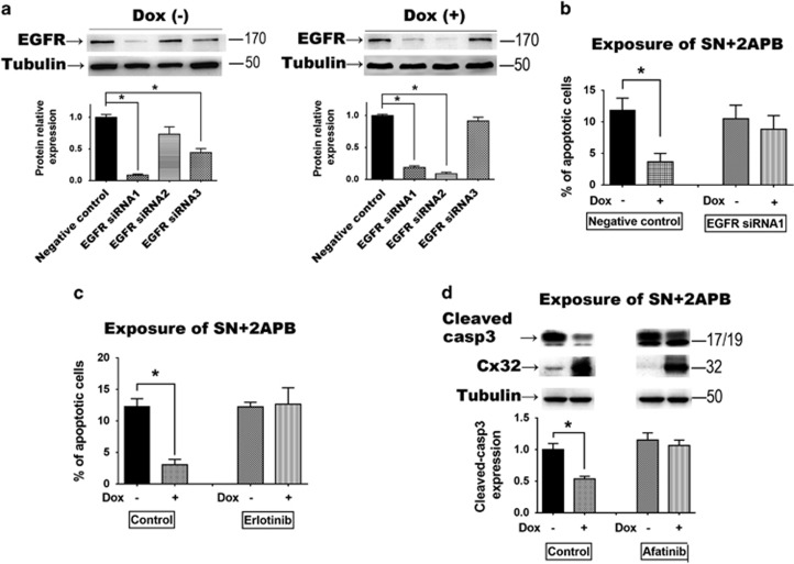
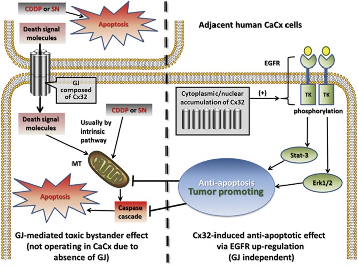
The role of connexin proteins (Cx), which form gap junctions (GJ), in progression and chemotherapeutic sensitivity of cervical cancer (CaCx), is unclear. Using cervix specimens (313 CaCx, 78 controls) and CaCx cell lines, we explored relationships among Cx expression, prognostic variables and mechanisms that may link them. In CaCx specimens, Cx32 was upregulated and cytoplasmically localized, and three other Cx downregulated, relative to controls. Cx32 expression correlated with advanced FIGO staging, differentiation and increased tumor size. In CaCx cell lines, Cx32 expression suppressed streptonigrin/cisplatin-induced apoptosis in the absence of functional GJ. In CaCx specimens and cell lines, expression of Cx32 upregulated epidermal growth factor receptor (EGFR) expression. Inhibition of EGFR signaling abrogated the anti-apoptotic effect of Cx32 expression. In conclusion, upregulated Cx32 in CaCx cells produces anti-apoptotic, pro-tumorigenic effects in vivo and vitro. Abnormal Cx32 expression/localization in CaCx appears to be both a mechanism and biomarker of chemotherapeutic resistance.


Conflict of interest statement

The authors declare no conflict of interest.

Figures

Figure 1
Figure 1
(a) Expression of Cx32, Cx26, Cx30 and Cx43 in normal cervix and CaCx samples. Expression of Cx26, Cx30 and Cx43 was decreased in the cancer samples relative to control, while expression of Cx32 was markedly increased. (b) Western blots showing that expression of expression of Cx32 correlated with increased FIGO score. Data are shown for five samples in each category (c) Immunohistochemistry showing that Cx32 and Cx43 in normal cervix tissue were localized to junctional regions. In CaCx cells, Cx32 aberrantly aggregated in para-nuclear cytoplasm and Cx43 was rarely expressed. (Scale bar: 20 μm). *P<0.05
Figure 2
Figure 2
(a) Western blot showing induction of Cx32 expression in the HeLa-Cx32 cell line after 48 h doxycycline treatment (n=5). (b) Demonstration of induction of GJIC following induction of Cx32 expression, and inhibition of GJIC with application of the GJ inhibitor 2APB (50 μM) (n=5). GJIC was assessed by parachute assay of dye coupling (c) Effects of functional gap junctions on apoptosis induced by streptonigrin (SN, 1 μM). In high-density cultures, which allow formation of GJ, SN-induced apoptosis was suppressed when GJ function was inhibited by 2APB (n=7). 2APB had no effect in the absence of induced Cx32 expression. Neither doxycycline (Dox, to induce Cx32 expression) nor 2APB (GJ inhibition) suppressed SN-induced apoptosis when applied alone. (d) When GJ formation was physically inhibited by low-density culture, expression of Cx32 suppressed SN-induced apoptosis (n=5). (e) Transient transfection of SiHa cells induced expression of Cx32 or Cx43 (n=3). (f) In transfected SiHa cells, both Cx32 and Cx43 mainly localized in cytoplasm (n=3, scale bar=10 μm). (g) Expression of Cx32, but not Cx43, in SiHa cells suppressed apoptosis induced by cisplatin (CDDP, 10 μM) under low-density culture (n=3). Error bar: standard error. *P<0.05; n=1 represents an independent cell culture
Figure 3
Figure 3
(a) C-33A cells endogenously express Cx32, unlike HeLa or SiHa cells (n=3). (b) The endogenous GJIC of C-33A cells was abolished by gap junction inhibitors 2APB and 18α-GA (n=3). (c) Western blot comparing effectiveness of three siRNAs in reducing expression of Cx32 in C-33A cells. (n=3). (d) Suppression of SN-induced apoptosis by treatment with 2APB or 18α-GA in C-33A cells (n=7). (e) The anti-apoptotic effect of 2APB and 18α-GA in C-33A cells was reversed by Cx32 RNA interference (n=5). Error bar: standard error. *P<0.05; n=1 represents an independent cell culture
Figure 4
Figure 4
(a) In 30 CaCx specimens, expression of EGFR was highly correlated with expression of Cx32 (r=0.604, P=0.00041). (b) In C-33A cells, when Cx32 expression was inhibited by Cx32 siRNAs, EGFR expression was significantly suppressed (n=3). (c) In HeLa-Cx32 cells, after induced Cx32 expression, EGFR was significantly increased and its downstream effectors p-Erk1/2 and p-Stat3 were upregulated (n=3). (d) In nuclear protein samples, Cx32 expression in CaCx cells (n=10) was higher than that in normal cervix cells (n=5, P=0.0152). Error bar: standard error. *P<0.05; n=1 represents an independent cell culture; r: Pearson correlation coefficient
Figure 5
Figure 5
(a) In HeLa-Cx32 cells, EGFR siRNA sequence 1 effectively inhibited EGFR expression with and without doxycycline treatment (n=3). (b) The anti-apoptotic effect of Cx32 expression was reversed by siRNA suppression of EGFR (n=3). (c and d) The anti-apoptotic effect of Cx32 expression was reversed by EGFR signaling inhibitors erlotinib and afatinib (n=3). Error bar: standard error. *P<0.05; n=1 represents an independent cell culture
Figure 6
Figure 6
Diagram of the inferred Cx32 anti-apoptotic mechanism. in vitro, Cx32 suppressed SN/CDDP-induced apoptosis only after GJ function was inhibited, either pharmacologically or physically. This suggests that upregulated expression of Cx32 per se has an anti-apoptotic effect, in contrast to its GJ-dependent pro-apoptotic effect, with exposure to these chemotherapeutic agents. Because of the upregulated Cx32 and its non-junctional localization in CaCx, the anti-apoptotic effect of Cx32 may dominate in cancer cells in vivo. Further, clinical-pathological data and in vitro findings indicate that the anti-apoptotic and tumor promoting effects involve the EGFR pathway in human CaCx cells. CDDP, Cisplatin; CaCx, cervical cancer; MT, mitochondria; SN, streptonigrin; TK, tyrosine kinase

References

    1. Kar R, Batra N, Riquelme MA, Jiang JX. Biological role of connexin intercellular channels and hemichannels. Arch Biochem Biophys 2012; 524: 2–15. - PMC - PubMed
    1. Kumar NM, Gilula NB. The gap junction communication channel. Cell 1996; 84: 381–388. - PubMed
    1. Mehta PP, Bertram JS, Loewenstein WR. Growth inhibition of transformed cells correlates with their junctional communication with normal cells. Cell 1986; 44: 187–196. - PubMed
    1. Loewenstein WR KY. Intercellular communication and the control of tissue growth: lack of communication between cancer cells. Nature 1966; 209: 1248–1249. - PubMed
    1. Kanczuga-Koda LKM, Sulkowski S, Wincewicz A, Zalewski B, Sulkowska M. Gradual loss of functional gap junction within progression of colorectal cancer_ a shift from membranous CX32 and CX43 expression to cytoplasmic pattern during colorectal. In Vivo 2010; 24: 101–107. - PubMed

MeSH terms