

Colposcopic imaging using visible-light optical coherence tomography
- PMID: 28492851
- PMCID: PMC5421648
- DOI: 10.1117/1.JBO.22.5.056003
Colposcopic imaging using visible-light optical coherence tomography
Abstract
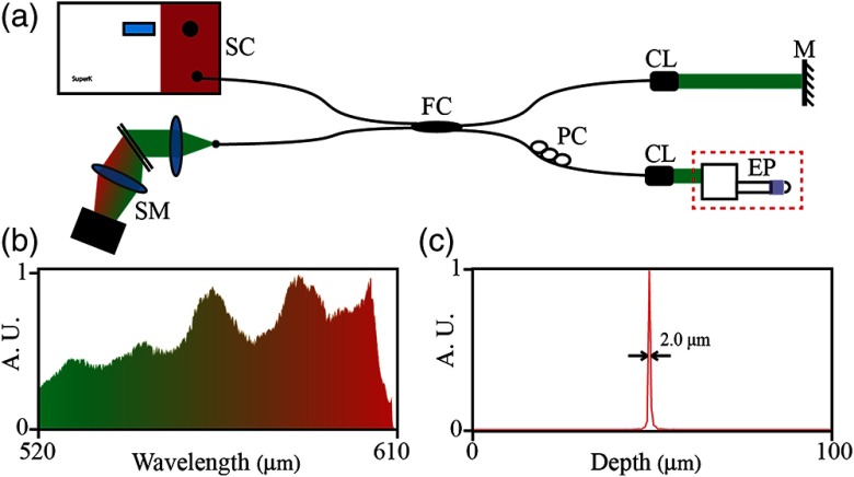
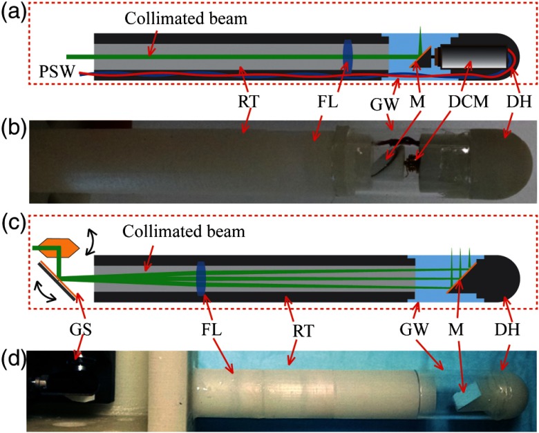
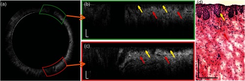
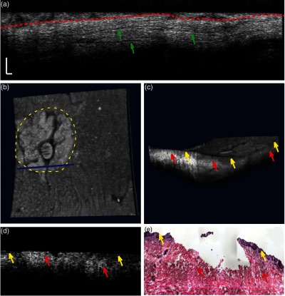
High-resolution colposcopic optical coherence tomography (OCT) provides key anatomical measures, such as thickness and minor traumatic injury of vaginal epithelium, of the female reproductive tract noninvasively. This information can be helpful in both fundamental investigations in animal models and disease screenings in humans. We present a fiber-based visible-light OCT and two probe designs for colposcopic application. One probe conducts circular scanning using a DC motor, and the other probe is capable of three-dimensional imaging over a 4.6 × 4.6 - mm 2 area using a pair of galvo scanners. Using this colposcopic vis-OCT with both probes, we acquired high-resolution images from whole isolated macaque vaginal samples and identified biopsy lesions.
Figures
Similar articles
-
Optical coherence tomography in biomedical research.Anal Bioanal Chem. 2011 Jul;400(9):2721-43. doi: 10.1007/s00216-011-5052-x. Epub 2011 May 12. Anal Bioanal Chem. 2011. PMID: 21562739 Review.
-
Miniature probe combining optical-resolution photoacoustic microscopy and optical coherence tomography for in vivo microcirculation study.Appl Opt. 2013 Mar 20;52(9):1928-31. doi: 10.1364/AO.52.001928. Appl Opt. 2013. PMID: 23518738
-
Preclinical evaluation and intraoperative human retinal imaging with a high-resolution microscope-integrated spectral domain optical coherence tomography device.Retina. 2013 Jul-Aug;33(7):1328-37. doi: 10.1097/IAE.0b013e3182831293. Retina. 2013. PMID: 23538579 Free PMC article.
-
Mirau-based line-field confocal optical coherence tomography for three-dimensional high-resolution skin imaging.J Biomed Opt. 2022 Aug;27(8):086002. doi: 10.1117/1.JBO.27.8.086002. J Biomed Opt. 2022. PMID: 35962466 Free PMC article.
-
Ultrahigh-resolution optical coherence tomography.J Biomed Opt. 2004 Jan-Feb;9(1):47-74. doi: 10.1117/1.1629679. J Biomed Opt. 2004. PMID: 14715057 Review.
Cited by
-
Visible-light optical coherence tomography: a review.J Biomed Opt. 2017 Dec;22(12):1-14. doi: 10.1117/1.JBO.22.12.121707. J Biomed Opt. 2017. PMID: 29218923 Free PMC article. Review.
-
Designing visible-light optical coherence tomography towards clinics.Quant Imaging Med Surg. 2019 May;9(5):769-781. doi: 10.21037/qims.2019.05.01. Quant Imaging Med Surg. 2019. PMID: 31281773 Free PMC article.
-
Visible Light Optical Coherence Tomography: Technology and Biomedical Applications.Bioengineering (Basel). 2025 Jul 17;12(7):770. doi: 10.3390/bioengineering12070770. Bioengineering (Basel). 2025. PMID: 40722462 Free PMC article. Review.
-
Optical coherence tomography evaluation of vaginal epithelial thickness during CO2 laser treatment: A pilot study.J Biophotonics. 2022 Nov;15(11):e202200052. doi: 10.1002/jbio.202200052. Epub 2022 Aug 11. J Biophotonics. 2022. PMID: 35860856 Free PMC article.
-
PneumaOCT: Pneumatic optical coherence tomography endoscopy for targeted distortion-free imaging in tortuous and narrow internal lumens.Sci Adv. 2024 Aug 30;10(35):eadp3145. doi: 10.1126/sciadv.adp3145. Epub 2024 Aug 28. Sci Adv. 2024. PMID: 39196931 Free PMC article.
References
-
- Organization W. H., Manual for the Standardization of Colposcopy for the Evaluation of Vaginal Products, World Health Organization, Geneva: (2004).
MeSH terms
Grants and funding
LinkOut - more resources
Full Text Sources
Other Literature Sources
