. 2018 Jan;21(1):41-54.
doi: 10.1007/s10120-017-0721-x. Epub 2017 May 10.

MicroRNA-1 acts as a tumor suppressor microRNA by inhibiting angiogenesis-related growth factors in human gastric cancer

Affiliations

MicroRNA-1 acts as a tumor suppressor microRNA by inhibiting angiogenesis-related growth factors in human gastric cancer

Meng Xie et al. Gastric Cancer. 2018 Jan.

Abstract

Background: We recently reported that miR-1 was one of the most significantly downregulated microRNAs in gastric cancer (GC) patients from The Cancer Genome Atlas microRNA sequencing data. Here we aim to elucidate the role of miR-1 in gastric carcinogenesis.

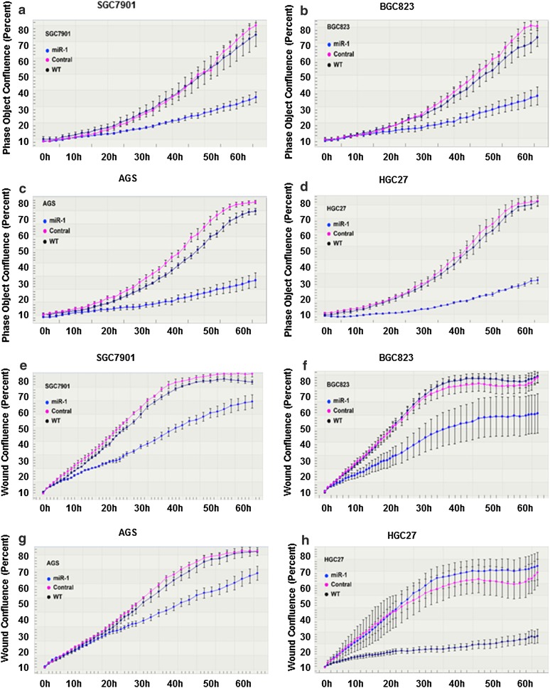
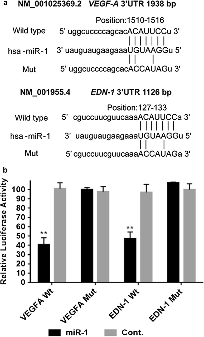
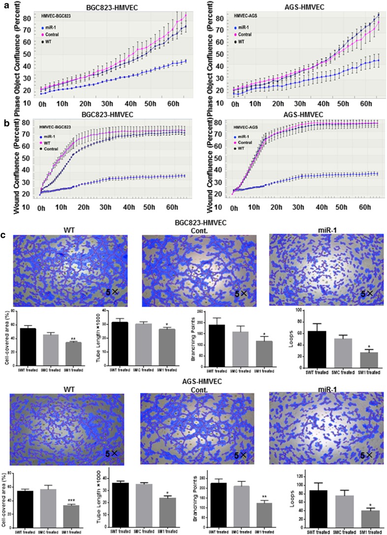
Methods: We measured miR-1 expression in human GC cell lines and 90 paired primary GC samples, and analyzed the association of its status with clinicopathological features. The effect of miR-1 on GC cells was evaluated by proliferation and migration assay. To identify the target genes of miR-1, bioinformatic analysis and protein array analysis were performed. Moreover, the regulation mechanism of miR-1 with regard to these predicted targets was investigated by quantitative PCR (qPCR), Western blot, ELISA, and endothelial cell tube formation. The putative binding site of miR-1 on target genes was assessed by a reporter assay.

Results: Expression of miR-1 was obviously decreased in GC cell lines and primary tissues. Patients with low miR-1 expression had significantly shorter overall survival compared with those with high miR-1 expression (P = 0.0027). Overexpression of miR-1 in GC cells inhibited proliferation, migration, and tube formation of endothelial cells by suppressing expression of vascular endothelial growth factor A (VEGF-A) and endothelin 1 (EDN1). Conversely, inhibition of miR-1 with use of antago-miR-1 caused an increase in expression of VEGF-A and EDN1 in nonmalignant GC cells or low-malignancy GC cells.

Conclusions: MiR-1 acts as a tumor suppressor by inhibiting angiogenesis-related growth factors in human gastric cancer. Downregulated miR-1 not only promotes cellular proliferation and migration of GC cells, but may activates proangiogenesis signaling and stimulates the proliferation and migration of endothelial cells, indicating the possibility of new strategies for GC therapy.

Keywords: Angiogenesis; Gastric cancer; Vascular endothelial growth factor A; miR-1.


Conflict of interest statement

Conflict of interest

The authors declare that they have no conflict of interest.

Research involving human participants and/or animals and informed consent

All samples were obtained with patients’ informed consent. The Ethics Committee of Beijing Cancer Hospital approved this study. This study does not involve animals.

Figures

Fig. 1
Fig. 1
MiR-1 expression in cultured gastric cancer (GC) cells and primary GC tissues samples, and its correlation with prognosis of GC patients a MiR-1 expression in GC cell lines compared with the immortalized gastric cell line GES-1. b Quantitative PCR levels showing reexpression of mature miR-1 in GC cell lines after 5-aza-2′-deoxycyridine (5-aza-dC) treatment. c Relative expression levels of miR-1 in primary gastric tumors and adjacent nontumor tissues (n = 90). The data represent 2−ΔΔCt expression values. The P value was calculated by a paired test. d Kaplan–Meier curves of overall survival for all patients with miR-1-high versus miR-1-low GC tissue. e Transfection efficiency of miR-1 in GC cells by qPCR . Mean ± standard deviation of three independent experiments. WT wild type, *P < 0.05, **P < 0.01, ***P < 0.001, ****P < 0.0001
Fig. 2
Fig. 2
Overexpression of miR-1 suppressed the proliferation and migration capability of GC cells. ad Cell growth curves. The y-axis is a label-free measure of cell confluence used for the IncuCyte ZOOM live cell imaging system to assess the cell growth. eh Wound-healing curves. Cell motility was monitored by the IncuCyte ZOOM live cell imaging system. WT wild type
Fig. 3
Fig. 3
MiR-1 inhibited angiogenesis-related factors at both the messenger RNA level and the protein level. a Quantitative reverse transcription PCR assay. P values were determined by an unpaired two-sided t test. b Western blotting. c Relative protein levels of vascular endothelial growth factor A (VEGF-A) in supernatant of gastric cancer cells transitorily transfected with pri-miR-1 plasmid determined by ELISA. Cont. control, WT wild type, *P < 0.05, **P < 0.01, ***P < 0.001, ****P < 0.0001
Fig. 4
Fig. 4
MiR-1 inhibits the expression of VEGF-A and EDN1 3′ untranlated region (UTR)-integrated luciferase reporter genes. a The target sites for miR-1 in the VEGF-A (NM_001025369.2), and EDN1 (NM_001955.4) 3′ UTR were identified with the TargetScan database. b Luciferase reporter assay using the vector encoding the full-length 3′ UTR of VEGF-A, and EDN1 3′ UTR messenger RNA. The Renilla luciferase values were used to normalize firefly luciferase values. Cont. control, Mut mutant, Wt wild type, *P < 0.05, **P < 0.01
Fig. 5
Fig. 5
Effects of miR-1 on the proliferation, migration and tube formation of human microvascular endothelial cells. After both BGC823 and AGS cells had been transfected with GV268-miR-1 or GV268-ctrl plasmid respectively, the conditioned medium was collected and the effects of conditioned medium on human microvascular endothelial cell proliferation (a), migration (b), and tube formation (c) were assessed. The cell-covered area, tube length, branching points, and loops of tube formation were determined by WimTube from Wimasis Image Analysis (n = 3). One-way analysis of variance and Dunnett’s test were applied for analysis. Cont. control, HMVEC human microvascular endothelial cell, WT wild type, *P < 0.05, **P < 0.01, ***P < 0.001

Similar articles

Cited by

References

    1. Torre LA, Bray F, Siegel RL, Ferlay J, Lortet-Tieulent J, Jemal A. Global cancer statistics, 2012. CA Cancer J Clin. 2015;65(2):87–108. doi: 10.3322/caac.21262. - DOI - PubMed
    1. Ferlay J, Soerjomataram I, Ervik M, Dikshit R, Eser S, Mathers C, Rebelo M, Parkin DM, Forman D, Bray, F. GLOBOCAN 2012 v1.0, cancer incidence and mortality worldwide: IARC CancerBase no. 11. Lyon: International Agency for Research on Cancer; 2013. Available from http://globocan.iarc.fr - PubMed
    1. Lim LP, Lau NC, Garrett-Engele P, Grimson A, Schelter JM, Castle J, et al. Microarray analysis shows that some microRNAs downregulate large numbers of target mRNAs. Nature. 2005;433(7027):769–773. doi: 10.1038/nature03315. - DOI - PubMed
    1. Vasudevan S, Tong Y, Steitz JA. Switching from repression to activation: microRNAs can up-regulate translation. Science. 2007;318(5858):1931–1934. doi: 10.1126/science.1149460. - DOI - PubMed
    1. Xie M, Dart DA, Owen S, Wen X, Ji J, Jiang W. Insights into roles of the miR-1, -133 and -206 family in gastric cancer (review) Oncol Rep. 2016;36(3):1191–1198. doi: 10.3892/or.2016.4908. - DOI - PubMed