Review
. 2017 May-Jun;37(3):855-870.
doi: 10.1148/rg.2017160116.

Shear-Wave Elastography: Basic Physics and Musculoskeletal Applications

Affiliations
Review

Shear-Wave Elastography: Basic Physics and Musculoskeletal Applications

Mihra S Taljanovic et al. Radiographics. 2017 May-Jun.

Abstract

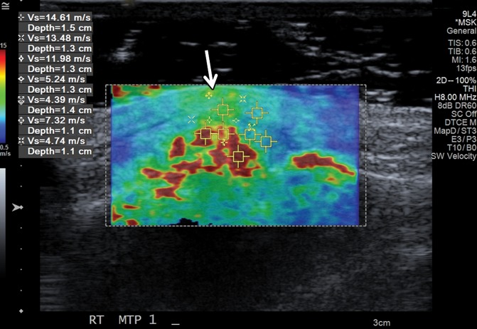
In the past 2 decades, sonoelastography has been progressively used as a tool to help evaluate soft-tissue elasticity and add to information obtained with conventional gray-scale and Doppler ultrasonographic techniques. Recently introduced on clinical scanners, shear-wave elastography (SWE) is considered to be more objective, quantitative, and reproducible than compression sonoelastography with increasing applications to the musculoskeletal system. SWE uses an acoustic radiation force pulse sequence to generate shear waves, which propagate perpendicular to the ultrasound beam, causing transient displacements. The distribution of shear-wave velocities at each pixel is directly related to the shear modulus, an absolute measure of the tissue's elastic properties. Shear-wave images are automatically coregistered with standard B-mode images to provide quantitative color elastograms with anatomic specificity. Shear waves propagate faster through stiffer contracted tissue, as well as along the long axis of tendon and muscle. SWE has a promising role in determining the severity of disease and treatment follow-up of various musculoskeletal tissues including tendons, muscles, nerves, and ligaments. This article describes the basic ultrasound physics of SWE and its applications in the evaluation of various traumatic and pathologic conditions of the musculoskeletal system. ©RSNA, 2017.


Figures

Figure 1a.
Figure 1a.
(a) Basic physics of SWE. In step 1, shear waves are generated using acoustic radiation force; they propagate perpendicularly to the primary US wave at a lower velocity. In step 2, fast plane wave excitation is used to track displacement and velocity as shear waves propagate, and tissue displacement is calculated using a speckle tracking algorithm. In step 3, tissue displacements are used to calculate shear-wave velocity (cs) and shear modulus (G). (b) Relationship between shear velocity and shear modulus expressed as a color bar, which assumes, in this case, a density equal to that of water (1 g/cm3). Actual density estimates will vary for different types of soft tissue and can also be found using values published in the literature.
Figure 1b.
Figure 1b.
(a) Basic physics of SWE. In step 1, shear waves are generated using acoustic radiation force; they propagate perpendicularly to the primary US wave at a lower velocity. In step 2, fast plane wave excitation is used to track displacement and velocity as shear waves propagate, and tissue displacement is calculated using a speckle tracking algorithm. In step 3, tissue displacements are used to calculate shear-wave velocity (cs) and shear modulus (G). (b) Relationship between shear velocity and shear modulus expressed as a color bar, which assumes, in this case, a density equal to that of water (1 g/cm3). Actual density estimates will vary for different types of soft tissue and can also be found using values published in the literature.
Figure 2a.
Figure 2a.
Normal fourth extensor compartment tendon of the wrist in a 21-year-old asymptomatic man. (a) Long-axis gray-scale US image of the dorsal aspect of the wrist shows a normal echogenic fibrillar appearance of the fourth extensor compartment tendon (arrow). L = lunate, R = radius. (b) SWE image (color elastogram) of the same region shows predominantly intermediate shear-wave velocity (6.66 m/sec) in the same tendon (arrow). Red = hard consistency (≤15 m/sec), blue = soft consistency (≥0.5 m/sec), and green and yellow = intermediate consistency. SWE data were collected using an Acuson S3000 US scanner (Siemens Healthineers, Erlangen, Germany) with a L9–4-MHz linear transducer.
Figure 2b.
Figure 2b.
Normal fourth extensor compartment tendon of the wrist in a 21-year-old asymptomatic man. (a) Long-axis gray-scale US image of the dorsal aspect of the wrist shows a normal echogenic fibrillar appearance of the fourth extensor compartment tendon (arrow). L = lunate, R = radius. (b) SWE image (color elastogram) of the same region shows predominantly intermediate shear-wave velocity (6.66 m/sec) in the same tendon (arrow). Red = hard consistency (≤15 m/sec), blue = soft consistency (≥0.5 m/sec), and green and yellow = intermediate consistency. SWE data were collected using an Acuson S3000 US scanner (Siemens Healthineers, Erlangen, Germany) with a L9–4-MHz linear transducer.
Figure 3.
Figure 3.
Normal Achilles tendon in a 69-year-old asymptomatic woman. Bottom image: Long-axis gray-scale US image shows normal echogenic fibrillar appearance of the Achilles tendon with an outlined region of interest (ROI). Top image: Color elastogram of the same region shows normal G of the examined tendon (177.4 kPa ± 42). SWE data were collected using an Aixplorer US scanner (Supersonic Imagine, Aix-en-Provence, France) with an L15–4-MHz linear transducer.
Figure 4.
Figure 4.
Tendinopathy and partial-thickness Achilles tendon tear in a 45-year-old man. Bottom image: Long-axis gray-scale US image shows thickening, heterogeneous echogenicity, and disorganized echotexture of the Achilles tendon fibers, with an outlined ROI. Note the partial-thickness tear (arrow) at the deep aspect of the tendon. Top image: Color elastogram of the same region shows a lower G, 41.2 kPa ± 17.6, for the tendon. Note the signal void (arrow) in the region of the partial-thickness tear. SWE data were collected using an Aixplorer US scanner with an L15–4-MHz linear transducer.
Figure 5a.
Figure 5a.
Normal subscapularis tendon in a 31-year-old woman with a history of seronegative polyarthritis. (a) Long-axis gray-scale US image of the subscapularis tendon shows a normal echogenic appearance (arrows). (b) Color elastogram of the same region shows predominantly intermediate shear-wave velocities (arrows) (mean, 7.50 m/sec) in the subscapularis tendon. SWE data were collected using an Acuson S3000 US scanner with an L9–4-MHz linear transducer.
Figure 5b.
Figure 5b.
Normal subscapularis tendon in a 31-year-old woman with a history of seronegative polyarthritis. (a) Long-axis gray-scale US image of the subscapularis tendon shows a normal echogenic appearance (arrows). (b) Color elastogram of the same region shows predominantly intermediate shear-wave velocities (arrows) (mean, 7.50 m/sec) in the subscapularis tendon. SWE data were collected using an Acuson S3000 US scanner with an L9–4-MHz linear transducer.
Figure 6a.
Figure 6a.
Massive supraspinatus tendon (SST) tear in a 73-year-old man. (a) Long-axis gray-scale US image of the expected SST region shows the deltoid muscle abutting the humeral head, related to a massive SST tear (arrows), with SST fibers not seen. (b) Color elastogram of the same region shows low shear-wave velocities (arrows) (mean, 3.15 m/sec) in the deltoid muscle. SWE data were collected using an Acuson S3000 US scanner with an L9–4-MHz linear transducer.
Figure 6b.
Figure 6b.
Massive supraspinatus tendon (SST) tear in a 73-year-old man. (a) Long-axis gray-scale US image of the expected SST region shows the deltoid muscle abutting the humeral head, related to a massive SST tear (arrows), with SST fibers not seen. (b) Color elastogram of the same region shows low shear-wave velocities (arrows) (mean, 3.15 m/sec) in the deltoid muscle. SWE data were collected using an Acuson S3000 US scanner with an L9–4-MHz linear transducer.
Figure 7a.
Figure 7a.
Tenosynovitis of the first extensor compartment tendons of the wrist in a 59-year-old woman with a history of rheumatoid arthritis. (a) Long-axis gray-scale US image shows mild hypoechogenicity of the extensor pollicis brevis (EPB) tendon fibers (white arrows) and a mildly heterogeneous synovial-fluid complex in the massively distended tendon sheath (black arrow). (b) Color elastogram of the same region shows predominantly intermediate shear-wave velocities (arrows) in the EPB tendon (mean, 8.65 m/sec) and low shear-wave velocities (mean, 3.04 m/sec) in the region of synovitis (black arrow) within the tendon sheath. SWE data were collected using an Acuson S3000 US scanner with an L9–4-MHz linear transducer. The right side of the images is proximal.
Figure 7b.
Figure 7b.
Tenosynovitis of the first extensor compartment tendons of the wrist in a 59-year-old woman with a history of rheumatoid arthritis. (a) Long-axis gray-scale US image shows mild hypoechogenicity of the extensor pollicis brevis (EPB) tendon fibers (white arrows) and a mildly heterogeneous synovial-fluid complex in the massively distended tendon sheath (black arrow). (b) Color elastogram of the same region shows predominantly intermediate shear-wave velocities (arrows) in the EPB tendon (mean, 8.65 m/sec) and low shear-wave velocities (mean, 3.04 m/sec) in the region of synovitis (black arrow) within the tendon sheath. SWE data were collected using an Acuson S3000 US scanner with an L9–4-MHz linear transducer. The right side of the images is proximal.
Figure 8a.
Figure 8a.
Normal medial head of gastrocnemius and soleus aponeurosis in the relaxed and contracted (maximal dorsiflexion of the ankle) states in an asymptomatic 48-year-old woman. (a) Long-axis gray-scale US image shows normal echotexture of the relaxed gastrocnemius (G) and soleus (S) muscles and of the aponeurosis (arrow). (b) Color elastogram of the same region shows low shear-wave velocities in the gastrocnemius muscle (mean, 2.89 m/sec) and intermediate velocities at the aponeurosis (arrow) (mean, 4.915 m/sec). (c) Long-axis gray-scale US image of the same region with maximal dorsiflexion of the ankle shows normal echotexture of the gastrocnemius (G) and soleus (S) muscles and of the aponeurosis (arrow). (d) Color elastogram of the same region shows increased shear-wave velocity of the contracted gastrocnemius muscle (6.79 m/sec) and at the aponeurosis (arrow) (mean, 10.83 m/sec). SWE data were collected using an Acuson S3000 US scanner with an L9–4-MHz linear transducer.
Figure 8b.
Figure 8b.
Normal medial head of gastrocnemius and soleus aponeurosis in the relaxed and contracted (maximal dorsiflexion of the ankle) states in an asymptomatic 48-year-old woman. (a) Long-axis gray-scale US image shows normal echotexture of the relaxed gastrocnemius (G) and soleus (S) muscles and of the aponeurosis (arrow). (b) Color elastogram of the same region shows low shear-wave velocities in the gastrocnemius muscle (mean, 2.89 m/sec) and intermediate velocities at the aponeurosis (arrow) (mean, 4.915 m/sec). (c) Long-axis gray-scale US image of the same region with maximal dorsiflexion of the ankle shows normal echotexture of the gastrocnemius (G) and soleus (S) muscles and of the aponeurosis (arrow). (d) Color elastogram of the same region shows increased shear-wave velocity of the contracted gastrocnemius muscle (6.79 m/sec) and at the aponeurosis (arrow) (mean, 10.83 m/sec). SWE data were collected using an Acuson S3000 US scanner with an L9–4-MHz linear transducer.
Figure 8c.
Figure 8c.
Normal medial head of gastrocnemius and soleus aponeurosis in the relaxed and contracted (maximal dorsiflexion of the ankle) states in an asymptomatic 48-year-old woman. (a) Long-axis gray-scale US image shows normal echotexture of the relaxed gastrocnemius (G) and soleus (S) muscles and of the aponeurosis (arrow). (b) Color elastogram of the same region shows low shear-wave velocities in the gastrocnemius muscle (mean, 2.89 m/sec) and intermediate velocities at the aponeurosis (arrow) (mean, 4.915 m/sec). (c) Long-axis gray-scale US image of the same region with maximal dorsiflexion of the ankle shows normal echotexture of the gastrocnemius (G) and soleus (S) muscles and of the aponeurosis (arrow). (d) Color elastogram of the same region shows increased shear-wave velocity of the contracted gastrocnemius muscle (6.79 m/sec) and at the aponeurosis (arrow) (mean, 10.83 m/sec). SWE data were collected using an Acuson S3000 US scanner with an L9–4-MHz linear transducer.
Figure 8d.
Figure 8d.
Normal medial head of gastrocnemius and soleus aponeurosis in the relaxed and contracted (maximal dorsiflexion of the ankle) states in an asymptomatic 48-year-old woman. (a) Long-axis gray-scale US image shows normal echotexture of the relaxed gastrocnemius (G) and soleus (S) muscles and of the aponeurosis (arrow). (b) Color elastogram of the same region shows low shear-wave velocities in the gastrocnemius muscle (mean, 2.89 m/sec) and intermediate velocities at the aponeurosis (arrow) (mean, 4.915 m/sec). (c) Long-axis gray-scale US image of the same region with maximal dorsiflexion of the ankle shows normal echotexture of the gastrocnemius (G) and soleus (S) muscles and of the aponeurosis (arrow). (d) Color elastogram of the same region shows increased shear-wave velocity of the contracted gastrocnemius muscle (6.79 m/sec) and at the aponeurosis (arrow) (mean, 10.83 m/sec). SWE data were collected using an Acuson S3000 US scanner with an L9–4-MHz linear transducer.
Figure 9a.
Figure 9a.
Acute tennis leg injury in a 51-year-old man. (a) Long-axis gray-scale US image shows partial avulsion of the medial head of the gastrocnemius muscle (G) from the soleus (S) aponeurosis (arrow). (b) Color elastogram of the same region shows low velocities in the injured avulsed soleus muscle (mean, 1.79 m/sec) with thickening and somewhat higher shear-wave velocities (mean, 2.32 m/sec) in the injured aponeurosis (arrow). Note that the shear-wave velocities in both the gastrocnemius muscle and at the aponeurosis are lower than in the healthy tissues in the same region in the patient in Figure 8. SWE data were collected using an Acuson S3000 US scanner with an L9–4-MHz linear transducer.
Figure 9b.
Figure 9b.
Acute tennis leg injury in a 51-year-old man. (a) Long-axis gray-scale US image shows partial avulsion of the medial head of the gastrocnemius muscle (G) from the soleus (S) aponeurosis (arrow). (b) Color elastogram of the same region shows low velocities in the injured avulsed soleus muscle (mean, 1.79 m/sec) with thickening and somewhat higher shear-wave velocities (mean, 2.32 m/sec) in the injured aponeurosis (arrow). Note that the shear-wave velocities in both the gastrocnemius muscle and at the aponeurosis are lower than in the healthy tissues in the same region in the patient in Figure 8. SWE data were collected using an Acuson S3000 US scanner with an L9–4-MHz linear transducer.
Figure 10a.
Figure 10a.
Normal ulnar nerve in the cubital tunnel in an asymptomatic 58-year-old woman. (a) Long-axis gray-scale US image shows a normal relatively hypoechoic ulnar nerve (arrows) in the cubital tunnel. (b) Color elastogram of the same region shows low shear-wave velocities (mean, 2.22 m/sec) in the ulnar nerve (arrows). SWE data were collected using an Acuson S3000 US scanner with an L9–4-MHz linear transducer. The right side of the image is distal.
Figure 10b.
Figure 10b.
Normal ulnar nerve in the cubital tunnel in an asymptomatic 58-year-old woman. (a) Long-axis gray-scale US image shows a normal relatively hypoechoic ulnar nerve (arrows) in the cubital tunnel. (b) Color elastogram of the same region shows low shear-wave velocities (mean, 2.22 m/sec) in the ulnar nerve (arrows). SWE data were collected using an Acuson S3000 US scanner with an L9–4-MHz linear transducer. The right side of the image is distal.
Figure 11a.
Figure 11a.
Abnormal median nerve in the carpal tunnel in a 34-year-old woman with suspected psoriatic arthritis and median neuropathy. (a) Long-axis gray-scale US image shows an enlarged relatively hypoechoic median nerve (arrows) in the carpal tunnel. (b) Color elastogram of the same region shows intermediate shear-wave velocities (mean, 5.21 m/sec) in the median nerve (arrows). SWE data were collected using an Acuson S3000 US scanner with an L9–4-MHz linear transducer. The right side of the image is distal.
Figure 11b.
Figure 11b.
Abnormal median nerve in the carpal tunnel in a 34-year-old woman with suspected psoriatic arthritis and median neuropathy. (a) Long-axis gray-scale US image shows an enlarged relatively hypoechoic median nerve (arrows) in the carpal tunnel. (b) Color elastogram of the same region shows intermediate shear-wave velocities (mean, 5.21 m/sec) in the median nerve (arrows). SWE data were collected using an Acuson S3000 US scanner with an L9–4-MHz linear transducer. The right side of the image is distal.
Figure 12a.
Figure 12a.
Normal anterior band of the medial ulnar collateral ligament (MUCL) of the elbow in a 21-year-old man. (a) Long-axis gray-scale US image shows a normal echogenic anterior band of the MUCL at the medial aspect of the elbow (arrows). (b) Color elastogram of the same region shows intermediate to high shear-wave velocities (mean, 8.69 m/sec) in the MUCL (arrows). SWE data were collected using an L9–4-MHz linear transducer.
Figure 12b.
Figure 12b.
Normal anterior band of the medial ulnar collateral ligament (MUCL) of the elbow in a 21-year-old man. (a) Long-axis gray-scale US image shows a normal echogenic anterior band of the MUCL at the medial aspect of the elbow (arrows). (b) Color elastogram of the same region shows intermediate to high shear-wave velocities (mean, 8.69 m/sec) in the MUCL (arrows). SWE data were collected using an L9–4-MHz linear transducer.
Figure 13a.
Figure 13a.
Tophaceous gout at the first metatarsophalangeal joint in a 49-year-old man. (a) Long-axis gray-scale US image along the medial aspect of the first metatarsophalangeal joint shows echogenic intra-articular gouty tophus (arrow) abutting the metatarsal head (MT). PP = proximal phalanx. (b) Color elastogram of the same region shows intermediate shear-wave velocity (arrow) (mean, 7.32 m/sec) in the gouty tophus. SWE data were collected using an Acuson S3000 US scanner with an L9–4-MHz linear transducer.
Figure 13b.
Figure 13b.
Tophaceous gout at the first metatarsophalangeal joint in a 49-year-old man. (a) Long-axis gray-scale US image along the medial aspect of the first metatarsophalangeal joint shows echogenic intra-articular gouty tophus (arrow) abutting the metatarsal head (MT). PP = proximal phalanx. (b) Color elastogram of the same region shows intermediate shear-wave velocity (arrow) (mean, 7.32 m/sec) in the gouty tophus. SWE data were collected using an Acuson S3000 US scanner with an L9–4-MHz linear transducer.
Figure 14a.
Figure 14a.
Intramuscular lipoma in a 48-year-old woman with a palpable painless lump in her forearm. (a) Sagittal T1-weighted magnetic resonance (MR) image shows a well-defined high-signal-intensity mass (arrow) in the distal aspect of the supinator muscle, consistent with an intramuscular lipoma. (b) Long-axis gray-scale US image in the region of palpable abnormality shows a nonspecific echogenic solid intramuscular mass (arrows) in the distal supinator muscle. (c) Color elastogram of the same region shows lower shear-wave velocities (mean, 1.74 m/sec) in the mass (arrows) than in the region of the pseudocapsule, which shows intermediate velocities (mean, 5.52 m/sec). SWE data were collected using an Acuson S3000 US scanner with an L9–4-MHz linear transducer.
Figure 14b.
Figure 14b.
Intramuscular lipoma in a 48-year-old woman with a palpable painless lump in her forearm. (a) Sagittal T1-weighted magnetic resonance (MR) image shows a well-defined high-signal-intensity mass (arrow) in the distal aspect of the supinator muscle, consistent with an intramuscular lipoma. (b) Long-axis gray-scale US image in the region of palpable abnormality shows a nonspecific echogenic solid intramuscular mass (arrows) in the distal supinator muscle. (c) Color elastogram of the same region shows lower shear-wave velocities (mean, 1.74 m/sec) in the mass (arrows) than in the region of the pseudocapsule, which shows intermediate velocities (mean, 5.52 m/sec). SWE data were collected using an Acuson S3000 US scanner with an L9–4-MHz linear transducer.
Figure 14c.
Figure 14c.
Intramuscular lipoma in a 48-year-old woman with a palpable painless lump in her forearm. (a) Sagittal T1-weighted magnetic resonance (MR) image shows a well-defined high-signal-intensity mass (arrow) in the distal aspect of the supinator muscle, consistent with an intramuscular lipoma. (b) Long-axis gray-scale US image in the region of palpable abnormality shows a nonspecific echogenic solid intramuscular mass (arrows) in the distal supinator muscle. (c) Color elastogram of the same region shows lower shear-wave velocities (mean, 1.74 m/sec) in the mass (arrows) than in the region of the pseudocapsule, which shows intermediate velocities (mean, 5.52 m/sec). SWE data were collected using an Acuson S3000 US scanner with an L9–4-MHz linear transducer.
Figure 15a.
Figure 15a.
Intramuscular lipoma in the left upper back of a 55-year-old woman with spuriously high shear-wave velocities. (a) Long-axis gray-scale US image in the region of palpable abnormality shows a relatively hypoechoic subcutaneous soft-tissue mass consistent with a lipoma (arrows). (b) Color elastogram of the same region shows spuriously high shear-wave velocities within the mass (arrows), which were due to an incorrect highest velocity setting at only 6.5 m/sec. SWE data were collected using an Acuson S3000 US scanner with an L9–4-MHz linear transducer.
Figure 15b.
Figure 15b.
Intramuscular lipoma in the left upper back of a 55-year-old woman with spuriously high shear-wave velocities. (a) Long-axis gray-scale US image in the region of palpable abnormality shows a relatively hypoechoic subcutaneous soft-tissue mass consistent with a lipoma (arrows). (b) Color elastogram of the same region shows spuriously high shear-wave velocities within the mass (arrows), which were due to an incorrect highest velocity setting at only 6.5 m/sec. SWE data were collected using an Acuson S3000 US scanner with an L9–4-MHz linear transducer.
Figure 16a.
Figure 16a.
Tender palpable palmar mass at the level of the mid-metacarpals centered at the ulnar aspect of the second digit flexor tendons in an 18-year-old man, consistent with a pathologically proven fibroma of the tendon sheath. (a) Short-axis power Doppler US image of the palmar aspect of the hand in the region of the palpable lump shows a hypoechoic mass (arrows) with associated moderate hyperemia. (b) Color elastogram of the same region shows intermediate shear-wave velocities (mean, 5.93 m/sec) within the mass (arrows). (c) Axial T2-weighted fat-saturated MR image of the same hand shows a high-signal-intensity mass centered at the ulnar aspect of the second digit flexor tendons (arrow). SWE data were collected using an Acuson S3000 US scanner with an L9–4-MHz linear transducer.
Figure 16b.
Figure 16b.
Tender palpable palmar mass at the level of the mid-metacarpals centered at the ulnar aspect of the second digit flexor tendons in an 18-year-old man, consistent with a pathologically proven fibroma of the tendon sheath. (a) Short-axis power Doppler US image of the palmar aspect of the hand in the region of the palpable lump shows a hypoechoic mass (arrows) with associated moderate hyperemia. (b) Color elastogram of the same region shows intermediate shear-wave velocities (mean, 5.93 m/sec) within the mass (arrows). (c) Axial T2-weighted fat-saturated MR image of the same hand shows a high-signal-intensity mass centered at the ulnar aspect of the second digit flexor tendons (arrow). SWE data were collected using an Acuson S3000 US scanner with an L9–4-MHz linear transducer.
Figure 16c.
Figure 16c.
Tender palpable palmar mass at the level of the mid-metacarpals centered at the ulnar aspect of the second digit flexor tendons in an 18-year-old man, consistent with a pathologically proven fibroma of the tendon sheath. (a) Short-axis power Doppler US image of the palmar aspect of the hand in the region of the palpable lump shows a hypoechoic mass (arrows) with associated moderate hyperemia. (b) Color elastogram of the same region shows intermediate shear-wave velocities (mean, 5.93 m/sec) within the mass (arrows). (c) Axial T2-weighted fat-saturated MR image of the same hand shows a high-signal-intensity mass centered at the ulnar aspect of the second digit flexor tendons (arrow). SWE data were collected using an Acuson S3000 US scanner with an L9–4-MHz linear transducer.
Figure 17a.
Figure 17a.
Pathologically proven, painless, palpable, epidermoid inclusion cyst at the anterior aspect of the proximal tibia in an 18-year-old woman. (a) Long-axis gray-scale US image in the region of palpable abnormality shows a homogeneously hypoechoic circumscribed oval subcutaneous mass (arrow) with increased through transmission. (b) Color elastogram of the same region shows low shear-wave velocities (mean, 2.76 m/sec) within the mass (arrows). SWE data were collected using an Acuson S3000 US scanner with an L9–4-MHz linear transducer.
Figure 17b.
Figure 17b.
Pathologically proven, painless, palpable, epidermoid inclusion cyst at the anterior aspect of the proximal tibia in an 18-year-old woman. (a) Long-axis gray-scale US image in the region of palpable abnormality shows a homogeneously hypoechoic circumscribed oval subcutaneous mass (arrow) with increased through transmission. (b) Color elastogram of the same region shows low shear-wave velocities (mean, 2.76 m/sec) within the mass (arrows). SWE data were collected using an Acuson S3000 US scanner with an L9–4-MHz linear transducer.
Figure 18a.
Figure 18a.
Large popliteal (Baker) cyst in a 66-year-old woman. (a) Long-axis gray-scale US image in the region of a palpable mass at the posterior aspect of the knee shows a large anechoic popliteal cyst (arrows). (b) Color elastogram of the same region shows low shear-wave velocities (white arrows) (mean, 2.8 m/sec) in the region of synovial proliferation at the periphery of the popliteal cyst, with a signal void centrally (black arrow) because the shear waves do not propagate though the fluid. SWE data were collected using an Acuson S3000 US scanner with an L9–4-MHz linear transducer.
Figure 18b.
Figure 18b.
Large popliteal (Baker) cyst in a 66-year-old woman. (a) Long-axis gray-scale US image in the region of a palpable mass at the posterior aspect of the knee shows a large anechoic popliteal cyst (arrows). (b) Color elastogram of the same region shows low shear-wave velocities (white arrows) (mean, 2.8 m/sec) in the region of synovial proliferation at the periphery of the popliteal cyst, with a signal void centrally (black arrow) because the shear waves do not propagate though the fluid. SWE data were collected using an Acuson S3000 US scanner with an L9–4-MHz linear transducer.
Figure 19a.
Figure 19a.
Enchondroma in a 13-year-old boy with Ollier disease and two large chondroid matrix lesions in the proximal humerus. The lesion at the anterior aspect of the humeral head was pathologically proven to be an enchondroma. (a) Axial contrast-enhanced T1-weighted fat-saturated MR image of the proximal humerus shows two heterogeneously enhancing chondroid matrix lesions at the anterior and posterior aspects of the humeral head (arrows). (b) Axial computed tomographic (CT) image of the same region obtained during biopsy of the anterior lesion shows two large expansile osteolytic lesions, centered at the anterior and posterior aspects of the humeral head, with inner sclerotic margins and subtle chondroid matrix in the anterior lesion (arrows). (c) Short-axis gray-scale US image at the anterior aspect of the humeral head (H) shows a heterogeneous relatively hypoechoic mass with multiple internal calcifications (arrows) and irregular margins with the underlying bone. (d) Color elastogram of the same region shows high shear-wave velocities (range, 9.21–15 m/sec or more) within the anterior humeral head (H) mass (arrows), indicating hard consistency. SWE data were collected using an Acuson S3000 US scanner with an L9–4-MHz linear transducer.
Figure 19b.
Figure 19b.
Enchondroma in a 13-year-old boy with Ollier disease and two large chondroid matrix lesions in the proximal humerus. The lesion at the anterior aspect of the humeral head was pathologically proven to be an enchondroma. (a) Axial contrast-enhanced T1-weighted fat-saturated MR image of the proximal humerus shows two heterogeneously enhancing chondroid matrix lesions at the anterior and posterior aspects of the humeral head (arrows). (b) Axial computed tomographic (CT) image of the same region obtained during biopsy of the anterior lesion shows two large expansile osteolytic lesions, centered at the anterior and posterior aspects of the humeral head, with inner sclerotic margins and subtle chondroid matrix in the anterior lesion (arrows). (c) Short-axis gray-scale US image at the anterior aspect of the humeral head (H) shows a heterogeneous relatively hypoechoic mass with multiple internal calcifications (arrows) and irregular margins with the underlying bone. (d) Color elastogram of the same region shows high shear-wave velocities (range, 9.21–15 m/sec or more) within the anterior humeral head (H) mass (arrows), indicating hard consistency. SWE data were collected using an Acuson S3000 US scanner with an L9–4-MHz linear transducer.
Figure 19c.
Figure 19c.
Enchondroma in a 13-year-old boy with Ollier disease and two large chondroid matrix lesions in the proximal humerus. The lesion at the anterior aspect of the humeral head was pathologically proven to be an enchondroma. (a) Axial contrast-enhanced T1-weighted fat-saturated MR image of the proximal humerus shows two heterogeneously enhancing chondroid matrix lesions at the anterior and posterior aspects of the humeral head (arrows). (b) Axial computed tomographic (CT) image of the same region obtained during biopsy of the anterior lesion shows two large expansile osteolytic lesions, centered at the anterior and posterior aspects of the humeral head, with inner sclerotic margins and subtle chondroid matrix in the anterior lesion (arrows). (c) Short-axis gray-scale US image at the anterior aspect of the humeral head (H) shows a heterogeneous relatively hypoechoic mass with multiple internal calcifications (arrows) and irregular margins with the underlying bone. (d) Color elastogram of the same region shows high shear-wave velocities (range, 9.21–15 m/sec or more) within the anterior humeral head (H) mass (arrows), indicating hard consistency. SWE data were collected using an Acuson S3000 US scanner with an L9–4-MHz linear transducer.
Figure 19d.
Figure 19d.
Enchondroma in a 13-year-old boy with Ollier disease and two large chondroid matrix lesions in the proximal humerus. The lesion at the anterior aspect of the humeral head was pathologically proven to be an enchondroma. (a) Axial contrast-enhanced T1-weighted fat-saturated MR image of the proximal humerus shows two heterogeneously enhancing chondroid matrix lesions at the anterior and posterior aspects of the humeral head (arrows). (b) Axial computed tomographic (CT) image of the same region obtained during biopsy of the anterior lesion shows two large expansile osteolytic lesions, centered at the anterior and posterior aspects of the humeral head, with inner sclerotic margins and subtle chondroid matrix in the anterior lesion (arrows). (c) Short-axis gray-scale US image at the anterior aspect of the humeral head (H) shows a heterogeneous relatively hypoechoic mass with multiple internal calcifications (arrows) and irregular margins with the underlying bone. (d) Color elastogram of the same region shows high shear-wave velocities (range, 9.21–15 m/sec or more) within the anterior humeral head (H) mass (arrows), indicating hard consistency. SWE data were collected using an Acuson S3000 US scanner with an L9–4-MHz linear transducer.
Figure 20a.
Figure 20a.
Pathologically proven telangiectatic osteosarcoma of the distal femur in a 20-year-old woman. F = femur. (a) Lateral radiograph of the knee shows a large, expansile osseous matrix lesion in the distal femur with associated aggressive periosteal reaction and a large soft-tissue mass (arrows). (b) Axial T2-weighted fat-saturated MR image of the distal femur shows an expansile heterogeneous osseous matrix tumor with associated cortical breakthrough and a large soft-tissue component (white arrows), with a secondary aneurysmal bone cyst containing multiple fluid levels (black arrows). (c) Short-axis gray-scale US image of the soft-tissue component of the lesion shows a heterogeneous relatively echogenic lesion (white arrows) with multiple fluid levels (black arrow), consistent with an aneurysmal bone cyst containing blood products. (d) Color elastogram of the same region shows low-to-intermediate shear cs (mean, 4.12 m/sec) within the soft-tissue component of the lesion (white arrows) with signal voids in the fluid components of the aneurysmal bone cyst (black arrow).
Figure 20b.
Figure 20b.
Pathologically proven telangiectatic osteosarcoma of the distal femur in a 20-year-old woman. F = femur. (a) Lateral radiograph of the knee shows a large, expansile osseous matrix lesion in the distal femur with associated aggressive periosteal reaction and a large soft-tissue mass (arrows). (b) Axial T2-weighted fat-saturated MR image of the distal femur shows an expansile heterogeneous osseous matrix tumor with associated cortical breakthrough and a large soft-tissue component (white arrows), with a secondary aneurysmal bone cyst containing multiple fluid levels (black arrows). (c) Short-axis gray-scale US image of the soft-tissue component of the lesion shows a heterogeneous relatively echogenic lesion (white arrows) with multiple fluid levels (black arrow), consistent with an aneurysmal bone cyst containing blood products. (d) Color elastogram of the same region shows low-to-intermediate shear cs (mean, 4.12 m/sec) within the soft-tissue component of the lesion (white arrows) with signal voids in the fluid components of the aneurysmal bone cyst (black arrow).
Figure 20c.
Figure 20c.
Pathologically proven telangiectatic osteosarcoma of the distal femur in a 20-year-old woman. F = femur. (a) Lateral radiograph of the knee shows a large, expansile osseous matrix lesion in the distal femur with associated aggressive periosteal reaction and a large soft-tissue mass (arrows). (b) Axial T2-weighted fat-saturated MR image of the distal femur shows an expansile heterogeneous osseous matrix tumor with associated cortical breakthrough and a large soft-tissue component (white arrows), with a secondary aneurysmal bone cyst containing multiple fluid levels (black arrows). (c) Short-axis gray-scale US image of the soft-tissue component of the lesion shows a heterogeneous relatively echogenic lesion (white arrows) with multiple fluid levels (black arrow), consistent with an aneurysmal bone cyst containing blood products. (d) Color elastogram of the same region shows low-to-intermediate shear cs (mean, 4.12 m/sec) within the soft-tissue component of the lesion (white arrows) with signal voids in the fluid components of the aneurysmal bone cyst (black arrow).
Figure 20d.
Figure 20d.
Pathologically proven telangiectatic osteosarcoma of the distal femur in a 20-year-old woman. F = femur. (a) Lateral radiograph of the knee shows a large, expansile osseous matrix lesion in the distal femur with associated aggressive periosteal reaction and a large soft-tissue mass (arrows). (b) Axial T2-weighted fat-saturated MR image of the distal femur shows an expansile heterogeneous osseous matrix tumor with associated cortical breakthrough and a large soft-tissue component (white arrows), with a secondary aneurysmal bone cyst containing multiple fluid levels (black arrows). (c) Short-axis gray-scale US image of the soft-tissue component of the lesion shows a heterogeneous relatively echogenic lesion (white arrows) with multiple fluid levels (black arrow), consistent with an aneurysmal bone cyst containing blood products. (d) Color elastogram of the same region shows low-to-intermediate shear cs (mean, 4.12 m/sec) within the soft-tissue component of the lesion (white arrows) with signal voids in the fluid components of the aneurysmal bone cyst (black arrow).

Similar articles

Cited by

References

    1. Jacobson JA. Musculoskeletal ultrasound: focused impact on MRI. AJR Am J Roentgenol 2009;193(3):619–627. - PubMed
    1. Klauser AS, Miyamoto H, Bellmann-Weiler R, Feuchtner GM, Wick MC, Jaschke WR. Sonoelastography: musculoskeletal applications. Radiology 2014;272(3):622–633. - PubMed
    1. Drakonaki EE, Allen GM, Wilson DJ. Ultrasound elastography for musculoskeletal applications. Br J Radiol 2012;85(1019):1435–1445. - PMC - PubMed
    1. Gao L, Yuan JS, Heden GJ, et al. . Ultrasound elasticity imaging for determining the mechanical properties of human posterior tibial tendon: a cadaveric study. IEEE Trans Biomed Eng 2015;62(4):1179–1184. - PMC - PubMed
    1. Arda K, Ciledag N, Aktas E, Aribas BK, Köse K. Quantitative assessment of normal soft-tissue elasticity using shear-wave ultrasound elastography. AJR Am J Roentgenol 2011;197(3):532–536. - PubMed

MeSH terms