. 2017 May 11;13(5):e1006788.
doi: 10.1371/journal.pgen.1006788. eCollection 2017 May.

Functional male accessory glands and fertility in Drosophila require novel ecdysone receptor

Affiliations

Functional male accessory glands and fertility in Drosophila require novel ecdysone receptor

Vandana Sharma et al. PLoS Genet. .

Erratum in

Abstract

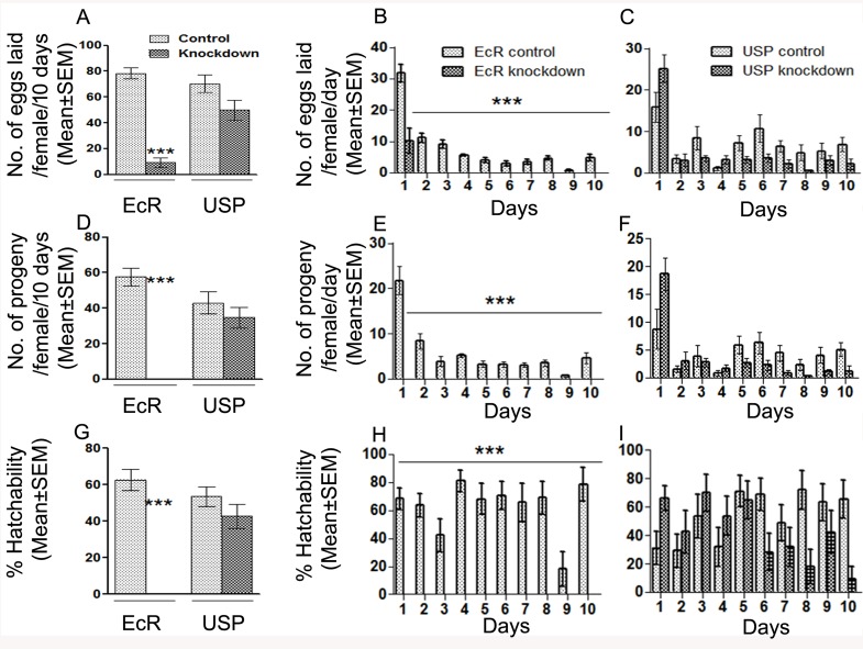
In many insects, the accessory gland, a secretory tissue of the male reproductive system, is essential for male fertility. Male accessory gland is the major source of proteinaceous secretions, collectively called as seminal proteins (or accessory gland proteins), which upon transfer, manipulate the physiology and behavior of mated females. Insect hormones such as ecdysteroids and juvenoids play a key role in accessory gland development and protein synthesis but little is known about underlying molecular players and their mechanism of action. Therefore, in the present study, we examined the roles of hormone-dependent transcription factors (Nuclear Receptors), in accessory gland development, function and male fertility of a genetically tractable insect model, Drosophila melanogaster. First, we carried out an RNAi screen involving 19 hormone receptors, individually and specifically, in a male reproductive tissue (accessory gland) for their requirement in Drosophila male fertility. Subsequently, by using independent RNAi/ dominant negative forms, we show that Ecdysone Receptor (EcR) is essential for male fertility due to its requirement in the normal development of accessory glands in Drosophila: EcR depleted glands fail to make seminal proteins and have dying cells. Further, our data point to a novel ecdysone receptor that does not include Ultraspiracle but is probably comprised of EcR isoforms in Drosophila male accessory glands. Our data suggest that this novel ecdysone receptor might act downstream of homeodomain transcription factor paired (prd) in the male accessory gland. Overall, the study suggests novel ecdysone receptor as an important player in the hormonal regulation of seminal protein production and insect male fertility.


Conflict of interest statement

The authors have declared that no competing interests exist.

Figures

Fig 1
Fig 1. The effect of knockdown of different hormone receptors on the fertility of mated females.
To identify the nuclear/hormone receptor involved in male fertility, 19 receptors were knocked down, individually, in accessory gland specific manner in the male reproductive tract and were allowed to mate with virgin females. Shown here is the number [Mean±Standard Error (SEM)] of progeny produced by females mated to knockdown or control males over a period of 10 days. Mates of met (Methoprene tolerant, a juvenile hormone receptor, **p<0.001) knockdown males produced significantly fewer progeny when compared to their controls and also those in strain background control (Jhe). Interestingly, females mated to EcR knockdown males failed to produce progeny (EcR; ***p<0.0001; Bonferroni corrected p value for significance is p<0.002). However, fertility of females mated to USP (p = 0.27) or the remaining 16 hormone receptor knockdown males was not significantly different from their respective controls. Number of females ranged from 15–45 depending on the hormone receptor analyzed.
Fig 2
Fig 2. Western blots showing the levels of EcR and USP in knockdown males compared to control males.
The EcR panel represents EcR levels in accessory glands from EcR control (+ lane, EcR), EcR knockdown (-lane, EcR), USP control (+ lane, USP) and USP knockdown (- lane, USP). Similarly, the USP panel represents the USP levels observed in accessory glands from above groups. Blots probed with α-tubulin antibodies (α-tubulin panels) served as controls for protein loading. Knockdowns were specific to the targeted hormone receptor. Further, the deficiency of EcR did not affect USP levels and vice-versa.
Fig 3
Fig 3. Reproductive performance of females mated to EcR or USP knockdown males over period of 10 days.
Panel A represents the overall fecundity (total no. of eggs laid/10 days) of mated females while Panels B and C represent day wise fecundity (no. of egg laid/day from day1-10) of mated females. The overall fecundity of EcR knockdown mates was significantly lower compared to controls (***p<0.0001). However, overall fecundity of USP knockdown mates was comparable to their controls. (p = 0.08). Further, EcR knockdown mates laid eggs for 24hr ASM, albeit at significantly fewer numbers (***p<0.0001) when compared to control and did not lay eggs from days 2–10. USP knockdown mates did not deviate from controls on day wise egg laying. Interestingly, there were no progeny from the eggs laid by EcR knockdown mates over period of 10 days (overall fertility, ***p<0.0001, Panel D; day-wise fertility, ***p<0.0001 Panel E). USP knockdown or control mates had comparable overall (Panel D; p = 0.3, USP) as well as day wise fertility (Panel F; p = 0.06 lowest) EcR control and knockdown mates differ significantly on total % hatchability (Panel G; EcR, ***p<0.0001) as well as % hatchability on day 1 (Panel H; ***p<0.0001). USP control and knockdown mates had significant differences in % hatchability on days 8 and 10 (Panel I; p = 0.01) but that did not have a significant bearing on the overall % hatchability (Panel H; p = 0.3). Values given here are Mean±SEM involving at least 15–30 females depending on the hormone receptor.
Fig 4
Fig 4. Analysis of sperm production in EcR knockdown males and their fate in mated females.
To evaluate the effect of EcR knockdown (EcR-miRNA) on sperm, we first generated control and knockdown males that express GFP labeled sperm (ProtamineB-EGFP). Subsequently, observation of seminal vesicles from these males under a confocal microscope revealed comparable levels of GFP tagged sperm (green) in both controls (Panels A&C) as well as knockdown (EcR, Panel B or USP, Panel D) males, suggesting that sperm production is normal in these males. To test if these males are able to transfer sperm to females during mating, males knockdown for EcR or USP in their accessory glands were allowed to mate with Oregon-R virgin females. At 2h ASM, reproductive tracts from mated females were isolated and observed under confocal microscope for the presence of GFP-labeled sperm. Reproductive tracts from females mated to (E) EcR control, (G) USP control or (H) knockdown males contained mating plug (MP, blue) in the uteri (U) and GFP-labeled sperm (green) in the uteri as well as sperm storage organs, namely seminal receptacle (SR) and spermathecae (SP). However, reproductive tracts of females mated to EcR knockdown males (F) contained mating plugs but had sperm only in the uterus but not in SR and SP. These observations suggest that knockdown of EcR or USP has no detectable effect on sperm transfer but the sperm transferred by EcR knockdown males fail to move towards sperm storage organs and are not stored at levels comparable to those in controls.
Fig 5
Fig 5. The effect of knockdown of EcR or USP on the structure of male accessory glands.
To assess the effect of depletion of EcR or USP, accessory glands were analyzed either at the ultrastructural level (panels at the top). Depicted at the top are the electron micrographs of male accessory glands from (A) EcR control (B) EcR knockdown (C) USP control and (D) knockdown males. Accessory glands from EcR control, USP control and USP knockdown males show normal protein filamentous structures (labeled as f) throughout in their lumen. However, glands of EcR knockdown have extreme vacuolization (v) and lack filamentous structures in the lumen. A minimum of five tissues from each group was used for ultrastructural analysis.
Fig 6
Fig 6. The effect of EcR knockdown on the cellular organization of the accessory glands.
Immunofluorescence panels shown here are the overlay images of accessory glands immunostained with α-Spectrin antibody (marking cell membrane, Green color) and labeled with nuclear stain DAPI (blue color). In EcR control glands, several polygonally shaped binucleate cells (the main cells) and a few large and spherical binucleate cells (the secondary cells, marked with arrow) interspersed between the polygonally shaped cells were observed at 400X (Panel A) and at a higher magnification of 630X (Panel A′). However, in EcR-deficient glands, cell membranes are highly disrupted (Panel B at 400X) and the nuclear distribution is distinctly different from that of control (Panel B′ at a total magnification of 630X). To assess the effect of depletion of EcR or USP, in accessory glands,western blots were probed for the secondary cell markers, Abd-B and ANCE. The glands lacked Abd-B (Panel C,—lane under EcR in Abd-B blot) while EcR control (Panel C, + lane under EcR), USP control (Panel C, + lane under USP) or USP knockdown (Panel C, -lane under USP) had detectable levels of Abd-B. In addition, EcR knockdown glands did not contain ANCE (Panel D, -lane under EcR in ANCE blot) while the same could be detected in controls, suggesting the disruption of secondary cells due to depletion of EcR.
Fig 7
Fig 7. Main cell or secondary cell derived Acps in EcR or USP deficient male accessory glands.
Depicted here are the western blots of accessory gland protein extracts from EcR control (+ lane under EcR), EcR knockdown (- lane under EcR), USP control (+ lane under USP) and USP knockdown (- lane under USP) males. All the Acps known to be derived from main cells (Ovulin, SP, Acp36DE, CG10586, and Acp62F) or secondary cells (CG1652, CG1656, CG17575) were detectable in EcR control, USP control and USP knockdown glands but not in EcR knockdown glands. α-tubulin was used as control for protein loading.
Fig 8
Fig 8. Analysis of cleaved Caspase 3 immunoreactivity in accessory glands of EcR control and knockdown males.
To examine if loss of EcR leads to induction of apoptosis in accessory glands, tissues were immunostained with antibodies for cleaved Caspase 3, which react with initiator and effector caspases in Drosophila. Control tissues show well organized nuclei (Blue in color; DAPI, Panel A) and lack of detectable cleaved Caspase 3 immunoreactivity (Panel B) the overlay (Panel C) shows only the nuclei, in contrast, glands from EcR knockdown contain distorted as well as disorganized nuclei (DAPI, Panel D) and high levels of cleaved Caspase 3 immunoreactivity (Panel E). The overlay (Panel F) shows distorted nuclei (blue) and cleaved Caspase 3 labeling (red). (G) Females mated to EcR knockdown males over expressing P35 (EcR↓+P35↑) produced progeny at control levels as opposed to sterility in EcR knockdown mates (EcR↓, ***p<0.0001) indicating that over-expression of P35 rescued the fertility of EcR knockdown males. (H) EcR knockdown males overexpressing Diap1 (EcR↓+Diap1↑) were fertile as opposed to sterility in EcR knockdown mates (EcR↓, ***p<0.0001) indicating that overexpression of Diap1 rescued the fertility of EcR knockdown males. (I) Westerns blots of accessory gland proteins to confirm the knockdown status of EcR in males over expressing P35 (EcR↓+P35↑) or Diap1 (EcR↓+ Diap1↑) in EcR knockdown background. Blots were probed with anti-β-actin antibody as control for protein loading.
Fig 9
Fig 9. Reproductive performance of females mated to males expressing dominant negative isoforms of EcR.
Panels (A), (B) & (C) represent fecundity, fertility and % hatchability, respectively of females mated to males over expressing EcR-A, EcR-B1, or EcR-B2 over a period of 10 days. Females mated to any of these three laid significantly fewer eggs and produced fewer progeny (***p <0.0001) when compared to their respective controls. However, there was no significant effect on % hatchability (p>0.05). (D) Sperm stored in spermathecae (SP) or seminal receptacle (SR) and total sperm in storage (Total) of females mated to males over expressing EcR-A, EcR-B1, or EcR-B2 respectively over a period of 2 hrs and 4 days ASM. Significance is ascribed as ***p<0.001; *p<0.05. Values given here are Mean±SEM involving tissues from at least 15–20 females.
Fig 10
Fig 10. Morphology and secondary cells markers of accessory glands in males over-expressing dominant negative EcR isoforms.
The morphology of accessory glands from (A) control or males over expressing (B) EcR-A, (C) EcR-B1, or (D) EcR-B2 was observed under light microscopy. Morphology of accessory glands from males over expressing EcR-B1, or EcR-B2 is comparable to their controls. However, accessory glands from EcR-A appear slightly reduced in comparison to their controls but still not as extremely reduced as those in EcR-miRNA based knockdown males. (B) Western blots of accessory gland protein extracts depicting levels of Abd-B (Abd-B panel), ANCE (ANCE panel) proteins and cleaved Caspase 3 immunoreactivity (cleaved Caspase 3 panel) in males over expressing EcR-A, EcR-B1 and EcR-B2. Blots were probed with β-actin antibodies (β-actin panels) served as controls for protein loading.

Similar articles

Cited by

References

    1. Avila FW, Sirot LK, LaFlamme BA, Rubinstein CD, Wolfner MF (2011) Insect seminal fluid proteins: identification and function. Annu Rev Entomol 56: 21–40. doi: 10.1146/annurev-ento-120709-144823 - DOI - PMC - PubMed
    1. Sirot LK, LaFlamme BA, Sitnik JL, Rubinstein CD, Avila FW, et al. (2009) Molecular social interactions: Drosophila melanogaster seminal fluid proteins as a case study. Adv Genet 68: 23–56. doi: 10.1016/S0065-2660(09)68002-0 - DOI - PMC - PubMed
    1. Chapman T, Bangham J, Vinti G, Seifried B, Lung O, et al. (2003) The sex peptide of Drosophila melanogaster: female post-mating responses analyzed by using RNA interference. Proc Natl Acad Sci U S A 100: 9923–9928. doi: 10.1073/pnas.1631635100 - DOI - PMC - PubMed
    1. Liu H, Kubli E (2003) Sex-peptide is the molecular basis of the sperm effect in Drosophila melanogaster. Proc Natl Acad Sci U S A 100: 9929–9933. doi: 10.1073/pnas.1631700100 - DOI - PMC - PubMed
    1. Ravi Ram K, Wolfner MF (2007a) Seminal influences: Drosophila Acps and the molecular interplay between males and females during reproduction. Integr Comp Biol 47: 427–445. - PubMed

MeSH terms

LinkOut - more resources