. 2017 May 11;12(5):e0170792.
doi: 10.1371/journal.pone.0170792. eCollection 2017.

The effect of sphingosine-1-phosphate on colonic smooth muscle contractility: Modulation by TNBS-induced colitis

Affiliations

The effect of sphingosine-1-phosphate on colonic smooth muscle contractility: Modulation by TNBS-induced colitis

Aishah Al-Jarallah et al. PLoS One. .

Abstract

Aim: Increased levels of circulating sphingosine-1-phosphate (S1P) have been reported in ulcerative colitis. The objective of this study was to examine the effect of S1P on colonic smooth muscle contractility and how is it affected by colitis.

Methods: Colonic inflammation was induced by intrarectal administration of trinitrobenzene sulfonic acid. Five days later colon segments were isolated and used for contractility experiments and immunoblotting.

Results: S1P contracted control and inflamed colon segments and the contraction was significantly greater in inflamed colon segments. S1P-induced contraction was mediated by S1PR1 and S1PR2 in control and S1PR2 in inflamed colon segments. S1PR3 did not play a significant role in S1P-induced contractions in control or inflamed colon. S1PR1, S1PR2 and S1PR3 proteins were expressed in colon segments from both groups. The expression of S1PR1 and S1PR2 was significantly enhanced in control and inflamed colon segments, respectively. S1PR3 levels however were not significantly different between the two groups. Nifedipine significantly reduced S1P-induced contraction in control but not inflamed colon segments. Thapsigargin significantly reduced S1P-induced contraction of the inflamed colon. GF 109203X and Y-27632, alone abolished S1P-induced contraction of the control but not inflamed colon segments. Combination of GF 109203X, Y-27632 and thapsigargin abolished S1P-induced contraction of inflamed colon segments.

Conclusion: S1P contracted control colon via S1PR1 and S1PR2 and inflamed colon exclusively via S1PR2. Calcium influx (control) or release (inflamed) and calcium sensitization are involved in S1P-induced contraction. Exacerbated response to S1P in colitic colon segments may explain altered colonic motility reported in patients and experimental models of inflammatory bowel disease.


Conflict of interest statement

Competing Interests: The authors have declared that no competing interests exist.

Figures

Fig 1
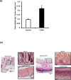
Fig 1. Characterization of ulcerative colitis.
(A) Colon weight to length ratio in control and colitic rats. (B) Representative eosin and hematoxylin stained colon sections from control and TNBS-treated rats. Data is mean ± SEM, *P < 0.05.
Fig 2
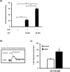
Fig 2. Effect of S1P on KCl-induced contractions in control and colitic colon segments.
After equilibration colonic segments from control rats were contracted twice with KCl (80m M) and washed immediately after reaching a peak contraction. Then colonic segments were incubated with BSA (40 μM) or S1P (10 or 40 μM) and allowed to reach a maximal response (A). Traces of S1P (40μM) induced contraction in control and colitic colon segments are shown in (B), arrows indicate S1P addition. Mean S1P (40μM) induced contraction in control and colitic colon segments is shown in (C). Data is mean ± SEM of S1P induced contraction expressed relative to the second KCl-induced contraction, * P < 0.05.
Fig 3
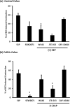
Fig 3. The Role of S1PR in S1P induced contraction in control and colitic colon.
Colonic segments from control (A) and colitic (B) rats were equilibrated in Krebs solution for 30 min and contracted twice with KCL (80mM) with consecutive washes after the maximal contraction was reached. Tissues were then preincubated with S1PR1 agonist (SEW2871, 1μM) S1PR1 antagonist (W146, 10μM), S1PR2 antagonist (JTE-013, 1μM) or with S1PR3 antagonist (CAY-10444, 10μM) for 30 min before the addition of S1P (40μM). Data is mean ± SEM of S1P induced contraction is expressed relative to the second KCl-induced contraction, * P < 0.05 relative to S1P induced contraction.
Fig 4
Fig 4. Immunoblotting of S1PR expression in control and colitic colon segments.
Representative images of total membrane preps (50 μg protein) from control and colitic colon tissues, from 8–12 independent rats, that were probed with S1PR1 (a), S1PR2 (b) and S1PR3 (c) antibodies. Band intensities were normalized to actin and expressed relative to the controls. Data is mean ± SEM, * P < 0.05 and NS indicates no significance.
Fig 5
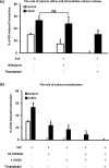
Fig 5. The Role of calcium and calcium sensitization pathways in S1P induced contraction in control and colitic colon.
Colonic segments from control (n = 3–6) and colitic (n = 3–7) rats were equilibrated in Krebs solution for 30 min and contracted twice with KCL with consecutive washes after the maximal contraction was reached. Tissues were then preincubated with L-type calcium channel blocker (nifedipine, 1μM) or SERCA antagonist (thapsigargin, 10μM) (A) or with PKC antagonist (GF 102903x, 1μM), ROCK inhibitor (Y-27632, 10μM) or inhibitor cocktails (B) for 30 min before the addition of S1P (40μM). Data is mean ± SEM of S1P induced contraction expressed relative to the second KCl-induced contraction, * P < 0.05 versus S1P induced contraction in corresponding group.
Fig 6
Fig 6. Proposed mechanism of S1P induced contraction in control and colitic colon.
In control colon S1P activates S1PR1 and S1PR2 and induce contractions that are dependent on calcium influx via L-type calcium channels, calcium release from the sarcoplasmic reticulum and calcium sensitization pathways including PKC and Rho kinase (A). In colitic colon however S1P induces larger contractions via S1PR2 that are independent of calcium influx through L-type calcium channels yet it involves intracellular calcium release and calcium sensitization pathways (B). The contribution of these pathways however appears to be different in control and inflamed colon segments. Whereas the blockade of a single pathway completely abolishes the response to S1P in control segments, S1P induced contractions appear to be salvaged by other pathways in inflamed colon segments. Eradication of S1P induced contraction in inflamed colon require simultaneous blockade of intracellular calcium release (SERCA) and calcium sensitization pathways (PKC and Rho kinase).

Similar articles

Cited by

References

    1. Zhang YZ, Li YY. Inflammatory bowel disease: pathogenesis. World J Gastroenterol. 2014;20(1):91–9. PubMed Central PMCID: PMC3886036. doi: 10.3748/wjg.v20.i1.91 - DOI - PMC - PubMed
    1. Coulie B, Camilleri M, Bharucha AE, Sandborn WJ, Burton D. Colonic motility in chronic ulcerative proctosigmoiditis and the effects of nicotine on colonic motility in patients and healthy subjects. Aliment Pharmacol Ther. 2001;15(5):653–63. - PubMed
    1. Sethi AK, Sarna SK. Colonic motor activity in acute colitis in conscious dogs. Gastroenterology. 1991;100(4):954–63. - PubMed
    1. Snape WJ Jr. The role of a colonic motility disturbance in ulcerative colitis. The Keio journal of medicine. 1991;40(1):6–8. - PubMed
    1. Bassotti G, de Roberto G, Chistolini F, Sietchiping-Nzepa F, Morelli O, Morelli A. Twenty-four-hour manometric study of colonic propulsive activity in patients with diarrhea due to inflammatory (ulcerative colitis) and non-inflammatory (irritable bowel syndrome) conditions. Int J Colorectal Dis. 2004;19(5):493–7. doi: 10.1007/s00384-004-0604-6 - DOI - PubMed

MeSH terms