. 2017 May 11;12(5):e0176396.
doi: 10.1371/journal.pone.0176396. eCollection 2017.

Characterization of a Bvg-regulated fatty acid methyl-transferase in Bordetella pertussis

Affiliations

Characterization of a Bvg-regulated fatty acid methyl-transferase in Bordetella pertussis

Alex Rivera-Millot et al. PLoS One. .

Abstract

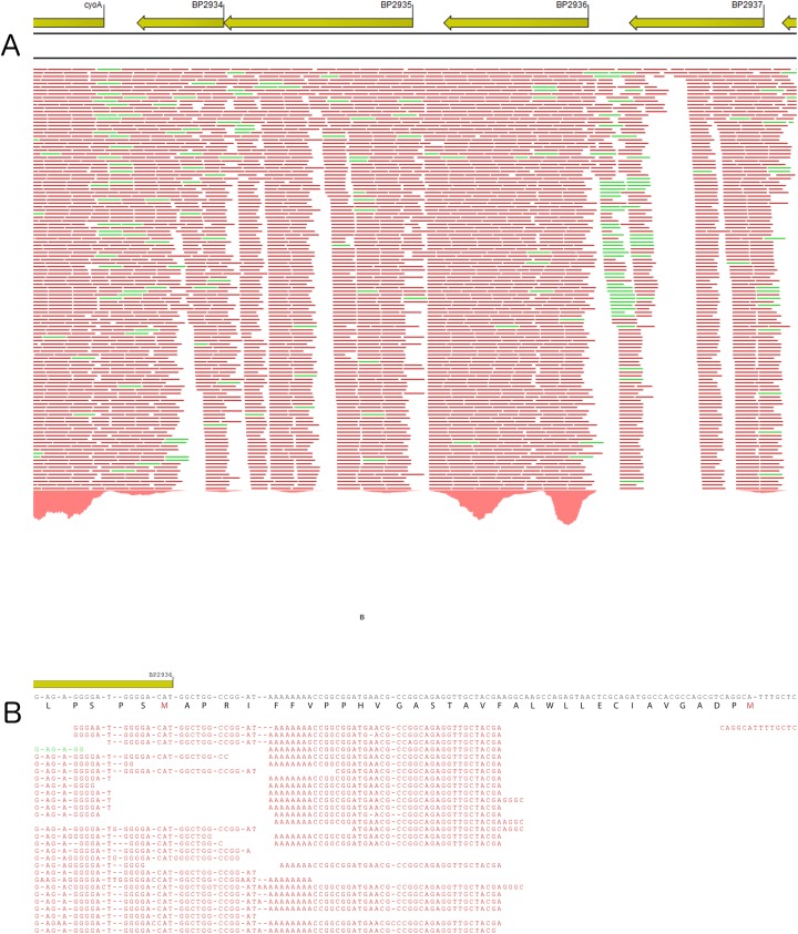
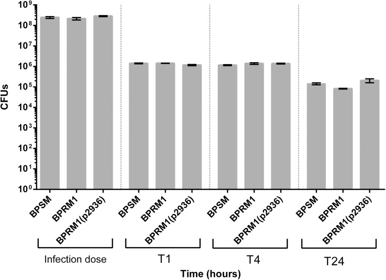
The whooping cough agent Bordetella pertussis controls the expression of its large virulence regulon in a coordinated manner through the two-component signal transduction system BvgAS. In addition to the genes coding for bona fide virulence factors, the Bvg regulon comprises genes of unknown function. In this work, we characterized a new Bvg-activated gene called BP2936. Homologs of BP2936 are found in other pathogenic Bordetellae and in several other species, including plant pathogens and environmental bacteria. We showed that the gene product of BP2936 is a membrane-associated methyl-transferase of free fatty acids. We thus propose to name it FmtB, for fatty acid methyl-transferase of Bordetella. The role of this protein was tested in cellular and animal models of infection, but the loss of BP2936 did not appear to affect host-pathogen interactions in those assays. The high level of conservation of BP2936 among B. pertussis isolates nevertheless argues that it probably plays a role in the life cycle of this pathogen.


Conflict of interest statement

Competing Interests: The authors have declared that no competing interests exist.

Figures

Fig 1
Fig 1. Characterization of the BP2936 locus by RNA seq.
A. Representation of the deep RNA sequencing results for the locus encompassing BP2936. BP2936 is encoded by the anti-sens strand of the genome. The red and green bars under the genes represent transcripts of the anti-sense and sense strands, respectively. B. Differential RNA sequencing was used to identify the transcription initiation site of BP2936. The consensus sequence is indicated. Translation of the coding DNA sequence is shown from right to left, indicating the two possible initiation Met. The primary transcripts identified by dRNA seq show that the transcription start site maps between these two codons, at nucleotide position 3125512 in the genome of Tohama I.
Fig 2
Fig 2. Detection of the product of BP2936 by immunoblot analyses.
(A), insoluble and soluble fractions of BPSM (wt control; lanes 1 and 4), BPRM1 (deletion mutant; lanes 2 and 5) and BPRM1(pBP2936) (mutant complemented on plasmid; lanes 3 and 6) were analyzed by immunoblotting with antibodies raised against the recombinant protein. (B), the same experiment was performed with the insoluble and soluble fractions of BPSM (lanes 1 and 3) and BPRM2 (mutant complemented on chromosome; lanes 2 and 4). (C), a comparison of the sizes of the three proteins, i.e., the 2936P protein produced by B. pertussis (lane 1), and the recombinant proteins rec2936P-sh (lane 2) and rec2936P-lg (lane 3) produced in E. coli, was performed. Note that 2936P migrates slightly faster than the shorter recombinant protein, most likely because of the purification tag added to the latter.
Fig 3
Fig 3. Structure of the protein coded by the B. parapertussis ortholog of BP2936.
The coordinates found in the RCSB Protein Data Bank (Nr 3OJC) correspond to the B. parapertussis ortholog (97% sequence identity with 2936P). (A), the structure of the protein coded by BPP1064 is shown in cartoon representation. The palmitate molecule found in the cavity is shown in yellow stick representation. (B), A model of 2936P with palmitate and S-adenosyl homocysteine (SAH) in the cavity was constructed. Both SAH and the methyl palmitate are displayed in sticks. A zoom of the relevant region is presented in C. The sulfur atom of SAH, which carries the methyl group in SAM, is shown as a yellow ball. The conserved residues modified by site-directed mutagenesis (colored in slate blue) are indicated.
Fig 4
Fig 4. Detection of methyl fatty acids in B. pertussis.
The amounts of methyl fatty acids were determined in BPSM (wt control), BPRM1 (deletion mutant), BPRM2 (chromosomal complementation), BPRM1(pBP2936) (plasmid complementation) and BPRM1(pBP2936DA/YS) (complementation on plasmid with mutated BP2936). The relative abundance is defined as the ratio between the area of the C16 peak and the area of the internal standard peak, normalized to the number of cells. The experiments were performed on 3 to 5 independent samples for each strain. Means and standard errors of the means are shown. A Kruskal-Wallis test was used to analyze the data (*, p value < 0.05; ***, p value < 0.001).
Fig 5
Fig 5. Intracellular survival of B. pertussis in THP1 macrophages.
The BPSM, BPRM1 and BPRM1(pBP2936) strains were used in this experiment. The colony forming units were determined at each time point, expressed in hours after bacteria-macrophages contacts. The experiments were performed in triplicates, and the means and errors of the means are indicated.
Fig 6
Fig 6. Effect of BP2936 deletion on colonisation of mice lungs by B. pertussis.
BPSM, BPRM1 and BPRM2 were used to inoculate BalB/c mice intranasally. The mice were sacrificed at the indicated time points (in days), and the viable bacteria present in the lungs were counted. The dashed line indicates the lower limit for the counts of bacteria in the lungs. The means and errors of the means are indicated.
Fig 7
Fig 7. Colonisation of mice lungs by mutant B. pertussis strains.
BPSM, BPRA (ptx KO), BPGR4 (fhaB KO) strains and their respective derivatives in which BP2936 (fmtB) was deleted, were used in this experiment. Counts of viable bacteria found in the lungs were performed 3 hours (T0) and 5 days (T5) after infection. The means and errors of the means are indicated.

Similar articles

Cited by

References

    1. Melvin JA, Scheller EV, Miller JF, Cotter PA. Bordetella pertussis pathogenesis: current and future challenges. Nat Rev Microbiol. 2014; 12: 274–288. doi: 10.1038/nrmicro3235 - DOI - PMC - PubMed
    1. Libster Edwards CA. Re-emergence of pertussis: what are the solutions? Expert Rev Vaccines. 2012; 11: 1331–1346. doi: 10.1586/erv.12.118 - DOI - PubMed
    1. Rohani P, Zhong X, King AA. Contact network structure explains the changing epidemiology of pertussis. Science. 2010; 330: 982–985. doi: 10.1126/science.1194134 - DOI - PubMed
    1. Lavine JS, King AA, Bjornstad ON. Natural immune boosting in pertussis dynamics and the potential for long-term vaccine failure. Proc Natl Acad Sci U S A. 2011; 108: 7259–7264. doi: 10.1073/pnas.1014394108 - DOI - PMC - PubMed
    1. Cotter PA, Jones AM. Phosphorelay control of virulence gene expression in Bordetella. Trends Microbiol. 2003; 11: 367–373. - PubMed

MeSH terms

Substances