. 2017 May 11;12(5):e0177557.
doi: 10.1371/journal.pone.0177557. eCollection 2017.

Pannexin 3 regulates proliferation and differentiation of odontoblasts via its hemichannel activities

Affiliations

Pannexin 3 regulates proliferation and differentiation of odontoblasts via its hemichannel activities

Tsutomu Iwamoto et al. PLoS One. .

Abstract

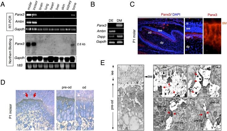
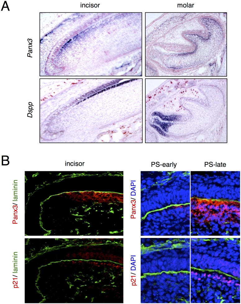
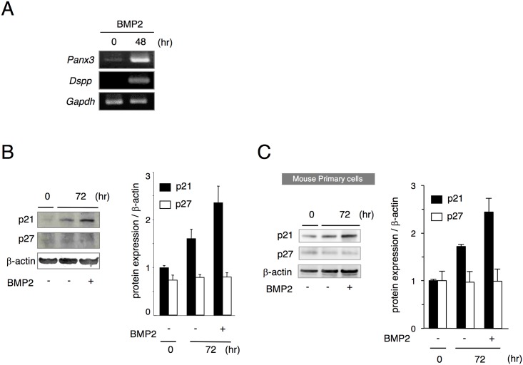
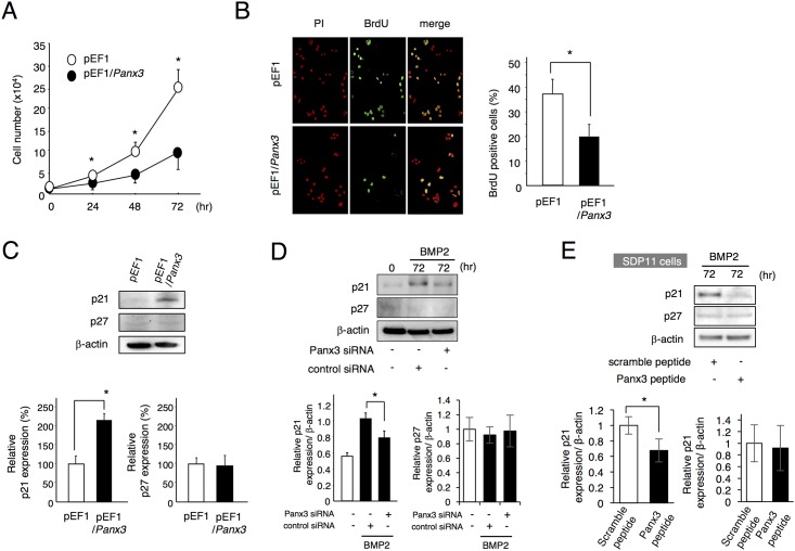
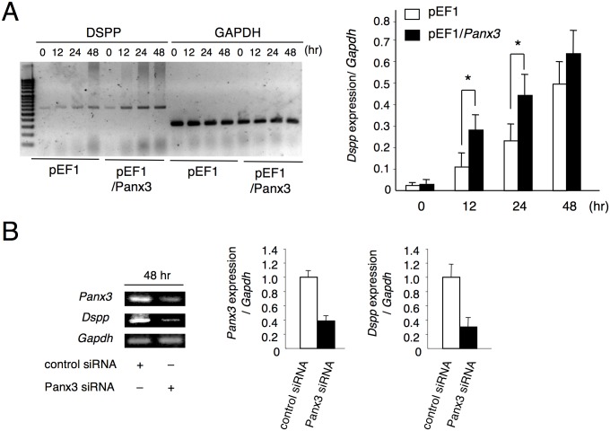
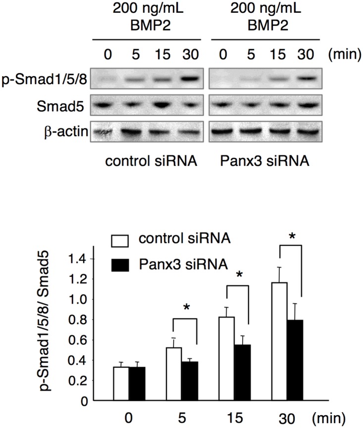
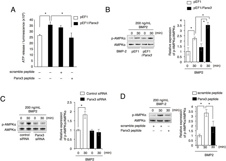
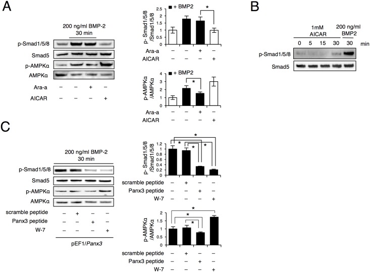
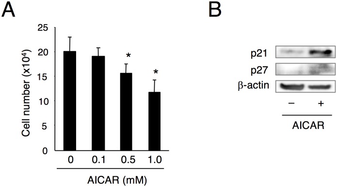
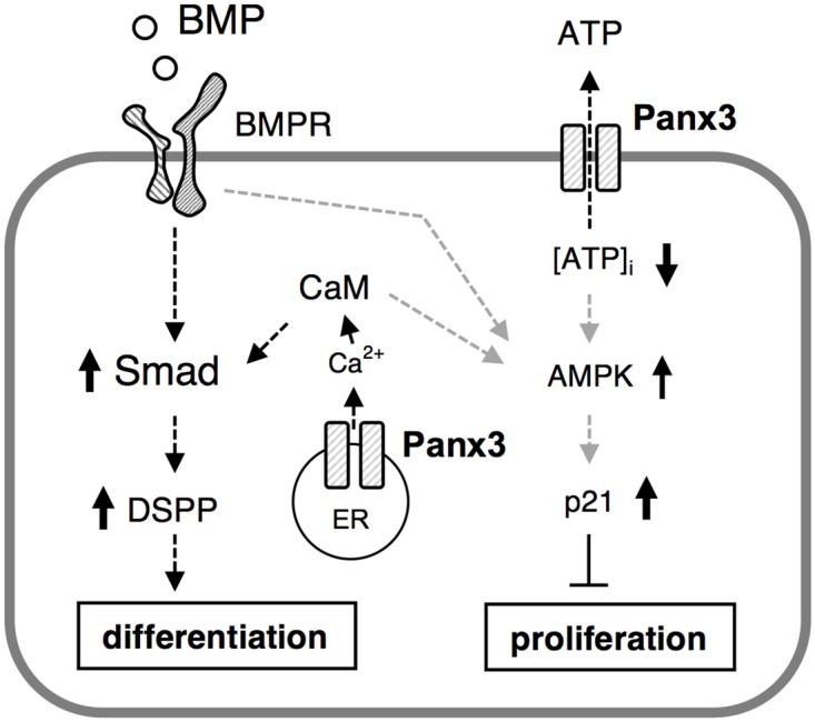
Highly coordinated regulation of cell proliferation and differentiation contributes to the formation of functionally shaped and sized teeth; however, the mechanism underlying the switch from cell cycle exit to cell differentiation during odontogenesis is poorly understood. Recently, we identified pannexin 3 (Panx3) as a member of the pannexin gap junction protein family from tooth germs. The expression of Panx3 was predominately localized in preodontoblasts that arise from dental papilla cells and can differentiate into dentin-secreting odontoblasts. Panx3 also co-localized with p21, a cyclin-dependent kinase inhibitor protein, in preodontoblasts. Panx3 was expressed in primary dental mesenchymal cells and in the mDP dental mesenchymal cell line. Both Panx3 and p21 were induced during the differentiation of mDP cells. Overexpression of Panx3 in mDP cells reduced cell proliferation via up-regulation of p21, but not of p27, and promoted the Bone morphogenetic protein 2 (BMP2)-induced phosphorylation of Smad1/5/8 and the expression of dentin sialophosphoprotein (Dspp), a marker of differentiated odontoblasts. Furthermore, Panx3 released intracellular ATP into the extracellular space through its hemichannel and induced the phosphorylation of AMP-activated protein kinase (AMPK). 5-Aminoimidazole-4-carboxamide-ribonucleoside (AICAR), an activator of AMPK, reduced mDP cell proliferation and induced p21 expression. Conversely, knockdown of endogenous Panx3 by siRNA inhibited AMPK phosphorylation, p21 expression, and the phosphorylation of Smad1/5/8 even in the presence of BMP2. Taken together, our results suggest that Panx3 modulates intracellular ATP levels, resulting in the inhibition of odontoblast proliferation through the AMPK/p21 signaling pathway and promotion of cell differentiation by the BMP/Smad signaling pathway.


Conflict of interest statement

Competing Interests: The authors have declared that no competing interests exist.

Figures

Fig 1
Fig 1. Expression of Panx3 in tooth germ.
(A) RT-PCR analysis (upper three panels) and northern blotting analysis (lower three panels) using RNA from postnatal day 1 (P1) mouse tissues (molar, incisor, brain, lung, heart, liver, skin, kidney, and bone). (B) RT-PCR analysis using the dental epithelium (DE) and mesenchyme (DM), dissected from P1 mouse tooth germ. (C) Immnostaining with anti-Panx3 antibody (red) and DAPI nuclear staining (blue). (D) Light microscopy images of semi-thin sections stained with methylene blue for immunoelectron microscopy. Panx3 is present in the preodontoblasts (pre-od) but not in the odontoblasts (od). (E) Immunoelectron microscopy images of the Panx3 protein in preodontoblasts from P1 incisors showing labeling in the preodontoblasts (upper panels) and at the cell-cell contact sites (lower panels). dp; dental papilla, iee; inner enamel epithelium, si; stratum intermedium, BM; basement membrane.
Fig 2
Fig 2. Expression of Panx3 mRNA and protein in tooth germ.
(A) In situ hybridization with Panx3 and Dspp probes in postnatal day 1 (P1) incisor or molar semi-serial sections. (B) immunostaining with anti-Panx3 or anti-p21 antibodies (red) or laminin (green) in P1 incisor semi-serial sections. Blue signifies DAPI nuclear staining. PS; presecretory stage.
Fig 3
Fig 3. Expression of Panx3 and p21 in differentiating mDP cells and mouse primary dental papilla cells.
(A) mRNA expression in differentiating mDP cells. mDP cells were cultured with 200 ng/mL BMP2. Total RNA was extracted from the cells at the indicated time points following BMP2 treatment and analyzed by RT-PCR. Gapdh, glyceraldehyde-3-phosphate dehydrogenase, was used as a control. p21 expression in differentiating mDP cells (B) and mouse primary dental papilla cells (C). Both mDP cells and mouse primary dental papilla cells were cultured with or without 200 ng/mL BMP2 for 72 h and cell extracts were analyzed by western blotting using anti-p21 and anti-p27 antibodies. β-actin was used as a control. Data were pooled from three independent experiments with error bars designating standard deviation of the mean.
Fig 4
Fig 4. Inhibition of cell proliferation by Panx3.
(A) mDP cells were stably transfected with the empty control vector (pEF1) or with the Panx3 expression vector (pEF1/Panx3). The cells were cultured for 72 h and cell numbers were counted. The number of Panx3-transfected cells was reduced compared to the control cells. (B) Cells were cultured for 48 h and BrdU incorporation after 30 min was analyzed using a fluorescence microscope. (C) Cell extracts were analyzed by western blotting using anti-p21 and anti-p27 antibodies. β-actin was used as a control. (D) mDP cells transfected with either control siRNA or Panx3 siRNA, were cultured with 200 ng/ml BMP2 for 72 h. Western blotting was performed for p21 and p27 expressions. (E) SDP11 cells were cultured with 200 ng/mL BMP2 and 0.5 μg/ml Panx3 inhibitory peptide or control peptide for 72 h. Western blotting was performed for p21 and p27 expressions. All experiments were repeated at least three times with similar results. Data were pooled from three independent experiments with error bars designating standard deviation of the mean. Statistical analysis was performed using analysis of variance (*P < 0.01).
Fig 5
Fig 5. Increase in BMP2-mediated Dspp induction in mDP cells due to Panx3.
(A) mDP cells stably transfected with either control pEF1 or pEF1/Panx3 were cultured with 200 ng/mL of BMP2 for the indicated time. Total RNA was then extracted and analyzed by RT-PCR. (B) mDP cells transfected with either control siRNA or Panx3 siRNA were cultured with the addition of 200 ng/mL BMP2 for 48 h. RT-PCR was performed to examine Panx3 and Dspp expression. All experiments were repeated at least five times with similar results. Data were pooled from three independent experiments with error bars designating standard deviation of the mean. Statistical analysis was performed using analysis of variance (*P < 0.01).
Fig 6
Fig 6. Decrease in BMP2-induced Smad1/5/8 phosphorylation due to Panx3 siRNA.
Following transfection with either control siRNA or Panx3 siRNA for 48 h, cells were treated with 200 ng/mL BMP2 for the time indicated. Protein extracts were analyzed by western blotting using anti-phospho-Smad1/5/8, anti-Smad5, and anti-β-actin antibodies. Image J 1.33u was used to quantify the protein bands. Data were pooled from three independent experiments with error bars designating standard deviation of the mean. Statistical analysis was performed using analysis of variance (*P < 0.01).
Fig 7
Fig 7. Increased ATP efflux and AMPK phosphorylation in Panx3-transfected mDP cells.
(A) Cells were plated at ~50% confluency and ATP levels in the media were measured by luminometry. Statistical analysis was performed using analysis of variance (*P < 0.01). (B) Time course of AMPK phosphorylation in control pEF1 or pEF1/Panx3-transfected mDP cells following treatment with or without BMP2 was analyzed by western blotting using the anti-phospho-AMPK antibody. (C) Western blotting was performed using the anti-phospho-AMPK antibody for mDP cells transfected with either control siRNA or Panx3 siRNA. (D) mDP cells were incubated with the Panx3 peptide or control peptide for 30 min, and then AMPK phosphorylation, induced by BMP2, was analyzed by western blotting. Data were pooled from three independent experiments with error bars designating standard deviation of the mean. Statistical analysis was performed using analysis of variance (*P < 0.01).
Fig 8
Fig 8. The role of Panx3 in AMPK and Smad signaling.
(A) mDP cells were treated with 1 mM of Ara-a for 30 min before treatment with BMP2. 1 mM AICAR was tested Smad1/5/8 and AMPK phosphorylations by western blotting. Data were pooled from three independent experiments with error bars designating standard deviation of the mean. Statistical analysis was performed using analysis of variance (*P < 0.05). (B) The time course of Smad1/5/8 phosphorylation in mDP cells following treatment with AICAR was analyzed by western blotting using the anti-phospho-Smad1/5/8 antibody. (C) Panx3-transfected mDP cells were treated with either the Panx3 peptide or the calmodulin inhibitor W-7, and the phosphorylation of both Smad1/5/8 and AMPK was analyzed by western blotting. Data were pooled from three independent experiments with error bars designating standard deviation of the mean. Statistical analysis was performed using analysis of variance (*P < 0.05).
Fig 9
Fig 9. Inhibition of cell proliferation by AICAR.
mDP cells were cultured in the presence of 0, 0.1, 0.5, or 1.0 mM 5-Aminoimidazole-4-carboxamide-ribonucleoside (AICAR), an AMPK activator, for three days and cell proliferation was measured using the cell counting kit-8. Data were pooled from three independent experiments with error bars designating standard deviation of the mean. Statistical analysis was performed using analysis of variance (*P < 0.01). (B) mDP cells were treated with 1.0 mM AICAR for three days and analyzed by western blotting using the antibodies of p21 and p27. β-actin was used as a control.
Fig 10
Fig 10. Role of Panx3 in odontoblast proliferation and differentiation.
The Panx3 ER Ca2+ channel promotes BMP-Smad signaling for differentiation. Panx3 is expressed in preodontoblasts, and releases intracellular ATP to the extracellular space, which may result in an activation of AMPK and subsequent inhibition of cell proliferation.

Similar articles

Cited by

  • Unraveling the Role of the Apical Papilla During Dental Root Maturation.
    Driesen RB, Gervois P, Vangansewinkel T, Lambrichts I. Driesen RB, et al. Front Cell Dev Biol. 2021 May 6;9:665600. doi: 10.3389/fcell.2021.665600. eCollection 2021. Front Cell Dev Biol. 2021. PMID: 34026757 Free PMC article. Review.
  • ATP transporters in the joints.
    Larrañaga-Vera A, Marco-Bonilla M, Largo R, Herrero-Beaumont G, Mediero A, Cronstein B. Larrañaga-Vera A, et al. Purinergic Signal. 2021 Dec;17(4):591-605. doi: 10.1007/s11302-021-09810-w. Epub 2021 Aug 15. Purinergic Signal. 2021. PMID: 34392490 Free PMC article. Review.
  • Gelatinases Cleave Dentin Sialoprotein Intracellularly.
    Gou X, Xue Y, Zheng H, Yang G, Chen S, Chen Z, Yuan G. Gou X, et al. Front Physiol. 2020 Jun 25;11:686. doi: 10.3389/fphys.2020.00686. eCollection 2020. Front Physiol. 2020. PMID: 32670089 Free PMC article.
  • The functional effects of Piezo channels in mesenchymal stem cells.
    Huang Z, Huang Y, Ning X, Li H, Li Q, Wu J. Huang Z, et al. Stem Cell Res Ther. 2023 Aug 26;14(1):222. doi: 10.1186/s13287-023-03452-y. Stem Cell Res Ther. 2023. PMID: 37633928 Free PMC article. Review.
  • Pannexin 3 channels in health and disease.
    O'Donnell BL, Penuela S. O'Donnell BL, et al. Purinergic Signal. 2021 Dec;17(4):577-589. doi: 10.1007/s11302-021-09805-7. Epub 2021 Jul 12. Purinergic Signal. 2021. PMID: 34250568 Free PMC article. Review.

References

    1. Bennett MV, Verselis VK. Biophysics of gap junctions. Semin Cell Biol. 1992;3(1):29–47. Epub 1992/02/01. - PubMed
    1. Scemes E, Suadicani SO, Dahl G, Spray DC. Connexin and pannexin mediated cell-cell communication. Neuron Glia Biol. 2007;3(3):199–208. Epub 2008/07/19. 10.1017/S1740925X08000069 - DOI - PMC - PubMed
    1. Bruzzone R, Hormuzdi SG, Barbe MT, Herb A, Monyer H. Pannexins, a family of gap junction proteins expressed in brain. Proc Natl Acad Sci U S A. 2003;100(23):13644–9. Epub 2003/11/05. 10.1073/pnas.2233464100 - DOI - PMC - PubMed
    1. Bruzzone R, White TW, Paul DL. Connections with connexins: the molecular basis of direct intercellular signaling. Eur J Biochem. 1996;238(1):1–27. Epub 1996/05/15. - PubMed
    1. Lo CW, Cohen MF, Huang GY, Lazatin BO, Patel N, Sullivan R, et al. Cx43 gap junction gene expression and gap junctional communication in mouse neural crest cells. Dev Genet. 1997;20(2):119–32. Epub 1997/01/01. 10.1002/(SICI)1520-6408(1997)20:2<119::AID-DVG5>3.0.CO;2-A - DOI - PubMed

MeSH terms

LinkOut - more resources