. 2017 May 11;13(5):e1006374.
doi: 10.1371/journal.ppat.1006374. eCollection 2017 May.

Characterization of miR-122-independent propagation of HCV

Affiliations

Characterization of miR-122-independent propagation of HCV

Chikako Ono et al. PLoS Pathog. .

Abstract

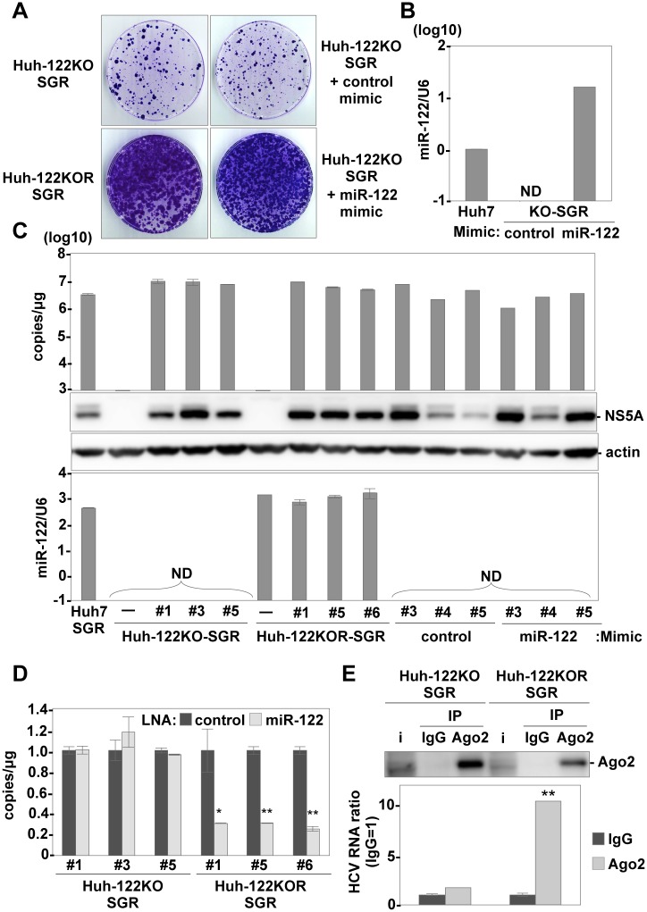
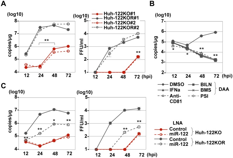
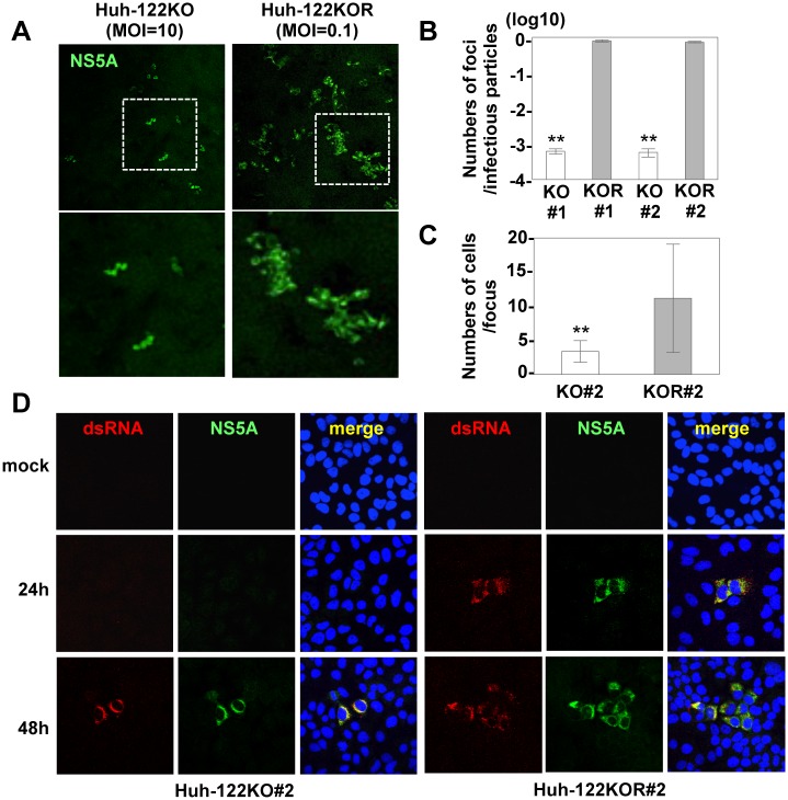
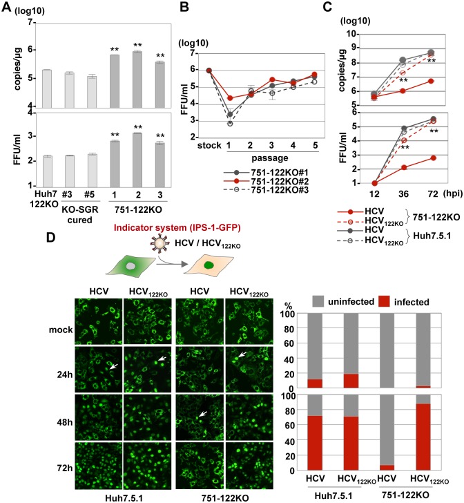
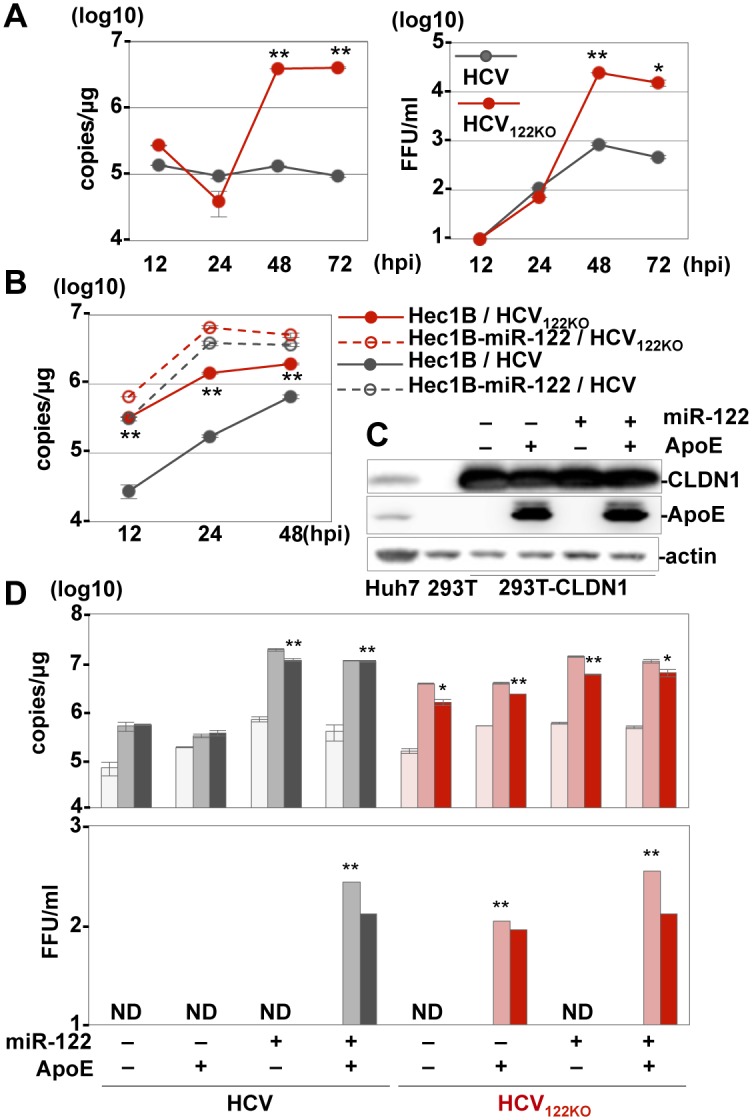
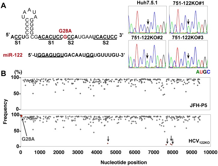
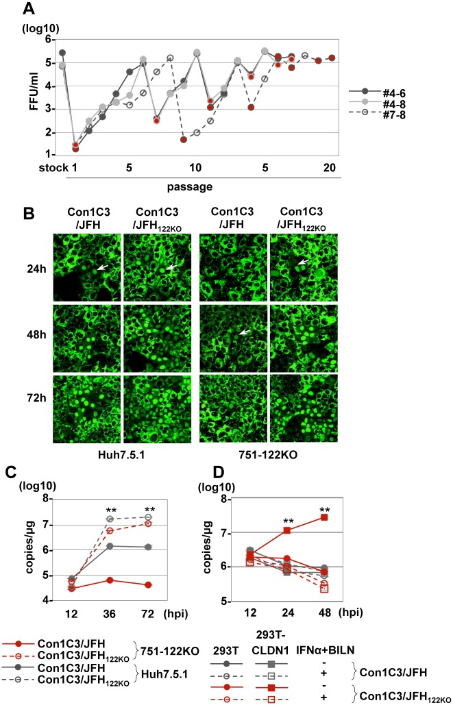
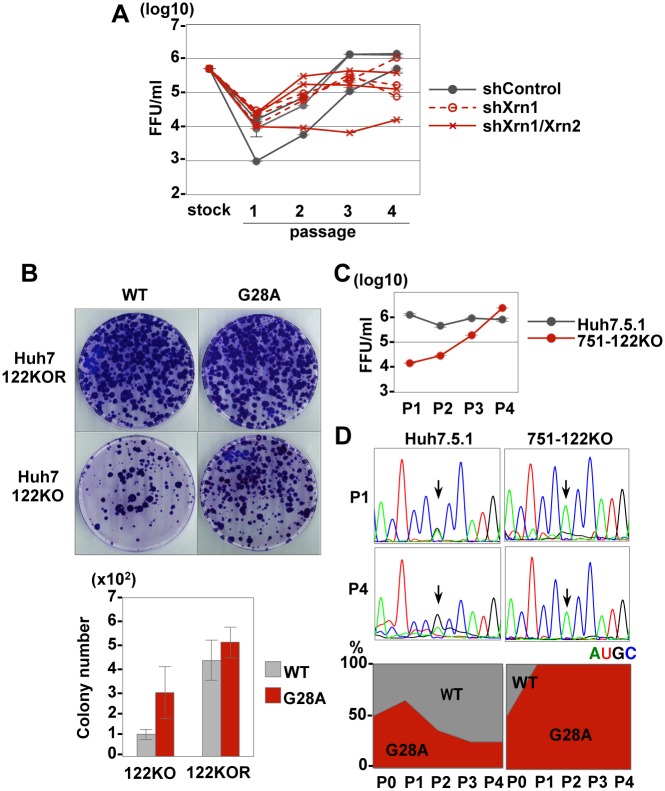
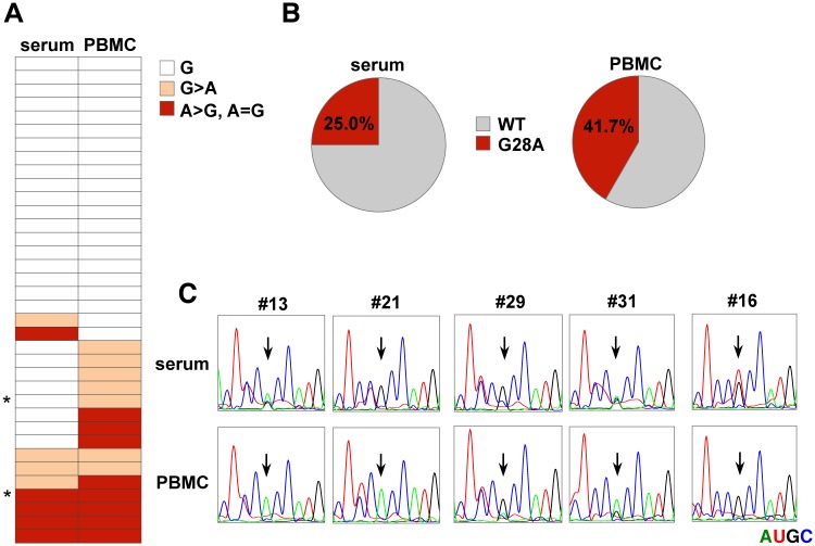
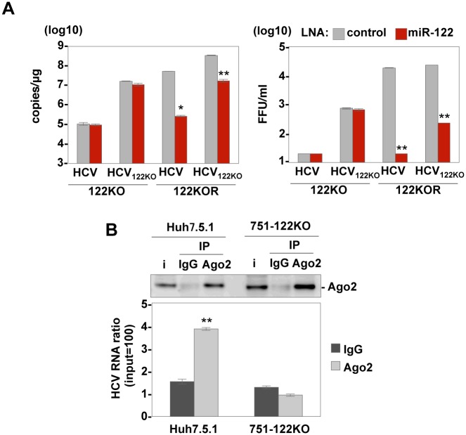
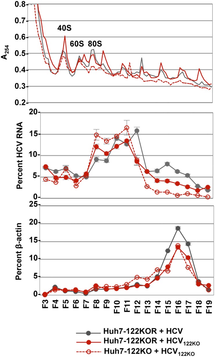
miR-122, a liver-specific microRNA, is one of the determinants for liver tropism of hepatitis C virus (HCV) infection. Although miR-122 is required for efficient propagation of HCV, we have previously shown that HCV replicates at a low rate in miR-122-deficient cells, suggesting that HCV-RNA is capable of propagating in an miR-122-independent manner. We herein investigated the roles of miR-122 in both the replication of HCV-RNA and the production of infectious particles by using miR-122-knockout Huh7 (Huh7-122KO) cells. A slight increase of intracellular HCV-RNA levels and infectious titers in the culture supernatants was observed in Huh7-122KO cells upon infection with HCV. Moreover, after serial passages of HCV in miR-122-knockout Huh7.5.1 cells, we obtained an adaptive mutant, HCV122KO, possessing G28A substitution in the 5'UTR of the HCV genotype 2a JFH1 genome, and this mutant may help to enhance replication complex formation, a possibility supported by polysome analysis. We also found the introduction of adaptive mutation around miR-122 binding site in the genotype 1b/2a chimeric virus, which originally had an adenine at the nucleotide position 29. HCV122KO exhibited efficient RNA replication in miR-122-knockout cells and non-hepatic cells without exogenous expression of miR-122. Competition assay revealed that the G28A mutant was dominant in the absence of miR-122, but its effects were equivalent to those of the wild type in the presence of miR-122, suggesting that the G28A mutation does not confer an advantage for propagation in miR-122-rich hepatocytes. These observations may explain the clinical finding that the positive rate of G28A mutation was higher in miR-122-deficient PBMCs than in the patient serum, which mainly included the hepatocyte-derived virus from HCV-genotype-2a patients. These results suggest that the emergence of HCV mutants that can propagate in non-hepatic cells in an miR-122-independent manner may participate in the induction of extrahepatic manifestations in chronic hepatitis C patients.


Conflict of interest statement

The authors have declared that no competing interests exist.

Figures

Fig 1
Fig 1. Establishment of replicon cells derived from Huh7-122KO cells.
(A) Subgenomic HCV replicon RNA was electroporated into Huh7-122KO and Huh7-122KOR cells, or into Huh7-122KO cells together with control- or miR-122-mimic, and G418-resistant colonies were stained with crystal violet at 21 days post-transduction. (B) Expression of miR-122 in Huh7-122KO cells electroporated with either control-mimic or miR-122-mimic at 72 h post-electroporation. Relative expression of miR-122 was determined by qRT-PCR by using U6 snRNA as an internal control. (C) Each of the three clones derived from each type of replicon cells was subjected to qRT-PCR after extraction of total RNA (top) and to immunoblotting by using anti-NS5A and β-actin (middle). The relative expression of miR-122 was determined by qRT-PCR by using U6 snRNA as an internal control (bottom). (D) Intracellular HCV-RNA replication in Huh7-122KO-SGR cells (#1, #3, #5) and Huh7-122KOR-SGR cells (#1, #5, #6) in the presence of 20 nM of either LNA-control or LNA-miR122 was determined by qRT-PCR. (E) The Ago2 complex was immunoprecipitated from Huh7-122KO-SGR#1 and Huh7-122KOR-SGR#1 cells by using either anti-IgG or anti-Ago2 mouse antibody. The HCV-RNA associated with Ago2 was determined by qRT-PCR and Ago2 was detected by immunoblotting. Error bars indicate the standard deviation of the mean and asterisks indicate significant differences (*P < 0.05; **P < 0.01) versus the results for the control.
Fig 2
Fig 2. miR-122-independent HCV replication in Huh7-122KO cells.
(A) HCV was inoculated into Huh7-122KO and Huh7-122KOR cells at an MOI of 3, and intracellular HCV-RNA levels (left panel) and infectious titers in the culture supernatants (right panel) were determined by qRT-PCR and focus formation assay, respectively. (B) HCV-RNA replication was inhibited by the treatment with IFNα, BILN, BMS790052, PSI7977 and anti-CD81 antibody. (C) HCV replication in Huh7-122KO cells was resistant to treatment with an miR-122 inhibitor, LNA (HCV-RNA replication: left; infectious titer: right). Error bars indicate the standard deviation of the mean and asterisks indicate significant differences (*P < 0.05; **P < 0.01) versus the results for the control.
Fig 3
Fig 3. Focus formation in Huh7-122KO cells.
(A) Huh7-122KO (left) and Huh7-122KOR cells (right) infected with HCV at an MOI of 10 or 0.1, respectively, were fixed at 72 hpi and stained with antibodies to NS5A protein (green). The boxes in the top panels were magnified (bottom panel). Numbers of foci per infectious particle (B) and numbers of cells per focus (C, average of 75 foci) in Huh7-122KO (white bar) and Huh7-122KOR cells (gray bar) upon infection with HCV are shown. Error bars indicate the standard deviation of the mean and asterisks indicate significant differences (**P < 0.01) versus the results for the control. (D) Focus formation in Huh7-122KO (left) and Huh7-122KOR cells (right) infected with HCV at an MOI of 0.1 or 10, respectively. Each cell was fixed at the indicated time post-infection and stained with appropriate antibodies to dsRNA (red) and NS5A (green). Cell nuclei were stained with DAPI (blue).
Fig 4
Fig 4. Propagation of HCV122KO in 751-122KO cells.
(A) HCV was inoculated into Huh7-122KO (#2), Huh7-122KO-cured (#3 or #5), or 751-122KO (#1, #2 or #3) cells, and the levels of intracellular HCV-RNA replication (top) and infectious titers in the culture supernatants (bottom) were determined by qRT-PCR and focus formation assay, respectively, at 72 hpi. (B) Infectious titer in the culture medium on serial passage of each 751-122KO cell clone. (C) HCV and HCV122KO were inoculated into 751-122KO and Huh7.5.1 cells and the levels of intracellular HCV-RNA replication (top) and infectious titers in the culture supernatants (bottom) were determined at 72 hpi. Error bars indicate the standard deviation of the mean and asterisks indicate significant differences (*P < 0.05; **P < 0.01) versus the results for the control. (D) Nuclear translocation of IPS-GFP (arrows) in Huh7.5.1 and 751-122KO cells upon infection with HCV and HCV122KO (left panels). The numbers of cells having translocated GFP in their nuclei through propagation of HCV were counted and the infection ratios at 24 hpi (right top) and 72 hpi (right bottom) were determined.
Fig 5
Fig 5. miR-122-independent propagation of HCV122KO.
(A) Intracellular HCV-RNA levels (left panel) and infectious titers in the culture supernatants (right panel) of Hep3B cells infected with HCV or HCV122KO were determined. (B) Hec1B cells with or without exogenous expression of miR-122 were infected with HCV or HCV122KO and the levels of intracellular HCV-RNA were determined. (C) Immunoblotting of 293T-CLDN cells with exogenous expression of miR-122 and ApoE. (D) 293T-CLDN cells were infected with either HCV or HCV122KO and the levels of intracellular HCV-RNA (upper) and infectious titers in the culture supernatants (lower) were determined at 12, 36 and 72 hpi (horizontal). Error bars indicate the standard deviation of the mean and asterisks indicate significant differences (*P < 0.05; **P < 0.01) versus each result at 12 hpi.
Fig 6
Fig 6. Identification of adaptive mutation in HCV122KO.
(A) Mutation of G28A in the 5’UTR of HCV was identified in all independently isolated HCV propagated in the three 751-122KO cell clones (751-122KO#1~#3). Arrows indicate the position of nt28 in the 5’UTR of HCV. Each RNA base is represented as a colored peak: A, green; U, red; G, black; and C, blue. (B) Frequency and distribution of SNV in HCV independently cultured in Huh7.5.1 (JFH-P5; top) and 751-122KO cell clones (bottom). Six independently isolated HCV122KO viruses were obtained from three wells for each of two 751-122KO cell clones (751-122KO#1 and #2). Each sequence read was mapped to pHH-JFH1-E2p7NS2mt. Arrows indicate the detected substitutions.
Fig 7
Fig 7. Propagation of Con1C3/JFH122KO in 751-122KO cells.
(A) Infectious titer in the culture medium on serial passage of 751-122KO#1 or Huh7.5.1 cells. Red circles indicate the passage in 751-122KO cells, and the other circles indicate the passage in Huh7.5.1 cells. Three independent passages (#4–6, #4–8, #7–8) are shown. (B) Nuclear translocation of IPS-GFP (arrows) in Huh7.5.1 and 751-122KO cells upon infection with Con1C3/JFH and Con1C3/JFH122KO. (C) Con1C3/JFH and Con1C3/JFH122KO were inoculated into 751-122KO#1 and Huh7.5.1 cells, and the levels of intracellular HCV-RNA replication were determined. Error bars indicate the standard deviation of the mean and asterisks indicate significant differences (**P < 0.01) versus the results for the control. (D) 293T-CLDN cells infected with either Con1C3/JFH or Con1C3/JFH122KO were treated with IFNα and BILN and then the intracellular HCV-RNA level was determined at 12, 24 and 48 hpi. Error bars indicate the standard deviation of the mean and asterisks indicate significant differences (**P < 0.01) versus the results for the control.
Fig 8
Fig 8. Effects of G28A mutation in the 5’UTR on the propagation of HCV.
(A) Infectious titers in the culture media upon serial passage of three clones each of 751-122KO-shlacZ, 751-122KO-shXrn1 or 751-122KO-shXrn1/Xrn2 cells (#1~#3). (B) Colony formation in Huh7-122KO and Huh7-122KOR cells upon electroporation with the wild type and G28A-mutated JFH-SGR RNA (upper). The numbers of colonies of each cell type were quantified (bottom). Culture supernatants of 751-122KO and Huh7.5.1 cells co-electroporated with the wild type and G28A-mutated JFH1 HCV-RNA were harvested at each passage, and the infectious titers (C) and the sequences of viral RNA (D) were determined. Each RNA base is represented as a colored peak: A, green; U, red; G, black; and C, blue. Variations in the wild type and G28A mutant at passages 1 and 4 are shown (D, bottom). Error bars indicate the standard deviation of the mean and asterisks indicate significant differences (*P < 0.05; **P < 0.01) versus the results for the control.
Fig 9
Fig 9. Detection of G28A mutation in HCV-RNA from the serum or PBMCs of gt2 patients.
(A) Characterization of the nucleotide at nt28 from the serum and PBMCs of HCV gt2a patients. An asterisk indicates the samples from patients whose cases were complicated with hypothyroidism. (B) The ratio of samples between the WT and G28A from serum (left) or PBMCs (right). (C) Direct sequencing analysis. Viral RNA was purified from each PBMC or serum sample and subjected to sequencing analysis. Each RNA base is represented as a colored peak: A, green; U, red; G, black; and C, blue. Samples that included G28A (#13. #21, #29, #31) or G28U (#16) in either PBMCs or serum are shown.
Fig 10
Fig 10. G28A mutants can replicate efficiently in an Ago2-independent manner.
(A) Intracellular HCV-RNA levels (left panel) and infectious titers in the culture supernatants (right panel) of Huh7-122KO and Huh7-122KOR cells infected with either HCV or HCV122KO in the presence of either control-LNA or LNA-miR-122 were determined at 72 hpi. (B) Ago2 complexes in 751-122KO and Huh7.5.1 cells infected with HCV were immunoprecipitated by either anti-IgG or anti-Ago2 mouse antibody at 12 dpi. Levels of Ago2 and HCV-RNA in the precipitates were determined by immunoblotting and qRT-PCR, respectively. Error bars indicate the standard deviation of the mean and asterisks indicate significant differences (*P < 0.05; **P < 0.01) versus the results for the control.
Fig 11
Fig 11. Polysome analysis of lysates from HCV- or HCV122KO-infected cells.
Huh7 cells (5x10⁵ cells) were infected with HCV or HCV122KO and harvested at 72 hpi for polysome analysis. A254 absorbance (top), distribution of HCV-RNA (middle) and β-actin mRNA levels (bottom) were determined.

Similar articles

Cited by

References

    1. Maasoumy B, Wedemeyer H. Natural history of acute and chronic hepatitis C. Best Pract Res Clin Gastroenterol. 2012;26(4):401–12. 10.1016/j.bpg.2012.09.009 - DOI - PubMed
    1. Galossi A, Guarisco R, Bellis L, Puoti C. Extrahepatic manifestations of chronic HCV infection. J Gastrointestin Liver Dis. 2007;16(1):65–73. - PubMed
    1. Castillo I, Rodríguez-Iñigo E, Bartolomé J, de Lucas S, Ortíz-Movilla N, López-Alcorocho JM, et al. Hepatitis C virus replicates in peripheral blood mononuclear cells of patients with occult hepatitis C virus infection. Gut. 2005;54(5):682–5. 10.1136/gut.2004.057281 - DOI - PMC - PubMed
    1. Wilkinson J, Radkowski M, Laskus T. Hepatitis C virus neuroinvasion: identification of infected cells. J Virol. 2009;83(3):1312–9. 10.1128/JVI.01890-08 - DOI - PMC - PubMed
    1. Pawlotsky JM. New hepatitis C therapies: the toolbox, strategies, and challenges. Gastroenterology. 2014;146(5):1176–92. 10.1053/j.gastro.2014.03.003 - DOI - PubMed