

Evaluation of the tumor registration error in biopsy procedures performed under real-time PET/CT guidance
- PMID: 28494089
- PMCID: PMC6373450
- DOI: 10.1002/mp.12334
Evaluation of the tumor registration error in biopsy procedures performed under real-time PET/CT guidance
Abstract
Purpose: The purpose of this study is to quantify tumor displacement during real-time PET/CT guided biopsy and to investigate correlations between tumor displacement and false-negative results.
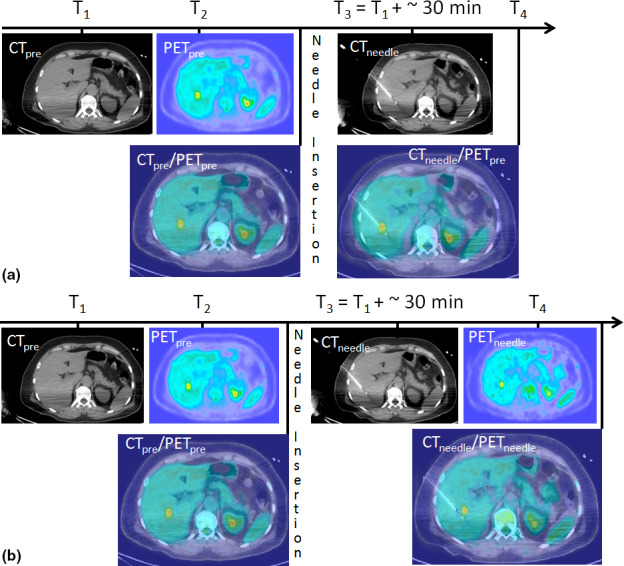
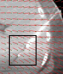
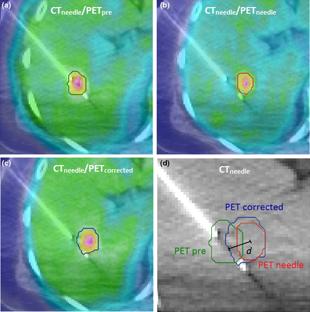
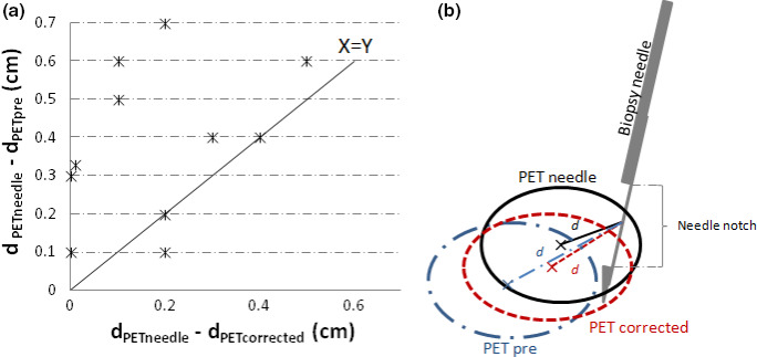
Methods: 19 patients who underwent real-time 18 F-FDG PET-guided biopsy and were found positive for malignancy were included in this study under IRB approval. PET/CT images were acquired for all patients within minutes prior to biopsy to visualize the FDG-avid region and plan the needle insertion. The biopsy needle was inserted and a post-insertion CT scan was acquired. The two CT scans acquired before and after needle insertion were registered using a deformable image registration (DIR) algorithm. The DIR deformation vector field (DVF) was used to calculate the mean displacement between the pre-insertion and post-insertion CT scans for a region around the tip of the biopsy needle. For 12 patients one biopsy core from each was tracked during histopathological testing to investigate correlations of the mean displacement between the two CT scans and false-negative or true-positive biopsy results. For 11 patients, two PET scans were acquired; one at the beginning of the procedure, pre-needle insertion, and an additional one with the needle in place. The pre-insertion PET scan was corrected for intraprocedural motion by applying the DVF. The corrected PET was compared with the post-needle insertion PET to validate the correction method.
Results: The mean displacement of tissue around the needle between the pre-biopsy CT and the postneedle insertion CT was 5.1 mm (min = 1.1 mm, max = 10.9 mm and SD = 3.0 mm). For mean displacements larger than 7.2 mm, the biopsy cores gave false-negative results. Correcting pre-biopsy PET using the DVF improved the PET/CT registration in 8 of 11 cases.
Conclusions: The DVF obtained from DIR of the CT scans can be used for evaluation and correction of the error in needle placement with respect to the FDG-avid area. Misregistration between the pre-biopsy PET and the CT acquired with the needle in place was shown to correlate with false negative biopsy results.
Keywords: PET/CT; biopsy; image registration; image-guidance; oncology.
© 2017 American Association of Physicists in Medicine.
Conflict of interest statement
The authors have no relevant conflicts of interest to disclose.
Figures
Similar articles
-
PET/CT-Guided Tissue Sampling in Patients With a Failed or Inconclusive CT-Guided Procedure: Outcomes and Contributing Factors.Clin Nucl Med. 2020 Aug;45(8):581-587. doi: 10.1097/RLU.0000000000003128. Clin Nucl Med. 2020. PMID: 32558712
-
PET/CT-guided biopsy with respiratory motion correction.Int J Comput Assist Radiol Surg. 2019 Dec;14(12):2187-2198. doi: 10.1007/s11548-019-02047-4. Epub 2019 Sep 11. Int J Comput Assist Radiol Surg. 2019. PMID: 31512193 Free PMC article.
-
Real-time intraprocedural 18F-FDG PET/CT-guided biopsy using automated robopsy arm (ARA) in the diagnostic evaluation of thoracic lesions with prior inconclusive biopsy results: initial experience from a tertiary health care centre.Br J Radiol. 2017 Dec;90(1080):20170258. doi: 10.1259/bjr.20170258. Epub 2017 Oct 27. Br J Radiol. 2017. PMID: 28937268 Free PMC article.
-
Advantages of percutaneous abdominal biopsy under PET-CT/ultrasound fusion imaging guidance: a pictorial essay.Abdom Imaging. 2014 Oct;39(5):1102-13. doi: 10.1007/s00261-014-0143-8. Abdom Imaging. 2014. PMID: 24777592 Review.
-
PET/CT Ultrasound Fusion for Percutaneous Biopsy: A Retrospective Single-Center Study and Review of the Literature.Clin Nucl Med. 2022 Aug 1;47(8):692-698. doi: 10.1097/RLU.0000000000004220. Epub 2022 Apr 22. Clin Nucl Med. 2022. PMID: 35452012 Review.
Cited by
-
[Progress in Image-planned and Real-time Image-guided Lung Cancer Biopsy in the Detection of Biomarkers].Zhongguo Fei Ai Za Zhi. 2023 Aug 20;26(8):630-638. doi: 10.3779/j.issn.1009-3419.2023.106.15. Zhongguo Fei Ai Za Zhi. 2023. PMID: 37752543 Free PMC article. Review. Chinese.
-
Tumor Region Location and Classification Based on Fuzzy Logic and Region Merging Image Segmentation Algorithm.J Healthc Eng. 2021 Oct 20;2021:1141619. doi: 10.1155/2021/1141619. eCollection 2021. J Healthc Eng. 2021. Retraction in: J Healthc Eng. 2023 Nov 1;2023:9756154. doi: 10.1155/2023/9756154. PMID: 34721822 Free PMC article. Retracted.
-
KRAS mutation effects on the 2-[18F]FDG PET uptake of colorectal adenocarcinoma metastases in the liver.EJNMMI Res. 2020 Nov 23;10(1):142. doi: 10.1186/s13550-020-00707-0. EJNMMI Res. 2020. PMID: 33226505 Free PMC article.
-
Augmenting CT-Guided Bone Biopsies Using 18F-FDG PET/CT Guidance.Cancers (Basel). 2024 Jul 29;16(15):2693. doi: 10.3390/cancers16152693. Cancers (Basel). 2024. PMID: 39123421 Free PMC article.
-
Practice and prospects for PET/CT guided interventions.Q J Nucl Med Mol Imaging. 2021 Mar;65(1):20-31. doi: 10.23736/S1824-4785.21.03291-X. Epub 2021 Jan 26. Q J Nucl Med Mol Imaging. 2021. PMID: 33494585 Free PMC article. Review.
References
-
- Kobayashi K, Bhargava P, Raja S, et al. Image‐guided biopsy: what the interventional radiologist needs to know about PET/CT. Radiographics. 2012;32:1483–1501. - PubMed
-
- Guralnik L, Rozenberg R, Frenkel A, Israel O, Keidar Z. Metabolic PET/CT‐guided lung lesion biopsies: impact on diagnostic accuracy and rate of sampling error. J Nucl Med. 2015;56:518–522. - PubMed
-
- Tatli S, Gerbaudo VH, Mamede M, Tuncali K, Shyn PB, Silverman SG. Abdominal masses sampled at PET/CT‐guided percutaneous biopsy: initial experience with registration of prior PET/CT images. Radiology. 2010;256:305–311. - PubMed
MeSH terms
Grants and funding
LinkOut - more resources
Full Text Sources
Other Literature Sources
