. 2017 May 10;21(5):611-618.e5.
doi: 10.1016/j.chom.2017.04.001.

Pseudomonas aeruginosa Effector ExoS Inhibits ROS Production in Human Neutrophils

Affiliations

Pseudomonas aeruginosa Effector ExoS Inhibits ROS Production in Human Neutrophils

Chairut Vareechon et al. Cell Host Microbe. .

Abstract

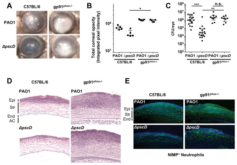
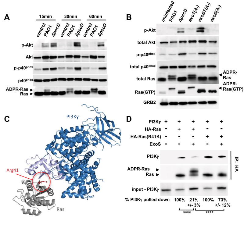
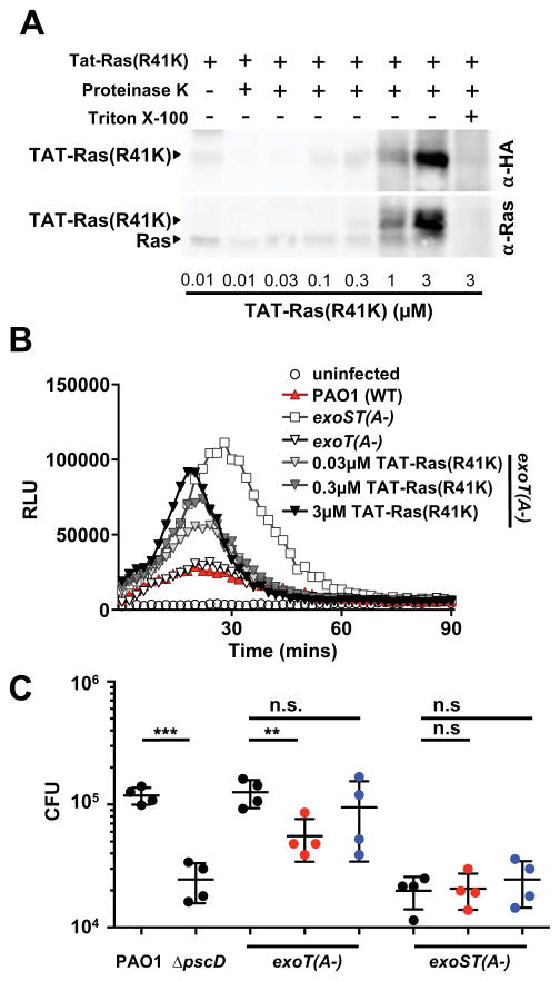
Neutrophils are the first line of defense against bacterial infections, and the generation of reactive oxygen species is a key part of their arsenal. Pathogens use detoxification systems to avoid the bactericidal effects of reactive oxygen species. Here we demonstrate that the Gram-negative pathogen Pseudomonas aeruginosa is susceptible to reactive oxygen species but actively blocks the reactive oxygen species burst using two type III secreted effector proteins, ExoS and ExoT. ExoS ADP-ribosylates Ras and prevents it from interacting with and activating phosphoinositol-3-kinase (PI3K), which is required to stimulate the phagocytic NADPH-oxidase that generates reactive oxygen species. ExoT also affects PI3K signaling via its ADP-ribosyltransferase activity but does not act directly on Ras. A non-ribosylatable version of Ras restores reactive oxygen species production and results in increased bacterial killing. These findings demonstrate that subversion of the host innate immune response requires ExoS-mediated ADP-ribosylation of Ras in neutrophils.

Keywords: T3SS; effector function; keratitis; type III secretion.


Figures

Figure 1
Figure 1. NADPH oxidase mediates ROS production by neutrophils and facilitates clearance of P. aeruginosa during bacterial keratitis
(A) Representative images of corneal opacification 24 h post-infection of C57BL/6 and gp91phox −/− (CGD) mice infected with 1x10⁵ CFU PAO1 (WT) or with the ΔpscD (T3SS null) mutant strain. (B) Quantification of corneal opacity by determining average pixel intensity of corneas described previously (Sun et al., 2012)(n = 5 mice). (C) Colony forming units (CFU) recovered from infected corneas 24 h post-infection (n = 9 mice). (D) Corneal sections were stained with hematoxylin and eosin, or (E) 4’,6-Diamidino-2-Phenylindole dye (DAPI, Blue) and an antibody to Ly6G (NIMP-R14, FITC, green) (Epi: epithelium, Str: stroma, End: corneal endothelium, AC: anterior chamber). B, C: Data points represent individual corneas. Median and interquartile range are indicated. Significance was calculated using the Kruskal-Wallis test, with Dunn’s multiple comparison correction. * p<0.05, **p<0.01, ***p<0.001, or n.s., not significant. See also Figure S1.
Figure 2
Figure 2. ExoS and ExoT ADPRT activities inhibit ROS production in human neutrophils
ROS production was measured using a chemiluminescent substrate (relative light units, RLU). Neutrophils were infected with: wild type (PAO1), a T3SS null mutant (ΔpscD), as well as: (A) a strain lacking the translocation apparatus (ΔpopBD), a strain lacking all 3 effectors (Δ3TOX), and a strain lacking exoS and exoT effector genes (ΔexoST). (B) a strain lacking exoSexoS), a strain lacking exoT(ΔexoT), or (C) strains with chromosomal point mutations inactivating the Rho-GAP (G-) or ADP-ribosyltransferase activities (A-) of ExoS and/or ExoT. A time course representative of at least three independent experiments is shown. See also Figure S2.
Figure 3
Figure 3. ExoS and ExoT ADP-ribosyltransferase activities interfere with PI3K signaling in neutrophils
(A) Cell lysates from uninfected human neutrophils, or from neutrophils infected with wild type PAO1, or a ΔpscD mutant strain were probed by Western blot for Akt, P-Akt (Thr308), p40phox, P-p40phox (Thr154), and Ras. The experiments were repeated 3 times with similar results. (B) Cell lysates from uninfected human neutrophils, or neutrophils infected for 30 minutes with PAO1, ΔpscD, or with strains in which the ADP-ribosyltransferase activity was inactivated in ExoS only (exoS(A-)), ExoT only (exoT(A-)), or both (exoST(A-)). P-Akt (Thr308), Akt, P-p40phox (Thr154), p40phox, total Ras, GTP-bound Ras, and Grb2 (loading control) were detected by western blot. The experiments were repeated 5 times with similar results. (C) Model of Ras (gray) bound to the Ras-binding domain (light blue) of PI3K (dark blue) based on the structure PDB:1he8 (Pacold et al., 2000). Residue Arg 41 of Ras is highlighted red. (D) Purified ExoS was used to ADP-ribosylate HA-tagged versions of Ras, or Ras(R41K), in vitro and subsequently mixed with purified PI3Kγ. The interaction between Ras and PI3Kγ was probed by immunoprecipitating Ras using an anti-HA-tag antibody. PI3Kγ, as well as unmodified and ADP-ribosylated Ras were detected by western blot. The experiments were repeated 3 times with similar results. Input and output levels of PI3Kγ were determined by densitometry. The input/output ratio for the untreated control sample was set to 100% and compared to the corresponding ExoS-treatment condition (mean and standard deviation of three independent replicates are noted below each lane). Results were compared by 1-way ANOVA with Bonferroni correction (**** p<0.0001). See also Figures S3.
Figure 4
Figure 4. Tat-Ras(R41K) rescues ROS production in human neutrophils, resulting in increased killing of P. aeruginosa.
(A) Human neutrophils were treated with increasing concentrations of Tat-Ras(R41K) for 30 minutes, and extracellular protein was degraded by proteinase K. Western blots of cell lysates were probed with antibodies to Ras (which detect endogenous and Tat-Ras(R41K)) or the HA tag (which detects only Tat-Ras(R41K)). (B) ROS production by human neutrophils infected with PAO1, exoT(A-), or exoST(A-) P. aeruginosa was measured by chemiluminescence. Neutrophils were incubated with increasing amounts of Tat-Ras (R41K) thirty minutes prior to infection with an exoT(A-) strain. The experiment was repeated 4 times with similar results. (C) Human neutrophils were incubated with Tat-Ras(R41K) (red) or Tat-Ras (blue) (3 μM final concentration) for 30 minutes prior to infection with PAO1, ΔpscD, exoS(A-), or exoST(A) 15 min (MOI30). Extracellular bacteria were killed with gentamicin for 30 minutes. Each point represents an individual human donor (n = 4 donors). Statistical significance was measured by 1-way ANOVA with Bonferroni correction. **p<0.01, ***p<0.001, n.s. not significant. See also Figure S4.

References

    1. Andersson K, Carballeira N, Magnusson KE, Persson C, Stendahl O, Wolf-Watz H, Fallman M. YopH of Yersinia pseudotuberculosis interrupts early phosphotyrosine signalling associated with phagocytosis. Molecular microbiology. 1996;20:1057–1069. - PubMed
    1. Andisi VF, Hinojosa CA, de Jong A, Kuipers OP, Orihuela CJ, Bijlsma JJ. Pneumococcal gene complex involved in resistance to extracellular oxidative stress. Infection and immunity. 2012;80:1037–1049. - PMC - PubMed
    1. Aussel L, Zhao W, Hebrard M, Guilhon AA, Viala JP, Henri S, Chasson L, Gorvel JP, Barras F, Meresse S. Salmonella detoxifying enzymes are sufficient to cope with the host oxidative burst. Molecular microbiology. 2011;80:628–640. - PubMed
    1. Barbieri JT, Sun J. Pseudomonas aeruginosa ExoS and ExoT. Reviews of physiology, biochemistry and pharmacology. 2004;152:79–92. - PubMed
    1. Bjur E, Eriksson-Ygberg S, Aslund F, Rhen M. Thioredoxin 1 promotes intracellular replication and virulence of Salmonella enterica serovar Typhimurium. Infect Immun. 2006;74:5140–5151. - PMC - PubMed

MeSH terms