Review
. 2017 Aug:12:946-955.
doi: 10.1016/j.redox.2017.04.030. Epub 2017 Apr 24.

Shotgun lipidomics in substantiating lipid peroxidation in redox biology: Methods and applications

Affiliations
Review

Shotgun lipidomics in substantiating lipid peroxidation in redox biology: Methods and applications

Changfeng Hu et al. Redox Biol. 2017 Aug.

Abstract

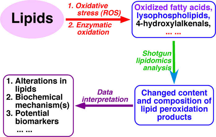
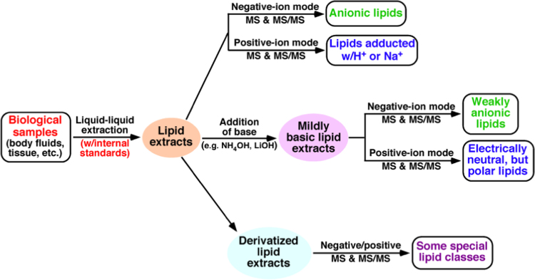
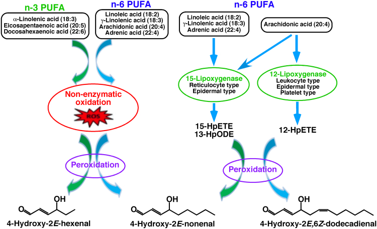
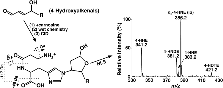
Multi-dimensional mass spectrometry-based shotgun lipidomics (MDMS-SL) has made profound advances for comprehensive analysis of cellular lipids. It represents one of the most powerful tools in analyzing lipids directly from lipid extracts of biological samples. It enables the analysis of nearly 50 lipid classes and thousands of individual lipid species with high accuracy/precision. The redox imbalance causes oxidative stress, resulting in lipid peroxidation, and alterations in lipid metabolism and homeostasis. Some lipid classes such as oxidized fatty acids, 4-hydroxyalkenal species, and plasmalogen are sensitive to oxidative stress or generated corresponding to redox imbalance. Therefore, accurate assessment of these lipid classes can provide not only the redox states, but also molecular insights into the pathogenesis of diseases. This review focuses on the advances of MDMS-SL in analysis of these lipid classes and molecular species, and summarizes their recent representative applications in biomedical/biological research. We believe that MDMS-SL can make great contributions to redox biology through substantiating the aberrant lipid metabolism, signaling, trafficking, and homeostasis under oxidative stress-related condition.

Keywords: 4-hydroxyalkenal species; Lipid peroxidation; Oxidative stress; Oxidized fatty acids; Plasmalogen; Shotgun lipidomics.


Figures

fx1
Graphical abstract
Fig. 1
Fig. 1
Schematic illustration of the workflow of MDMS-SL for analysis of cellular lipidomes directly from crude extracts of biological samples , .
Fig. 2
Fig. 2
Mechanisms of 4-hydroxyalkenal species production . PUFA abbreviates polyunsaturated fatty acid. 15-HpETE, 12-HpETE, and 13-HpODE stand for 15-hydroperoxyeicosatetraenoic acid, 12-hydroperoxyeicosatetraenoic acid, and 13-hydroperoxyoctadecadienoic acid.
Fig. 3
Fig. 3
Schematic illustration of the MDMS-SL method for analysis of 4-hydroxyalkenal species directly from lipid extracts of biological samples . D3−4-HNE is added as an internal standard (IS). 4-HHE, 4-HNDE, 4-HNE, and 4-HDTE stand for 4-hydroxy-2E-hexenal, 4-hydroxy-2E-nondienal, 4-hydroxy-2E-nonenal, and 4-hydroxy-dodecatrienal, respectively.
Fig. 4
Fig. 4
Schematic illustration of an MDMS-SL approach for analysis of oxidized fatty acids which contains at least one functional group of carboxylic acid . The amidation reaction is catalyzed by 1-ethyl-3-(3-dimethylaminopropyl)carbiodiimide (EDC).
Fig. 5
Fig. 5
Product ion ESI-MS analysis of hydroxyeicosatetraenoic acid (HETE) isomers after derivatization with N-(4-amiomethylphenyl)pyridinium (AMPP) . Product ion ESI-MS analysis of these derivatized HETE isomers at collision energy of 40 eV and collision gas pressure of 1 mTorr. The fragment ions at m/z 169 and 183 can be used for screening these isomers at the m/z position of molecular ions in the precursor-ion scans of m/z 169 and 189, respectively, and quantifying the total content of their mixture relative to a selected internal standard (e.g., a stable isotope labeled HETE). The fingerprints of the fragment ions between m/z 190 and 450 (i.e., m/z 239, 253, 267, 283, 293, 295, 307, 333, 335, 347, 375, 387) can be used to simulate a tandem MS spectrum of a mixture of the HETE isomers and determine their composition. Absolute amounts of individual HETE species can be derived from the total amount of the mixture and the composition.
Fig. 6
Fig. 6
Illustration of three major metabolic pathways responsible for plasmalogen reduction. These include peroxisomal dysfunction (Pathway 1), phospholipase A2 activation (Pathway 2), and oxidative stress (Pathway 3) . Determining the levels of the plasmalogen species and relative metabolites through MDMS-SL can readily identify the corresponding aberrant pathway. PLA2 and ROS stand for phospholipase A2 and reactive oxygen species, respectively.
Fig. 7
Fig. 7
Comparison of the contents of representative serum lipids between SLE patients and controls. Plasmalogen PE species (Panel A), and ethanolamine lysoglycerophospholipids (lysoPE) and 4-hydroxyalkenal species (Panel B) present in serum lipid extracts from SLE patients (n=30, red bar) and healthy controls (n=30, blue bar) were determined by MDMS-SL as previously described . The data represent means±SEM from different individuals (n=30). *p<0.05, **p<0.01, and ***p<0.001 compared with those in the control group. The prefix “p” in Panel A is used to abbreviate plasmalogen PE species. HHE, HNDE, and HNE stand for 4-hydroxy-2E-hexenal, 4-hydroxy-2E-nondienal, and 4-hydroxy-2E-nonenal, respectively. (For interpretation of the references to color in this figure legend, the reader is referred to the web version of this article).
Fig. 8
Fig. 8
MDMS-SL analysis determines the altered levels of lipids present in brain cortices of infant monkeys exposed to sevoflurane for 9 h, indicating the existence of oxidative stress . Panels A and B show the reduction of the mass levels of ethanolamine glycerophospholipid (PE) including many plasmalogen PE species (highlighted with red) by MS analysis. Panel C summarizes the total mass levels of the altered lipid classes induced by exposure to sevoflurane (Red bar) in comparison to controls (Blue bar) . *p<0.05; **p<0.01; and ***p<0.001. (For interpretation of the references to color in this figure legend, the reader is referred to the web version of this article).

Similar articles

Cited by

References

    1. Wymann M.P., Schneiter R. Lipid signalling in disease. Nat. Rev. Mol. Cell Biol. 2008;9:162–176. - PubMed
    1. Perry R.J., Samuel V.T., Petersen K.F., Shulman G.I. The role of hepatic lipids in hepatic insulin resistance and type 2 diabetes. Nature. 2014;510:84–91. - PMC - PubMed
    1. Zalba S., Hagen T.L. Ten. Cell membrane modulation as adjuvant in cancer therapy. Cancer Treat. Rev. 2017;52:48–57. - PMC - PubMed
    1. Mapstone M., Cheema A.K., Fiandaca M.S., Zhong X., Mhyre T.R., MacArthur L.H., Hall W.J., Fisher S.G., Peterson D.R., Haley J.M., Nazar M.D., Rich S.A., Berlau D.J., Peltz C.B., Tan M.T., Kawas C.H., Federoff H.J. Plasma phospholipids identify antecedent memory impairment in older adults. Nat. Med. 2014;20:415–418. - PMC - PubMed
    1. Han X. Lipid alterations in the earliest clinically recognizable stage of Alzheimer's disease: implication of the role of lipids in the pathogenesis of Alzheimer's disease. Curr. Alzheimer Res. 2005;2:65–77. - PubMed

Publication types

LinkOut - more resources