

Korean Thyroid Imaging Reporting and Data System features of follicular thyroid adenoma and carcinoma: a single-center study
- PMID: 28494523
- PMCID: PMC5621801
- DOI: 10.14366/usg.17020
Korean Thyroid Imaging Reporting and Data System features of follicular thyroid adenoma and carcinoma: a single-center study
Abstract
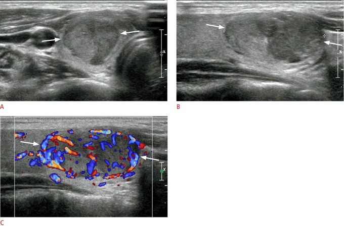
Purpose: This study aimed to assess the diagnostic efficacy of Korean Thyroid Imaging Reporting and Data System (K-TIRADS) features for distinguishing follicular thyroid adenoma (FTA) from follicular thyroid carcinoma (FTC).
Methods: From January 2013 to July 2016, 46 follicular neoplasms in 45 patients who underwent preoperative thyroid ultrasonography (US) and thyroid surgery were included. The US features of each thyroid nodule were retrospectively evaluated by a single radiologist using a picture archiving and communication system. The diagnostic indices of K-TIRADS for follicular neoplasms were calculated according to whether K-TIRADS category 4 lesions were excluded or classified as benign or malignant.
Results: Of the 46 follicular neoplasms (mean size, 3.1±1.6 cm), 37 were FTAs (mean size, 3.1±1.7 cm) and nine were FTCs (mean size, 3.0±1.5 cm). A statistically significant difference was found between FTAs and FTCs regarding the margin (P=0.035), while no significant differences were observed in the composition, echogenicity, shape, orientation, calcification, or vascularity of the lesions (P<0.05). The FTAs belonged to K-TIRADS categories 3 (n=22) and 4 (n=15), while the FTCs belonged to K-TIRADS categories 3 (n=4), 4 (n=4), and 5 (n=1). However, there was no statistically significant difference in the distribution of K-TIRADS categories between FTAs and FTCs (P=0.184).
Conclusion: K-TIRADS features were not helpful for distinguishing FTA from FTC, although follicular neoplasms showed a high prevalence of K-TIRADS categories 3 and 4.
Keywords: K-TIRADS; Neoplasms; Thyroid cancer, follicular; Thyroid nodule; Ultrasonography.
Conflict of interest statement
No potential conflict of interest relevant to this article was reported.
Figures

References
LinkOut - more resources
Full Text Sources
Other Literature Sources
