Review
. 2017 Nov 15;11(6):747-760.
doi: 10.5009/gnl16523.

Sessile Serrated Adenomas: How to Detect, Characterize and Resect

Affiliations
Review

Sessile Serrated Adenomas: How to Detect, Characterize and Resect

Michael X Ma et al. Gut Liver. .

Abstract

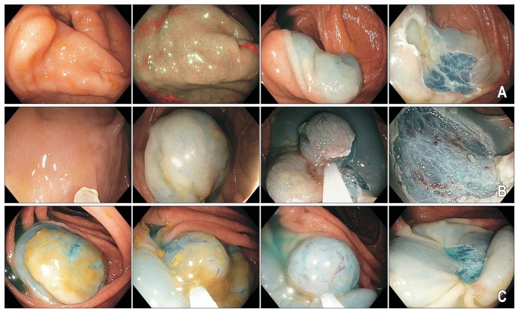
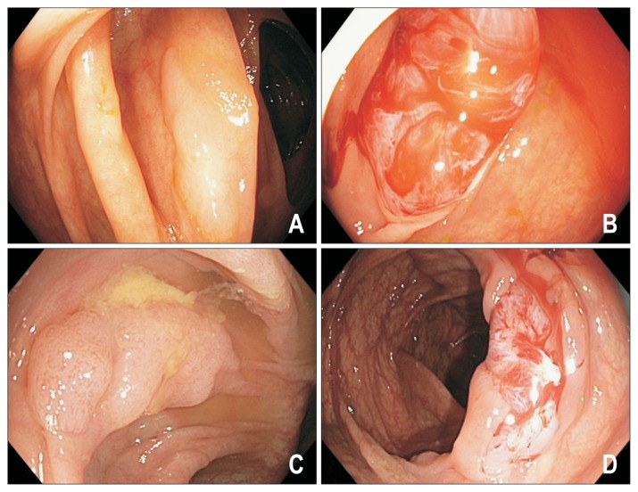
Serrated polyps are important contributors to the burden of colorectal cancers (CRC). These lesions were once considered to have no malignant potential, but currently up to 30% of all CRC are recognized to arise from the serrated neoplasia pathway. The primary premalignant lesions are sessile serrated adenomas/polyps (SSA/Ps), although traditional serrated adenomas are relatively uncommon. Compared to conventional adenomas, SSA/Ps are morphologically subtle with indistinct borders, may be difficult to detect endoscopically, are more prevalent than previously thought, are associated with synchronous and metachronous advanced neoplasia, and have a higher risk of incomplete resection. Although many lesions remain "dormant," progressive disease is associated with the development of dysplasia and more rapid progression to CRC. As a result, SSA/Ps are strongly implicated in the development of interval cancers. These factors represent unique challenges that require a meticulous approach to their management. In this review, we summarize the contemporary literature on the characterization, detection and resection of SSA/Ps.

Keywords: Detection; Endoscopic imaging; Endoscopic resection; Histology; Sessile serrated adenoma.


Conflict of interest statement

CONFLICTS OF INTEREST

No potential conflict of interest relevant to this article was reported.

Figures

Fig. 1
Fig. 1
Histologic features of sessile serrated adenomas/polyps (SSA/Ps). (A) A serrated adenoma (SSA/P) without dysplasia showing the classical features of broad bases and dilated crypts (arrow). H&E stained, low power magnification. (B) An SSA/P with mild dysplasia is shown in the right-side specimen (arrow). The glandular architecture and surface epithelium of the dysplastic component resembles a conventional adenoma. The left-sided specimen is nondysplastic. H&E stained, low power magnification.
Fig. 2
Fig. 2
(A–C) Endoscopic appearance of nondysplastic sessile serrated adenomas/polyps (SSA/Ps). SSA/Ps are often found in the right colon, are morphologically flat and pale, have a color similar to the surrounding mucosa and have indistinct borders (arrows). Detection requires good bowel preparation and a high index of suspicion.
Fig. 3
Fig. 3
Sessile serrated adenomas/polyp (SSA/P) before and after cleaning of the mucous cap. This nondysplastic SSA/P is covered by a tenacious mucous cap with a surrounding rim of stool (A, B). The lesion becomes less conspicuous (C) upon cleansing and can potentially be mistaken for a prominent mucosal fold.
Fig. 4
Fig. 4
Endoscopic appearance of sessile serrated adenomas/polyps (SSA/Ps) with dysplasia. A 20 mm SSA/P-D viewed under white light (A) and narrow band imaging (B) with and without the dysplastic (label D) and nondysplastic (label SSA) components outlined. The lesion has developed a raised, nodular component on the left-hand aspect with a type IV surface pit pattern indicative of dysplastic transformation (label D). The nondysplastic component of the lesion (label SSA) is pale with relatively hypovascular background surface markings and is covered by a thin layer of stool debris (arrowhead). Note there is an obvious transition zone from the nondysplastic flat SSA/P to the area of dysplasia (arrow). The lesion and a rim of normal tissue were removed en bloc by endoscopic mucosal resection; histology confirmed a completely resected SSA/P with mild dysplasia.
Fig. 5
Fig. 5
Endoscopic mucosal resection of sessile serrated adenomas/polyps (SSA/Ps). (A–C) Note the inconspicuous appearance of all three lesions despite their larger sizes. Submucosal chromogelofusine injection assists with delineating the peripheral extent of the lesion. A margin of normal tissue should be captured during mucosal resection. Thermal ablation of the resection margins with snare tip soft coagulation (effect 4, 80W; VIO 300D; Erbe) reduces the risk of lesion recurrence.
Fig. 6
Fig. 6
Piecemeal cold snare polypectomy of sessile serrated adenomas/polyp (SSA/P). Larger (10 to 15 mm) SSA/Ps (A, C) removed by piecemeal cold snare polypectomy (B, D).

References

    1. Bosman FT, Carneiro F, Hruban RH, Theise ND. WHO classification of tumours of the digestive system. Lyon: IARC Press; 2010.
    1. Rex DK, Ahnen DJ, Baron JA, et al. Serrated lesions of the colorectum: review and recommendations from an expert panel. Am J Gastroenterol. 2012;107:1315–1329. doi: 10.1038/ajg.2012.161. - DOI - PMC - PubMed
    1. Yang HM, Mitchell JM, Sepulveda JL, Sepulveda AR. Molecular and histologic considerations in the assessment of serrated polyps. Arch Pathol Lab Med. 2015;139:730–741. doi: 10.5858/arpa.2014-0424-RA. - DOI - PubMed
    1. IJspeert JE, Vermeulen L, Meijer GA, Dekker E. Serrated neoplasia-role in colorectal carcinogenesis and clinical implications. Nat Rev Gastroenterol Hepatol. 2015;12:401–409. doi: 10.1038/nrgastro.2015.73. - DOI - PubMed
    1. Obuch JC, Pigott CM, Ahnen DJ. Sessile serrated polyps: detection, eradication, and prevention of the evil twin. Curr Treat Options Gastroenterol. 2015;13:156–170. doi: 10.1007/s11938-015-0046-y. - DOI - PMC - PubMed

MeSH terms