. 2018 Jan;55(1):76-97.
doi: 10.1177/0300985817705174. Epub 2017 May 11.

Globule Leukocytes and Other Mast Cells in the Mouse Intestine

Affiliations

Globule Leukocytes and Other Mast Cells in the Mouse Intestine

Peter Vogel et al. Vet Pathol. 2018 Jan.

Abstract

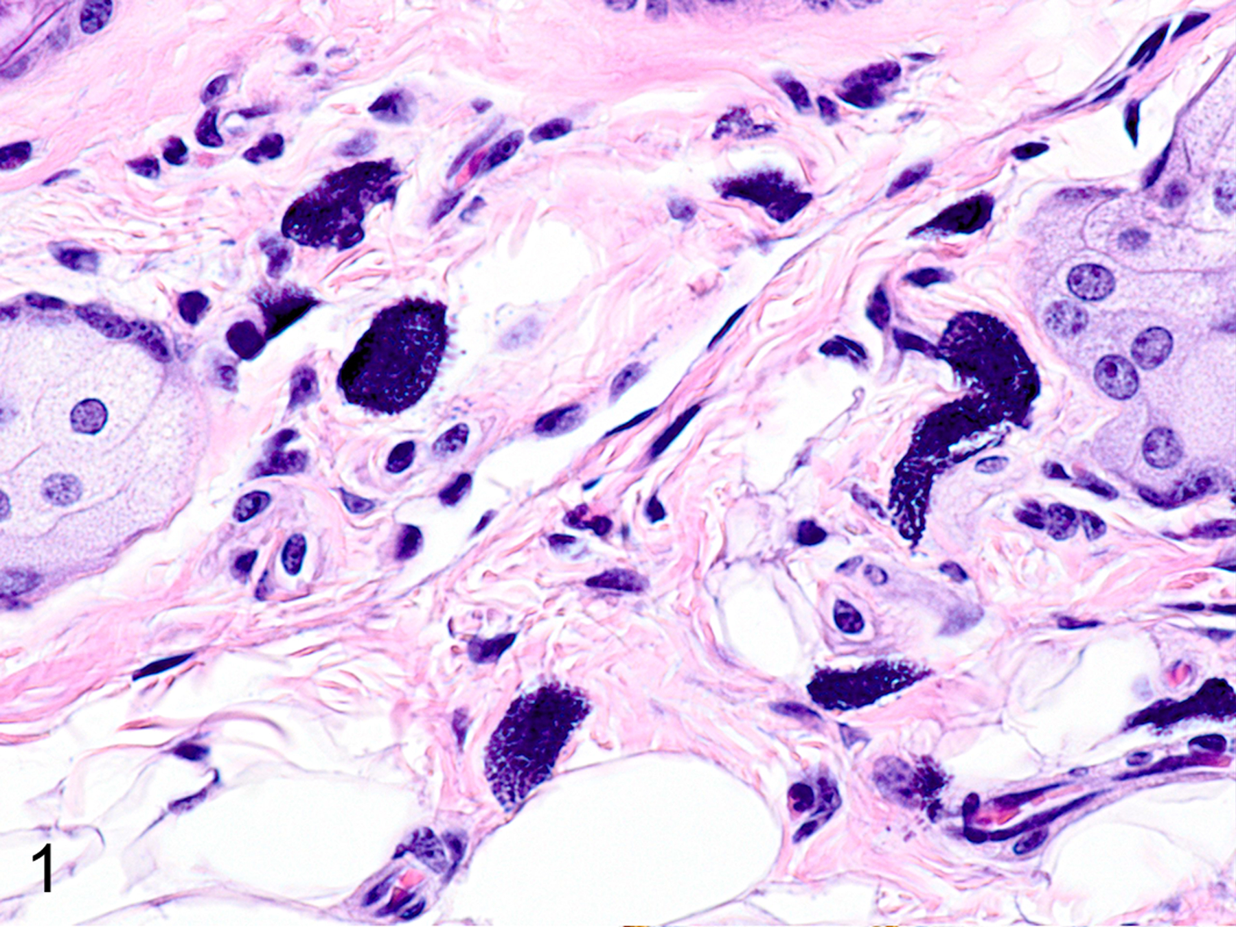
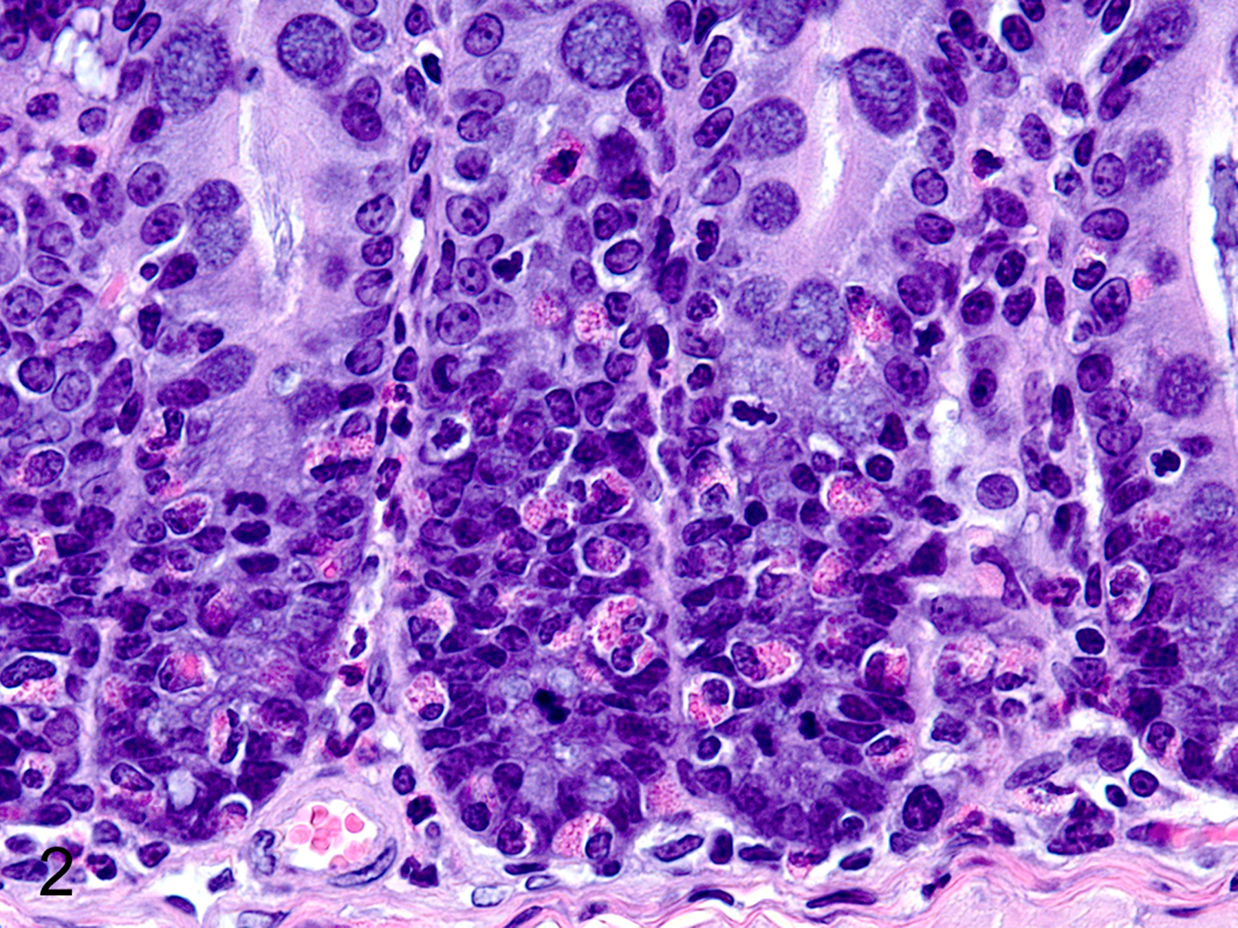
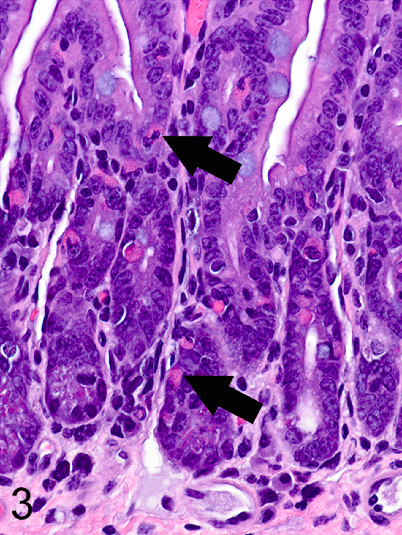
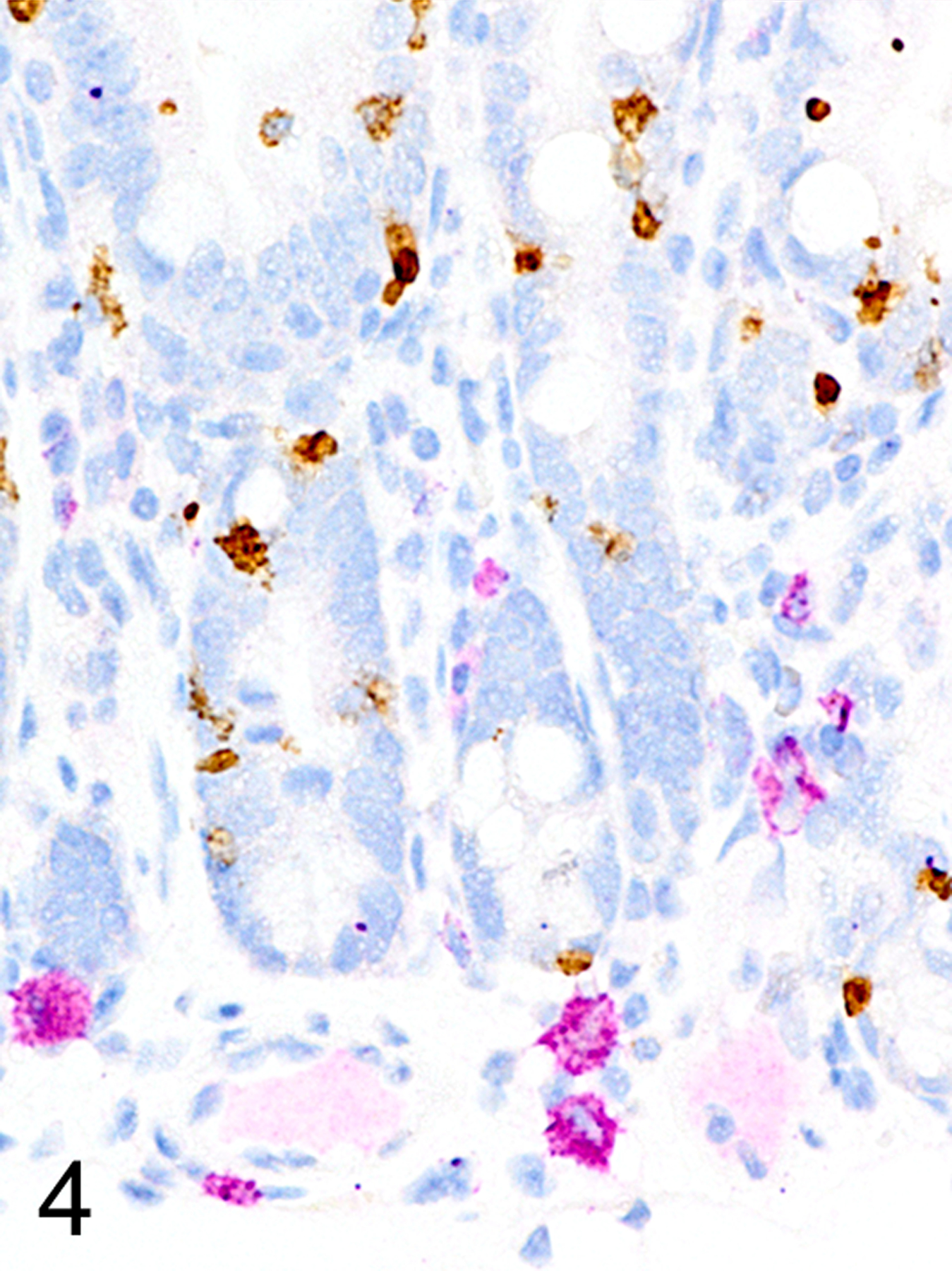
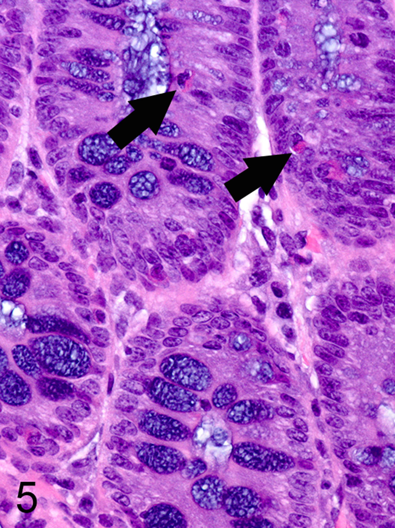
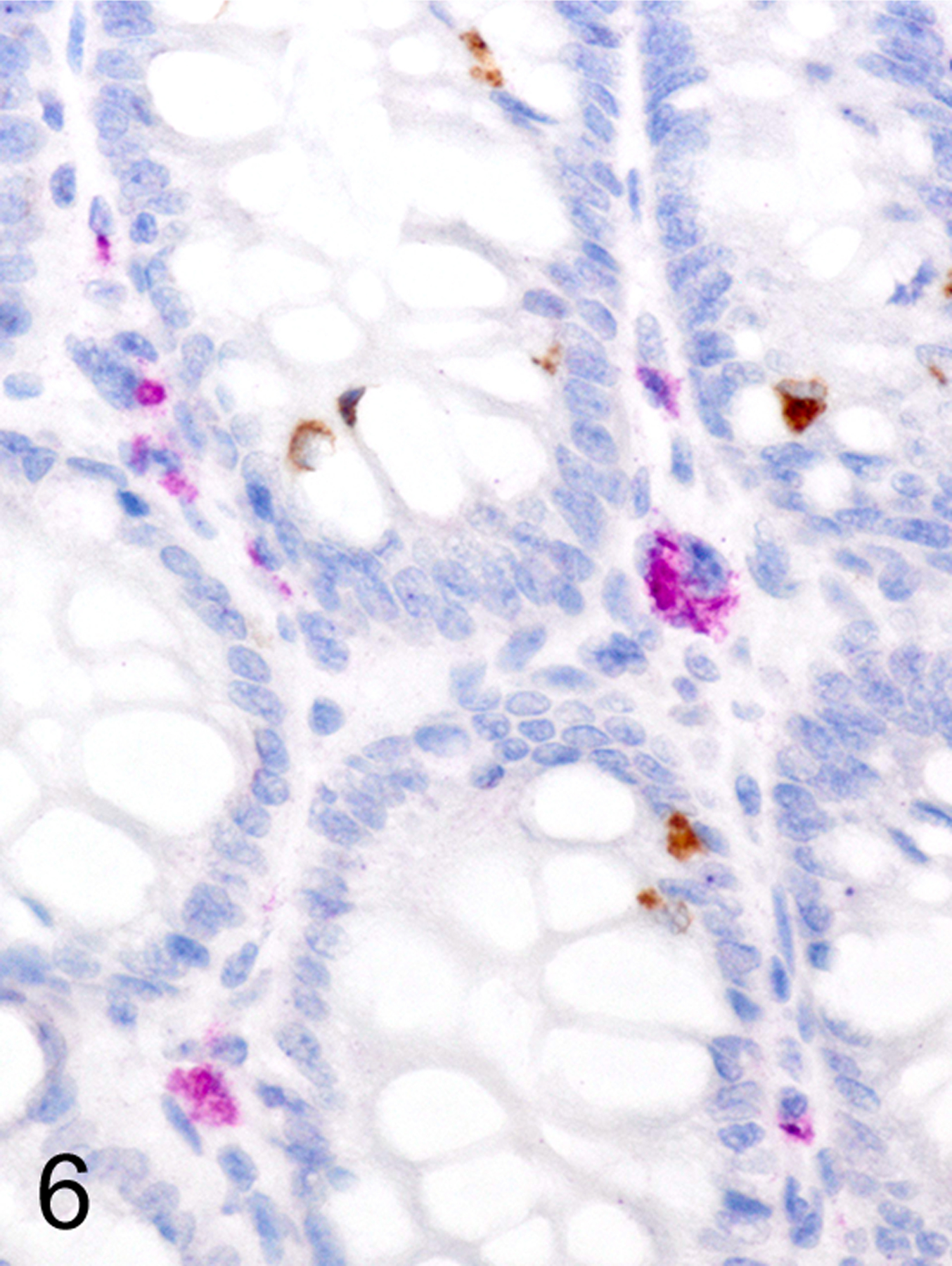
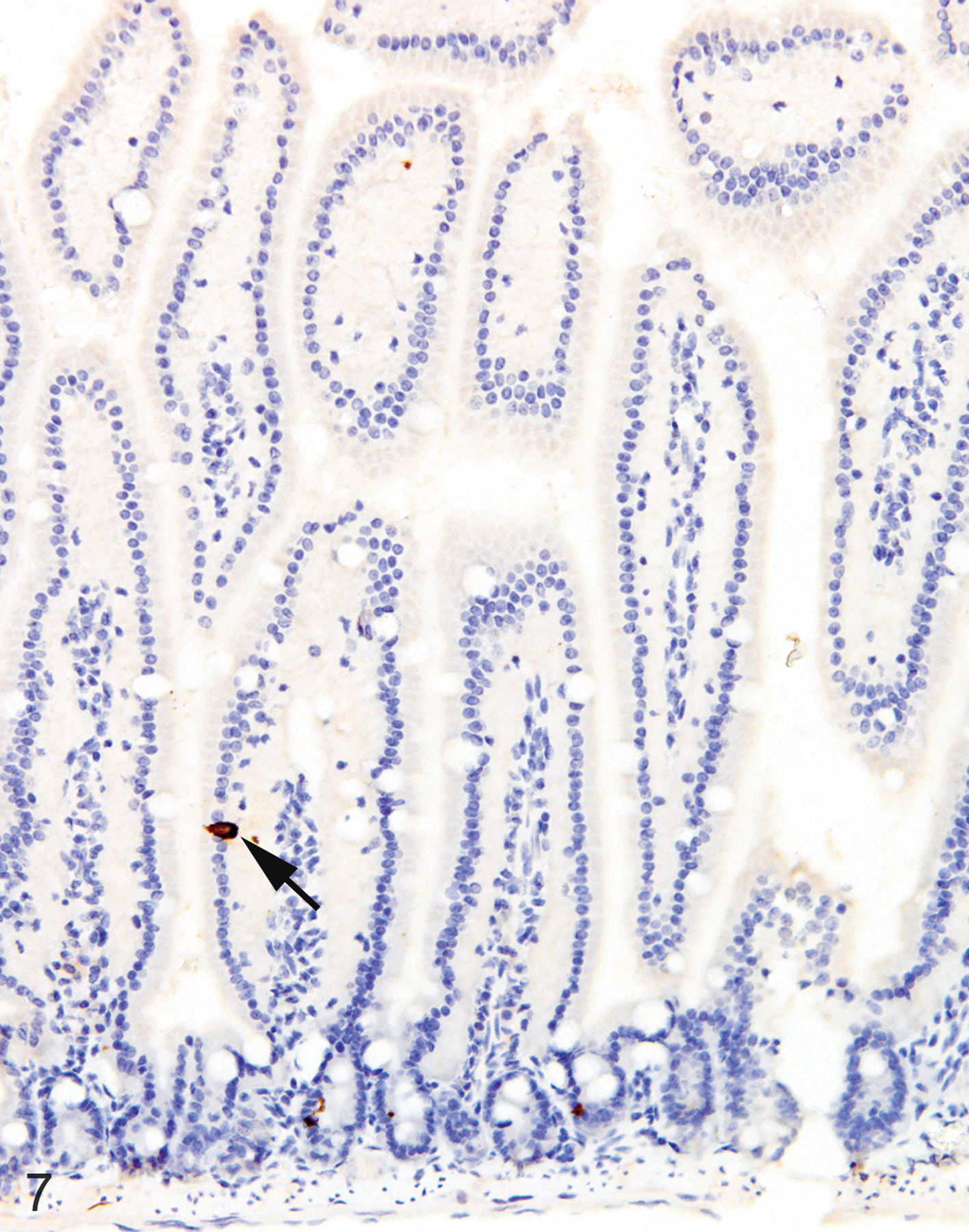
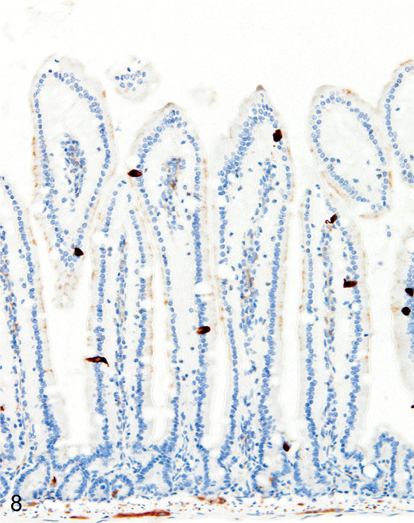
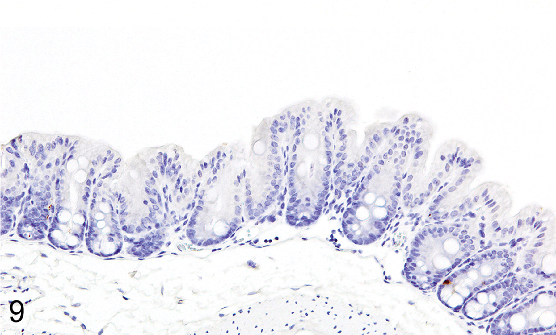
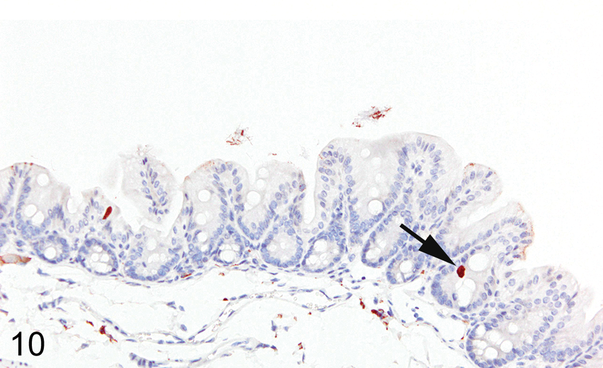
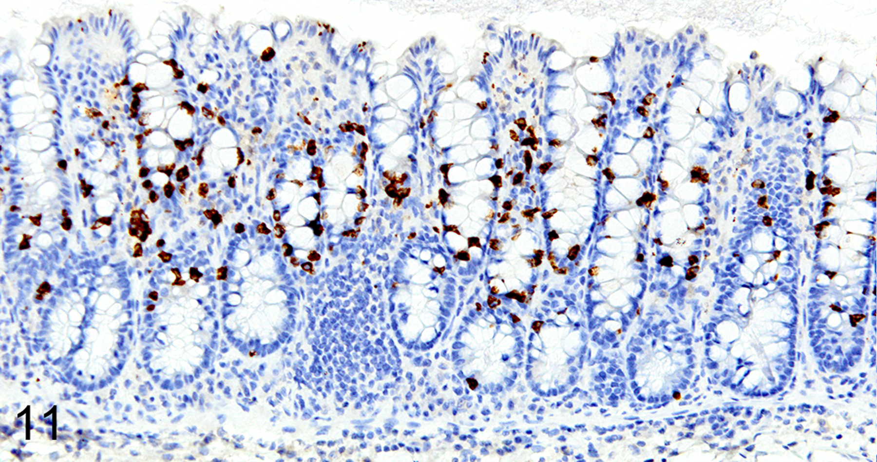
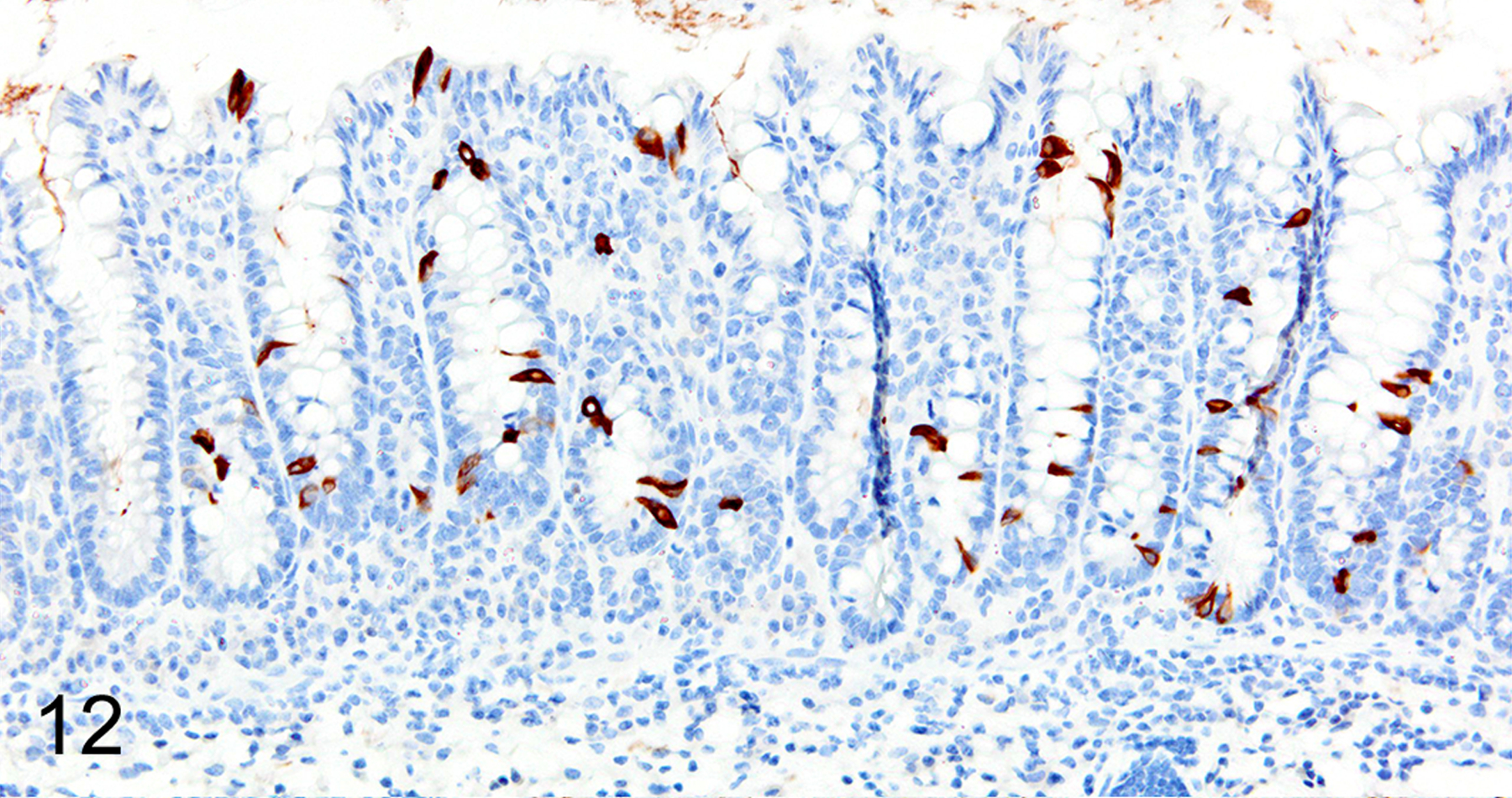
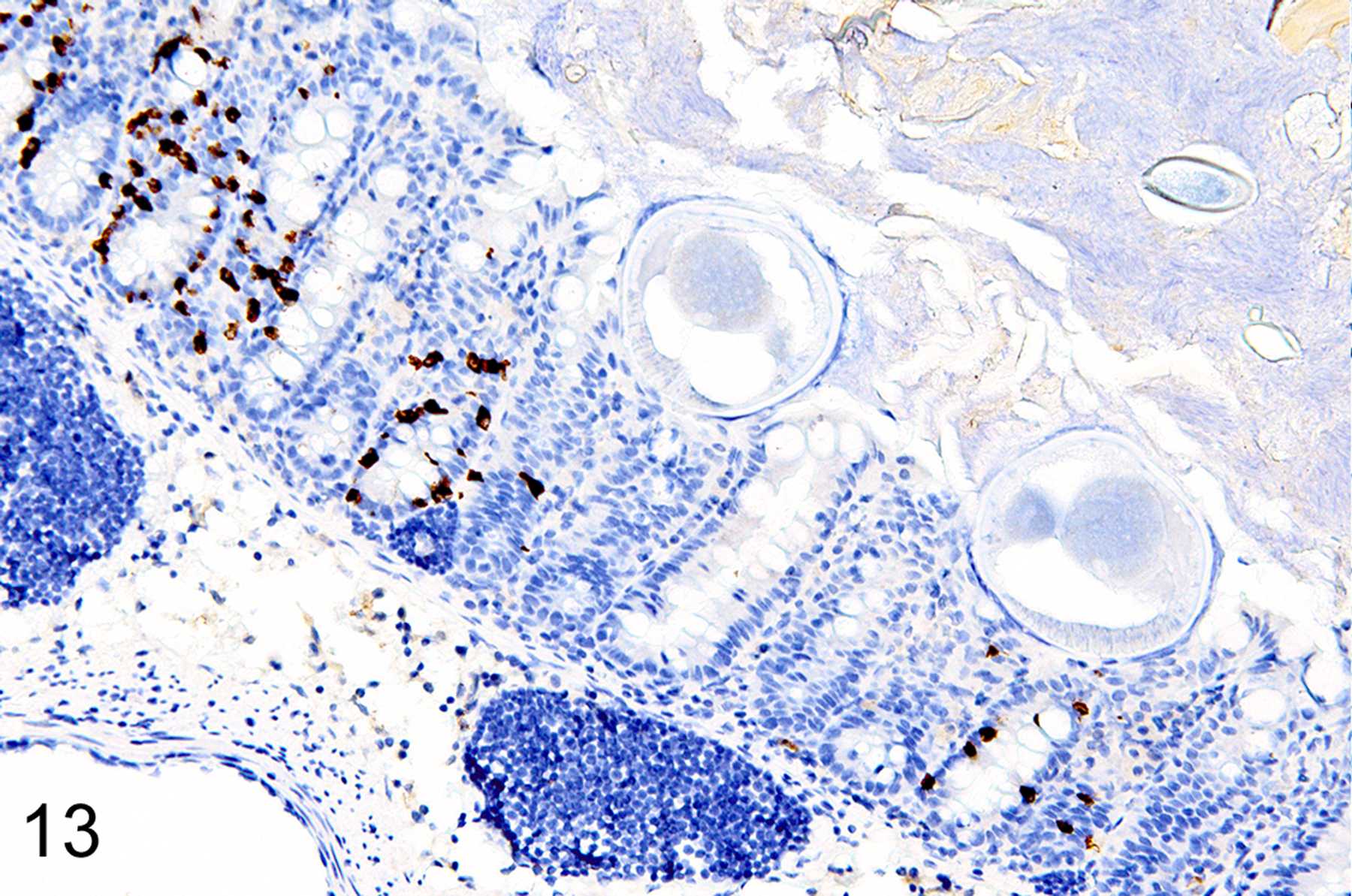
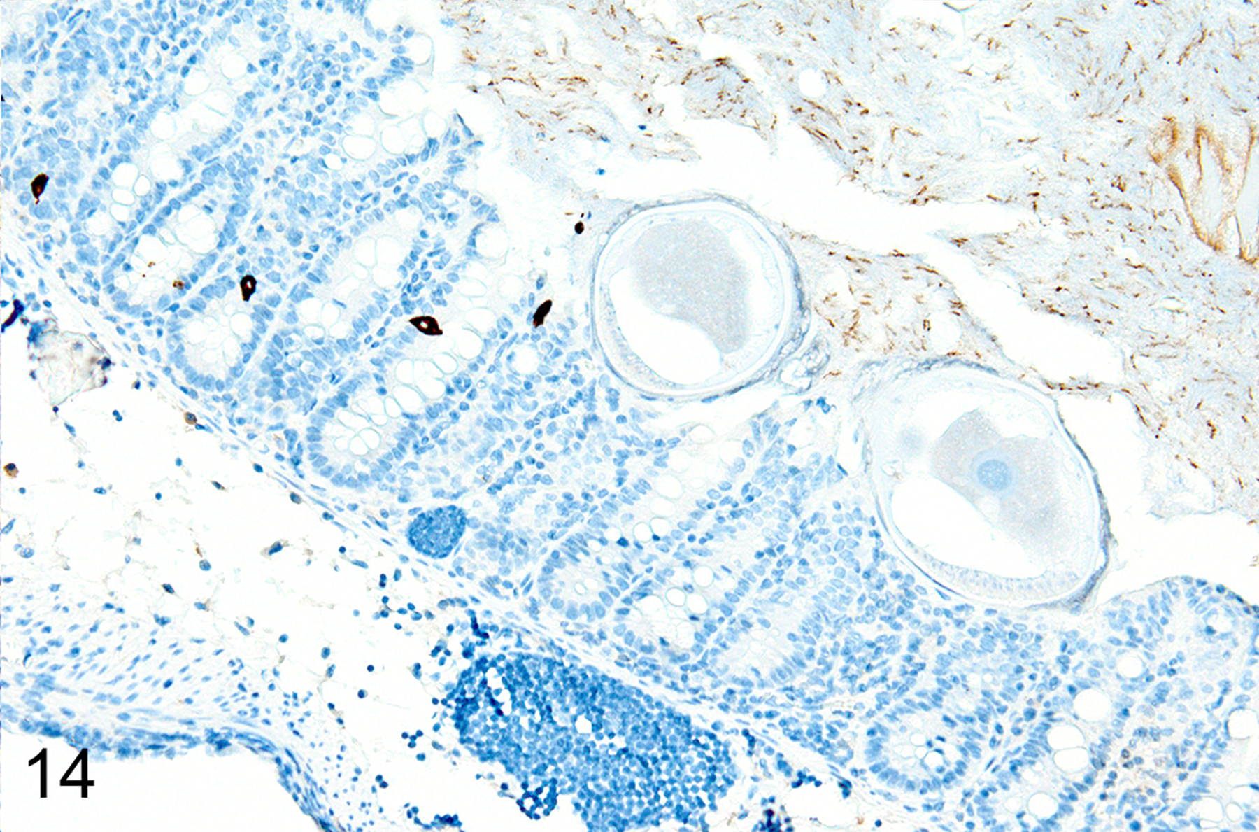
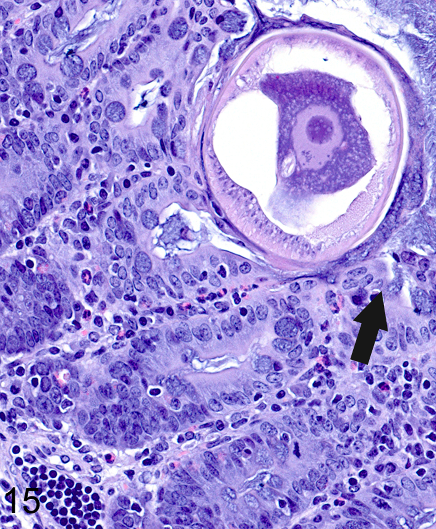
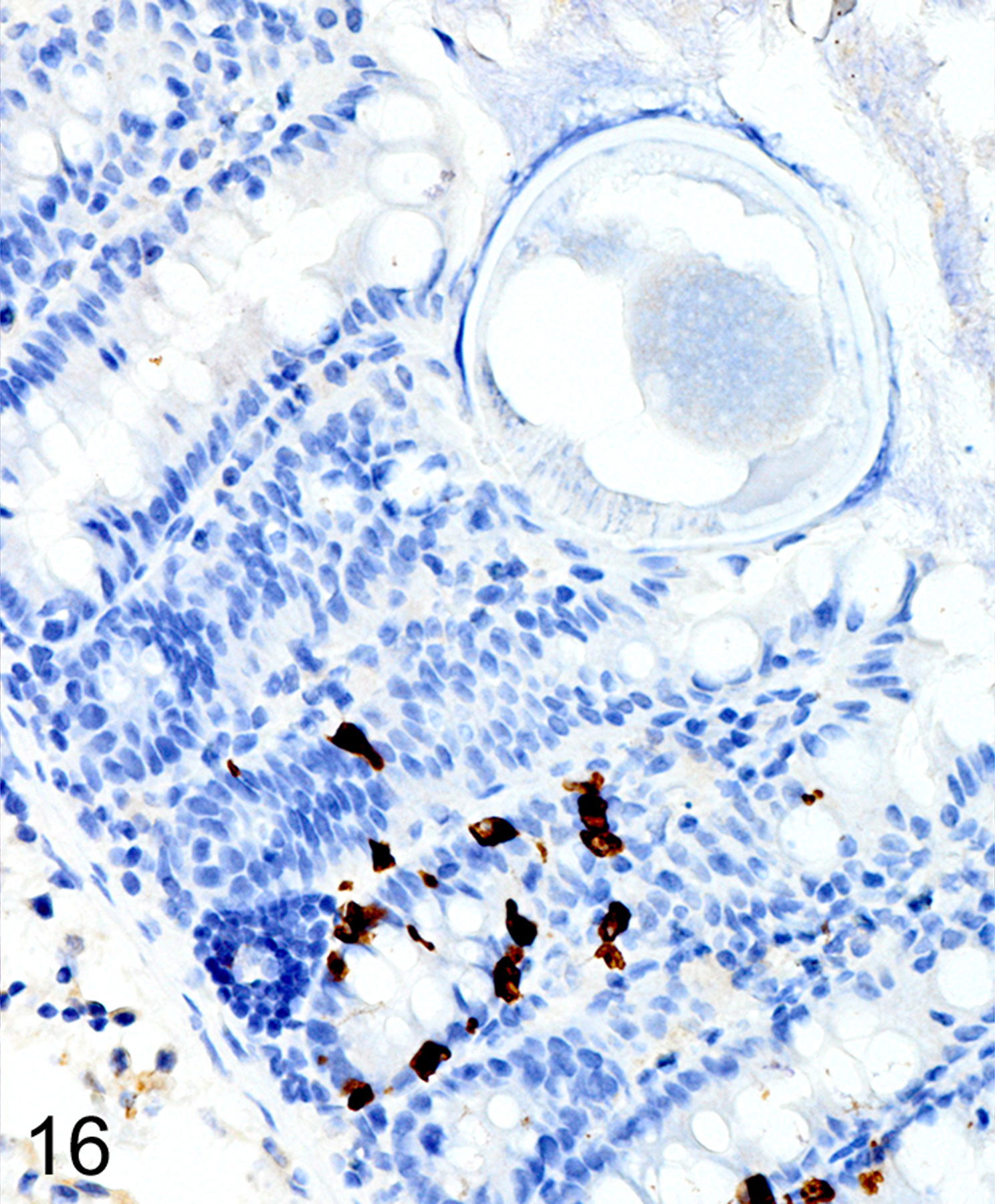
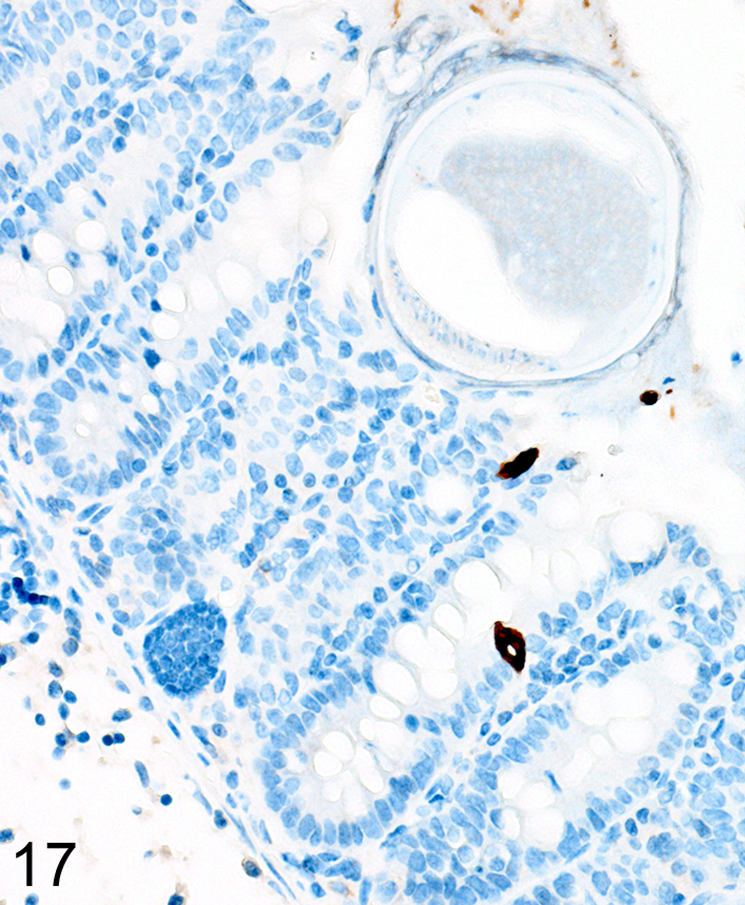
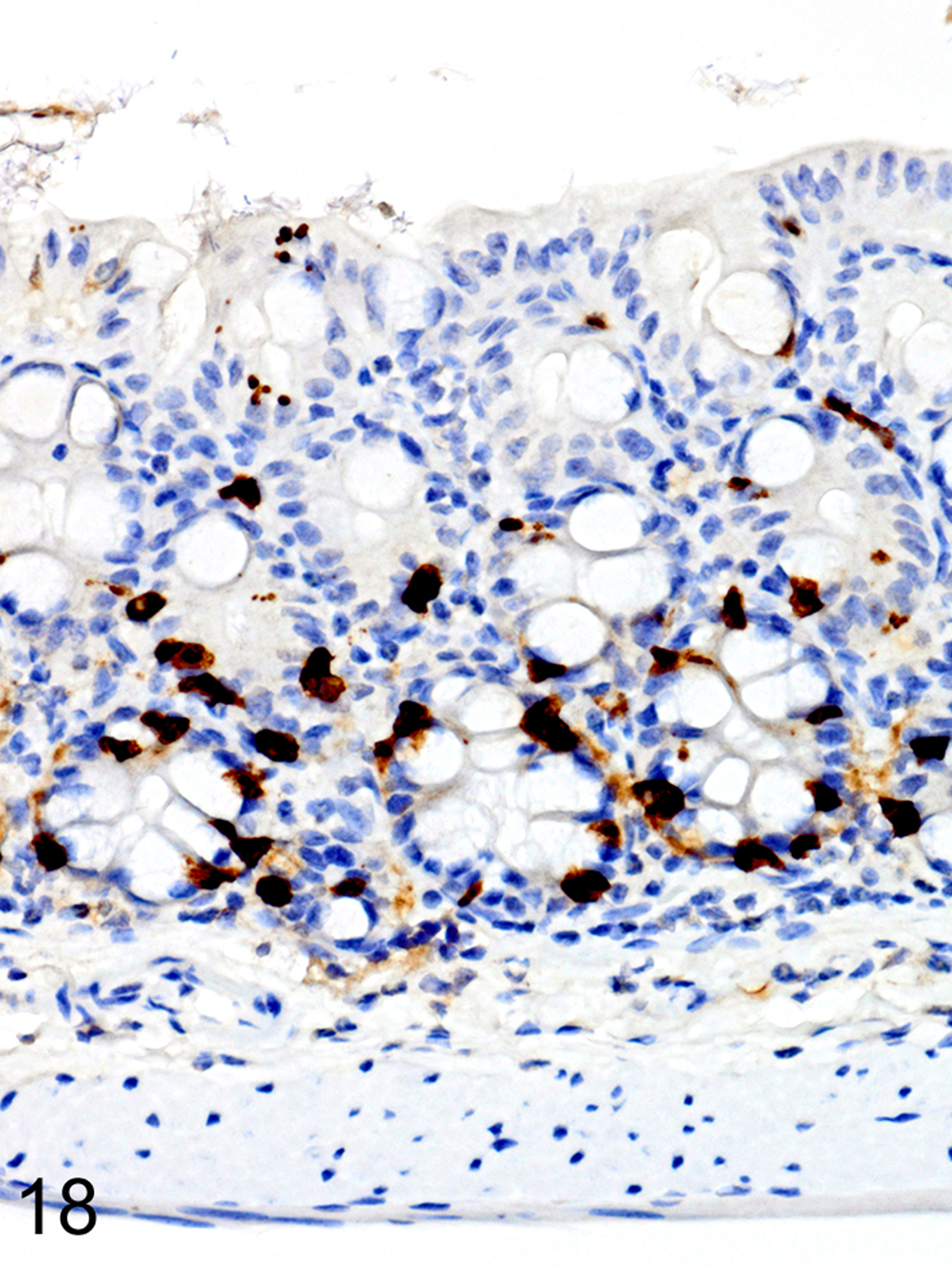
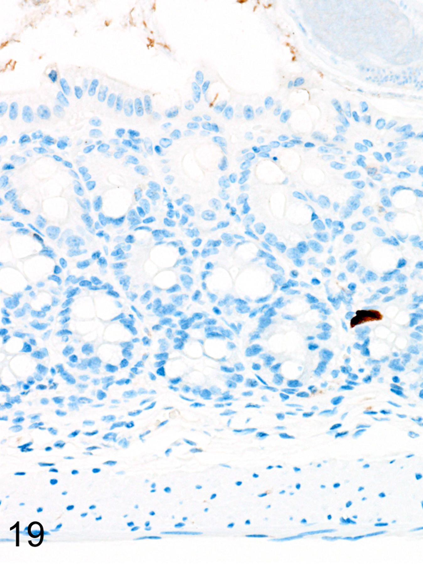
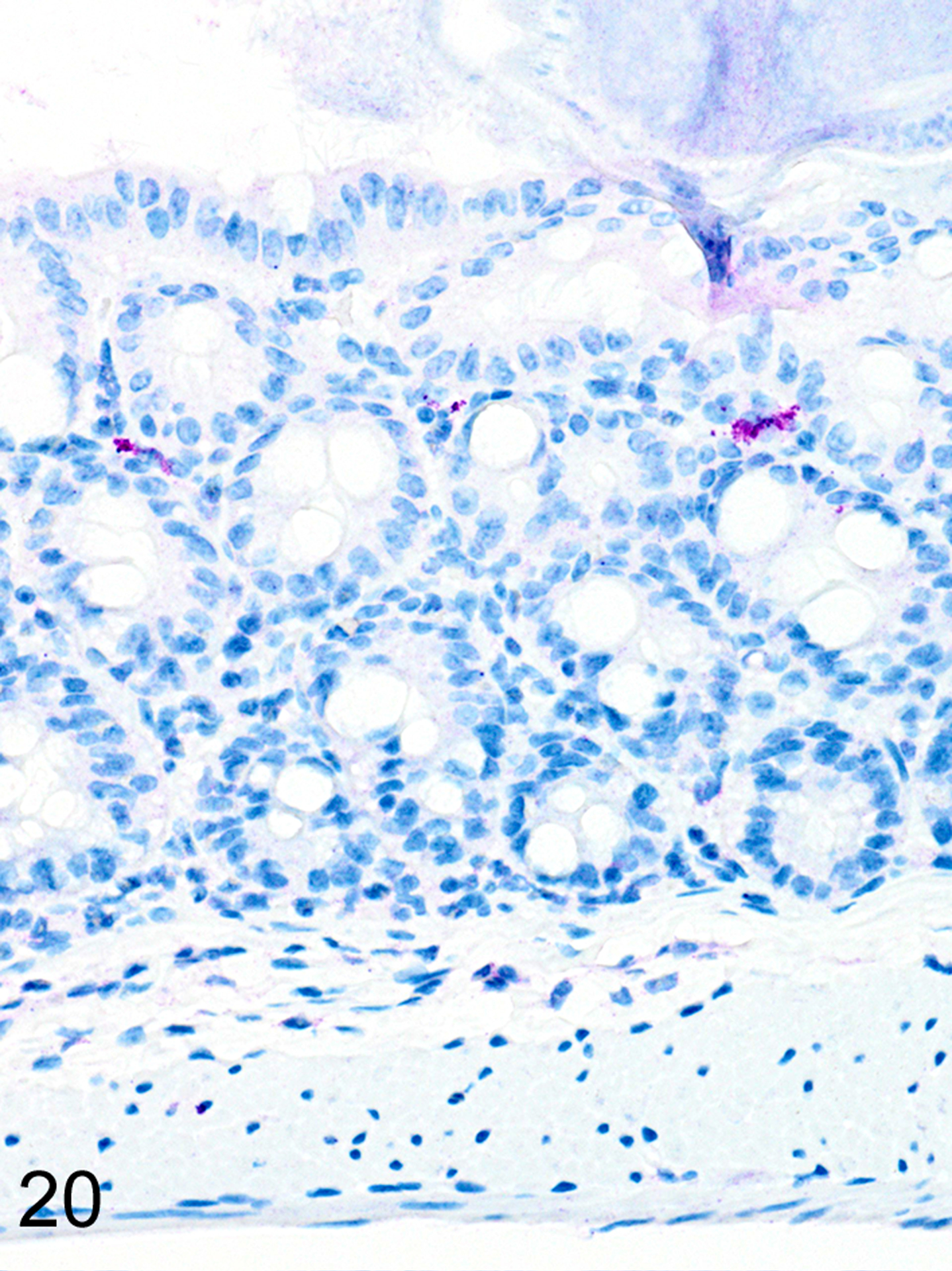
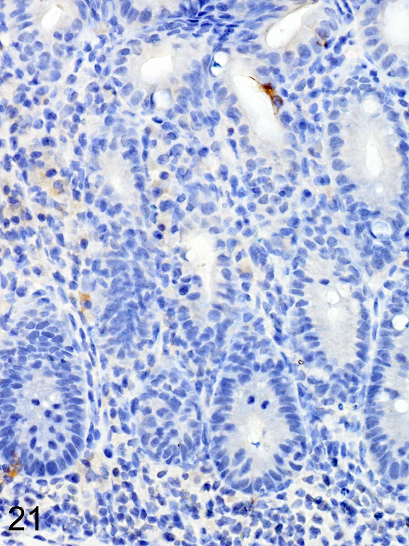
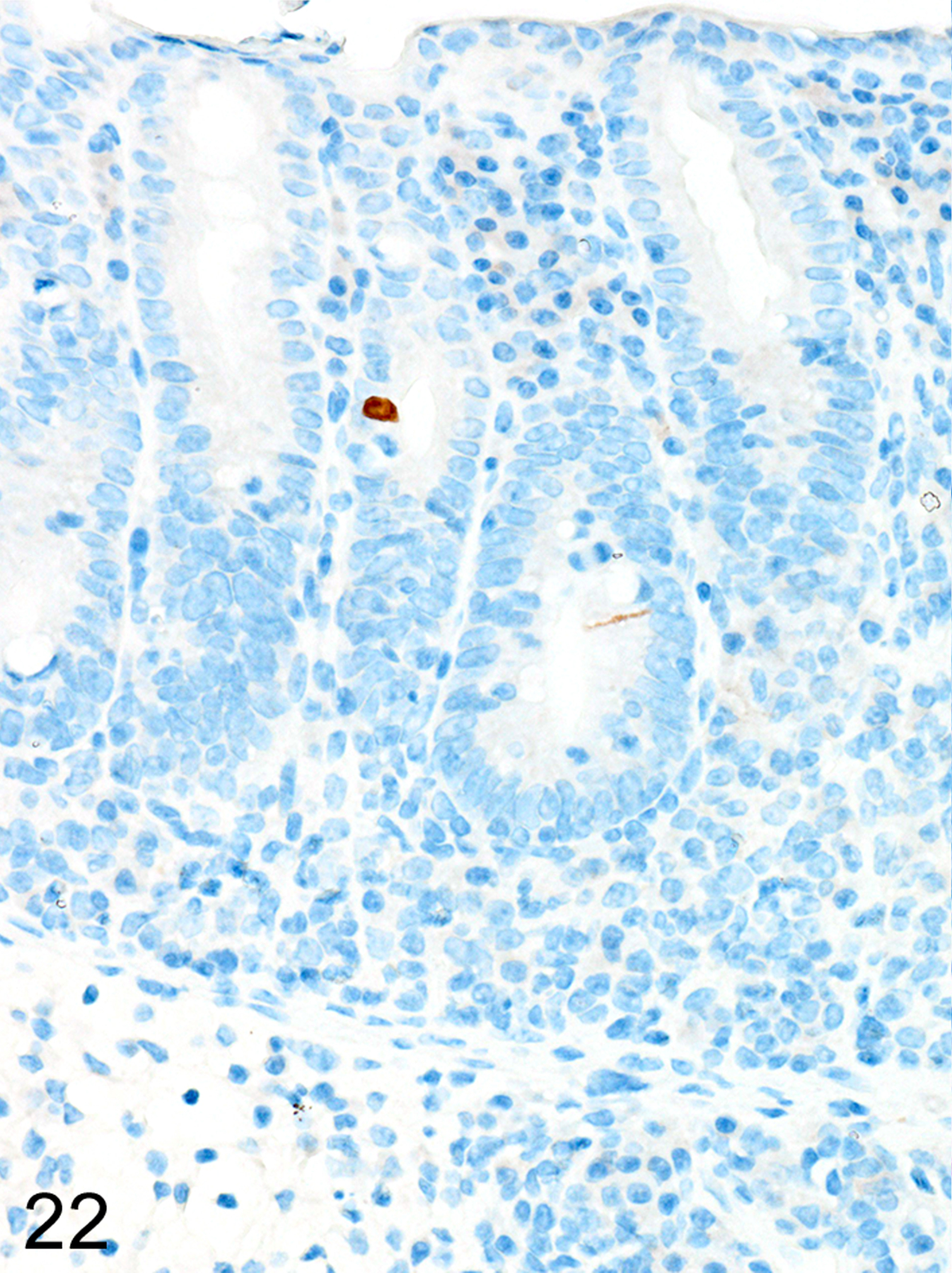
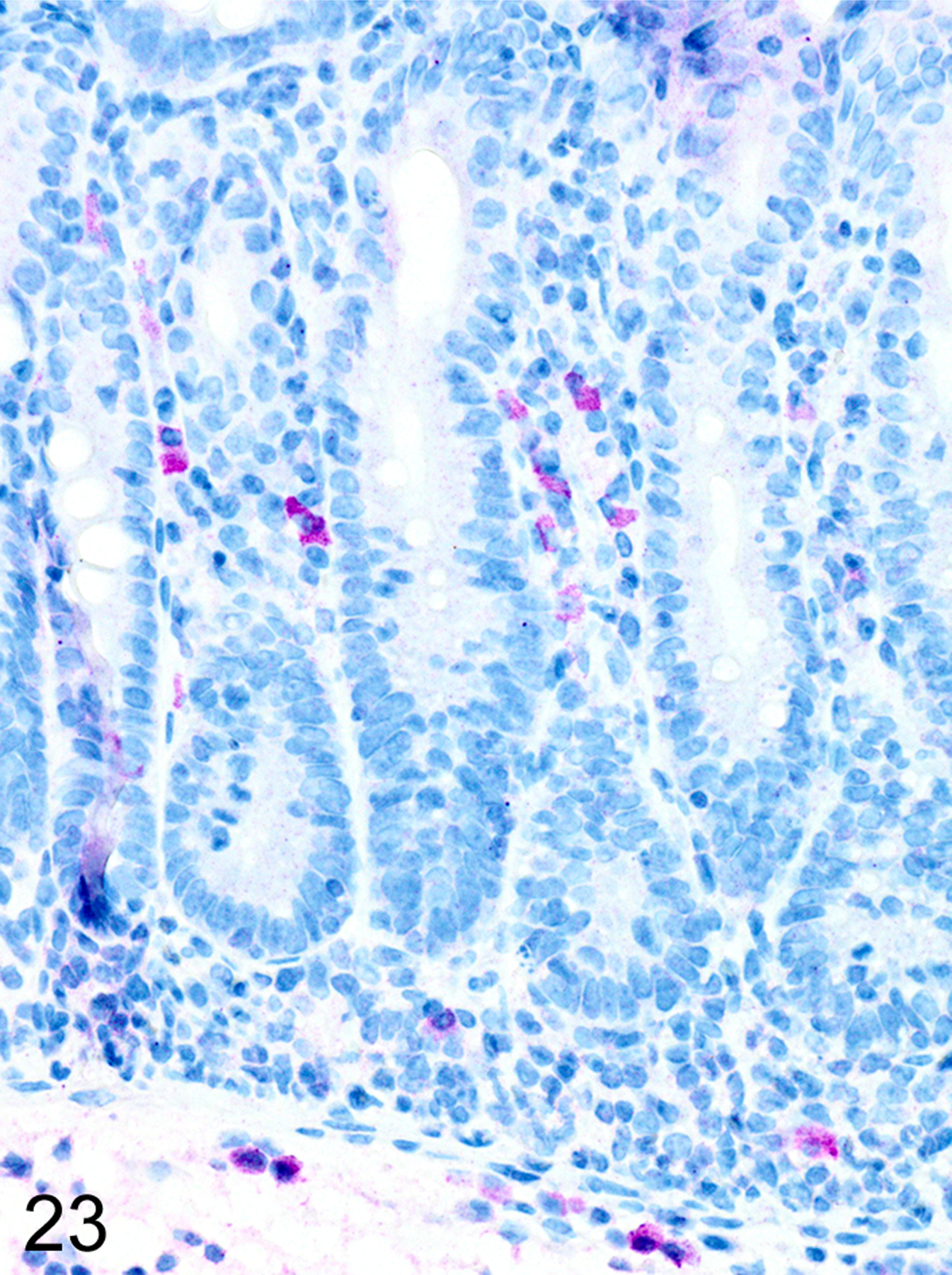
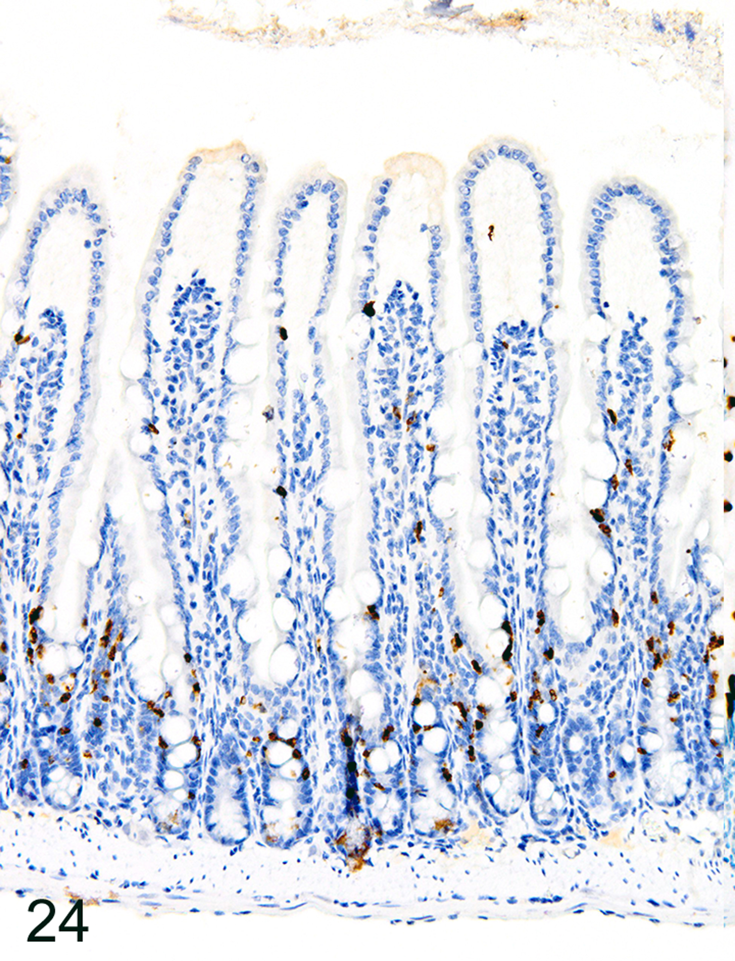
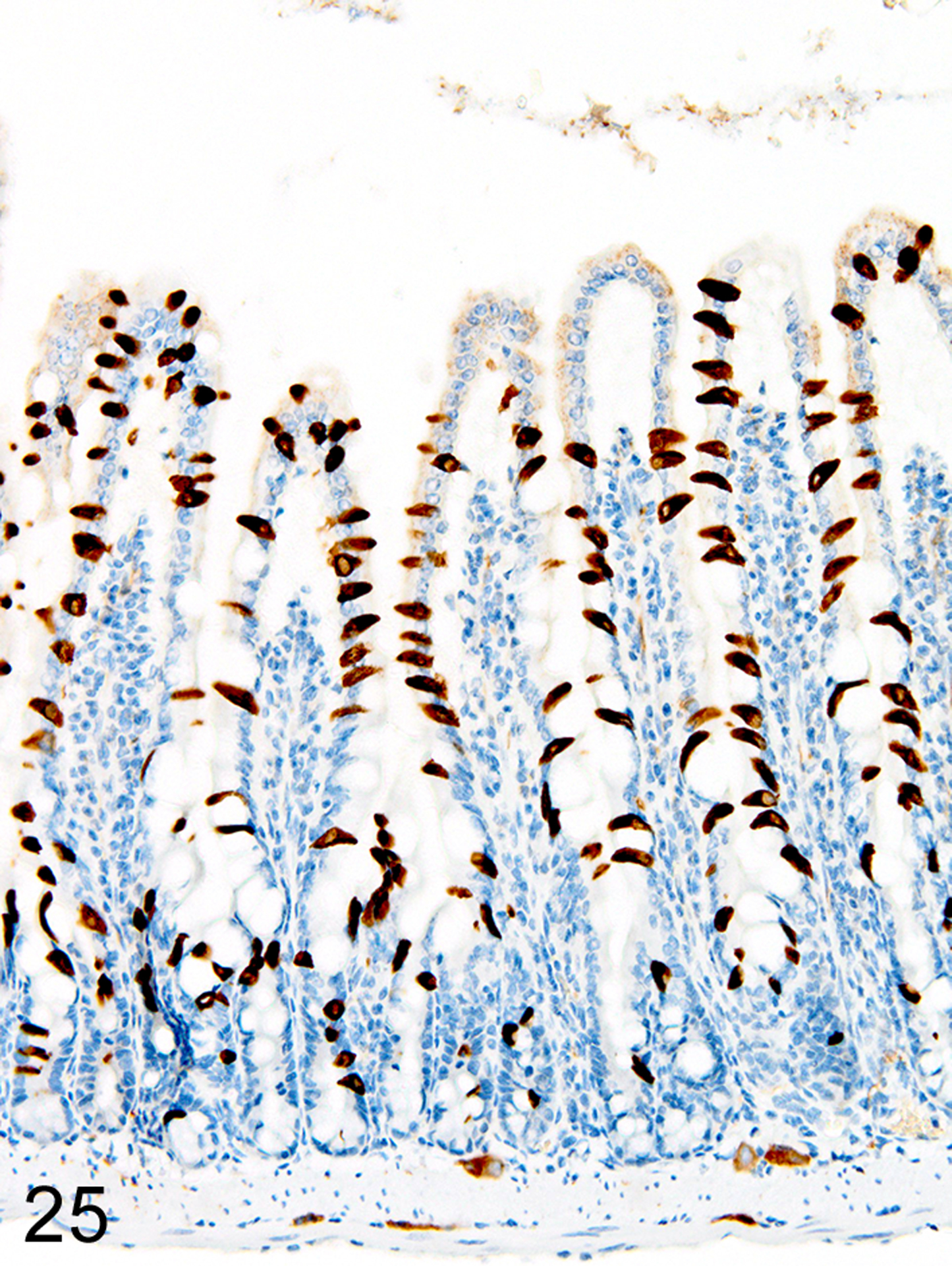
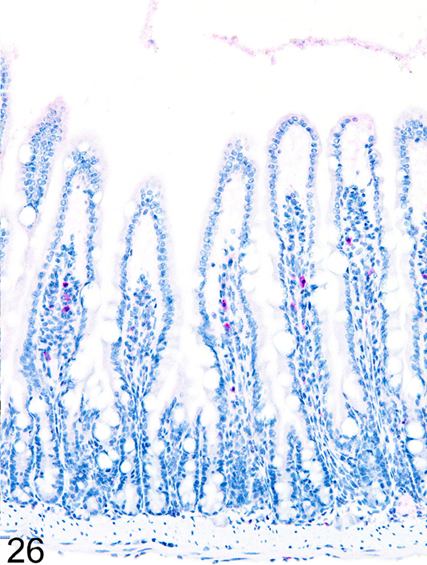
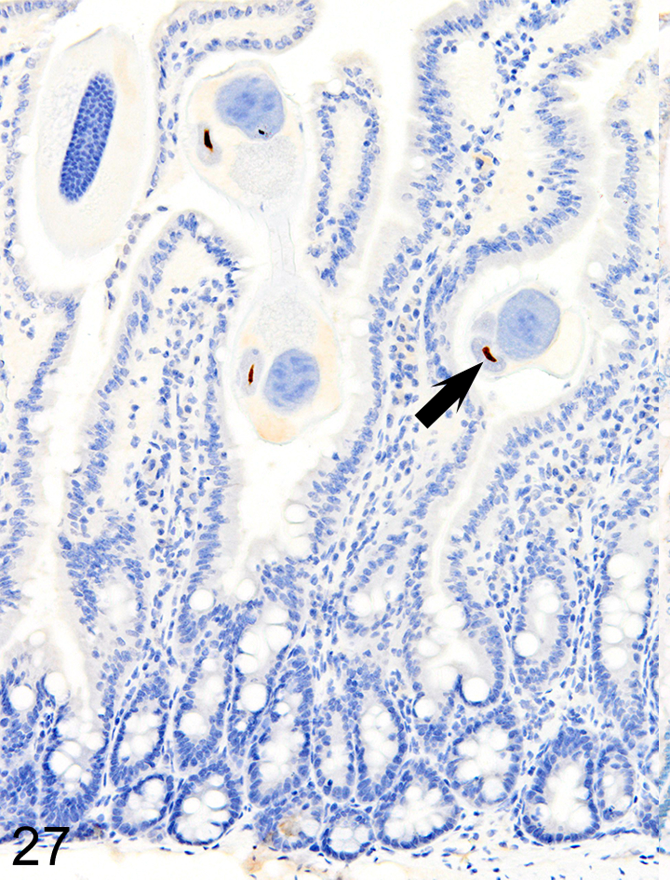
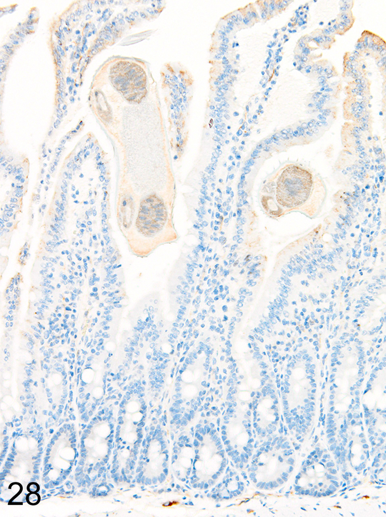
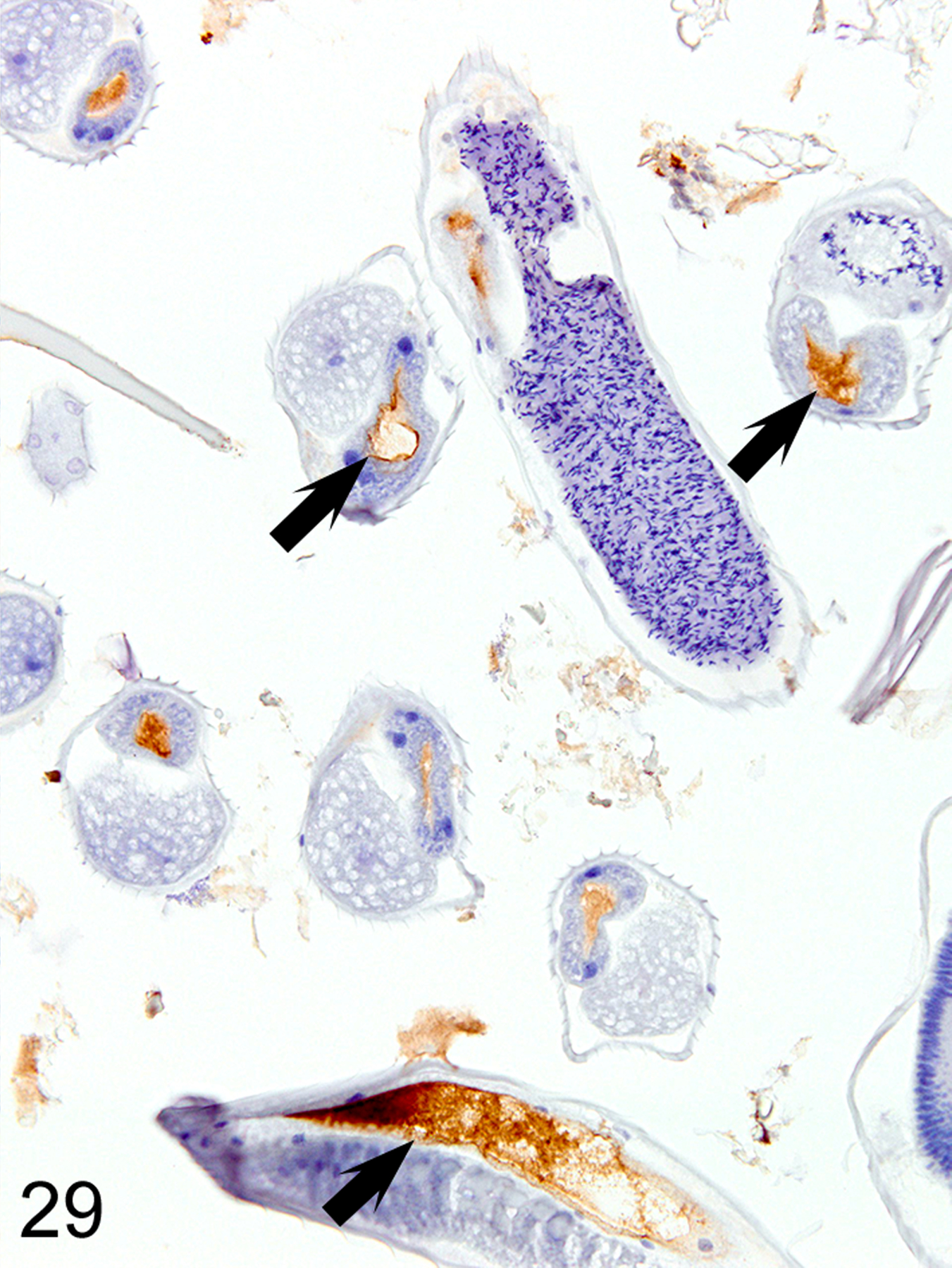
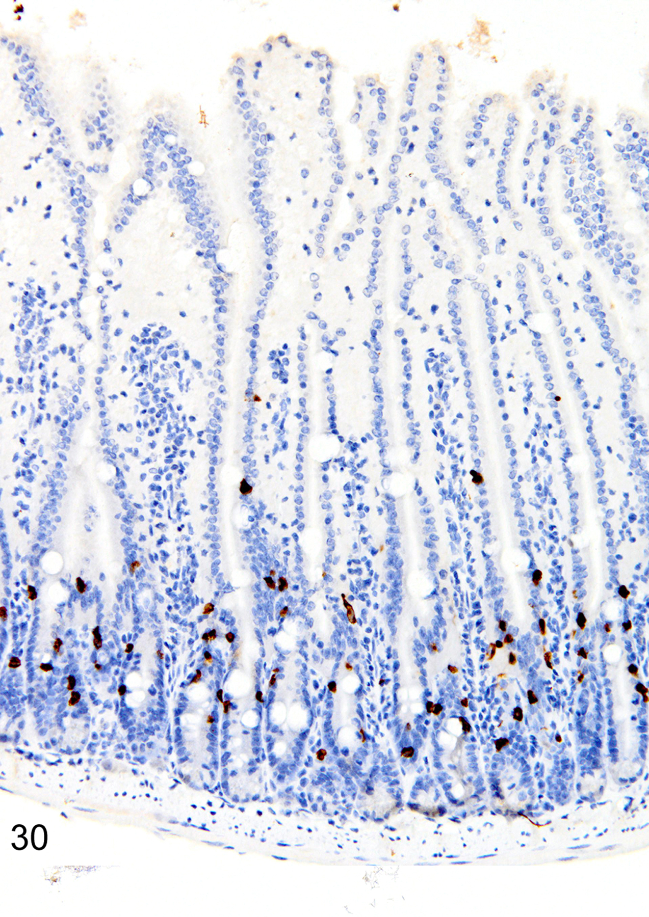
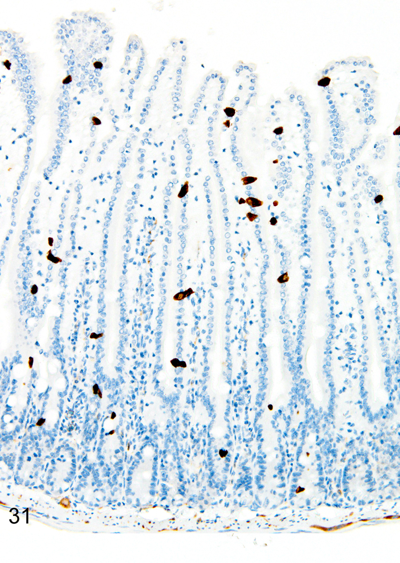
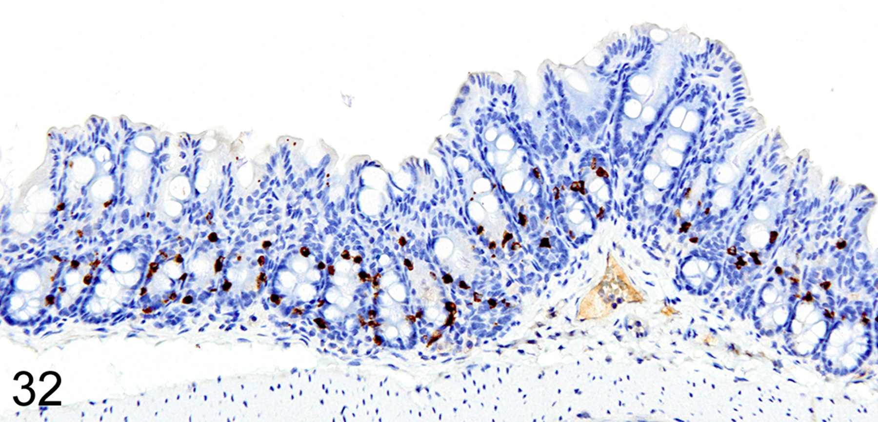
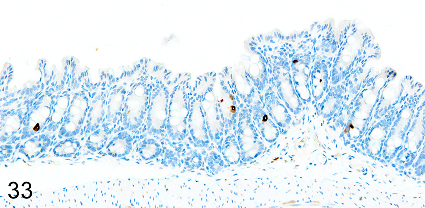
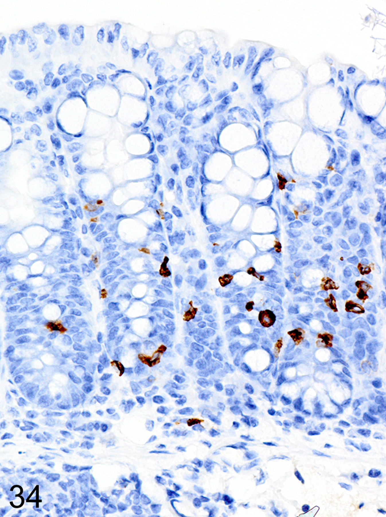
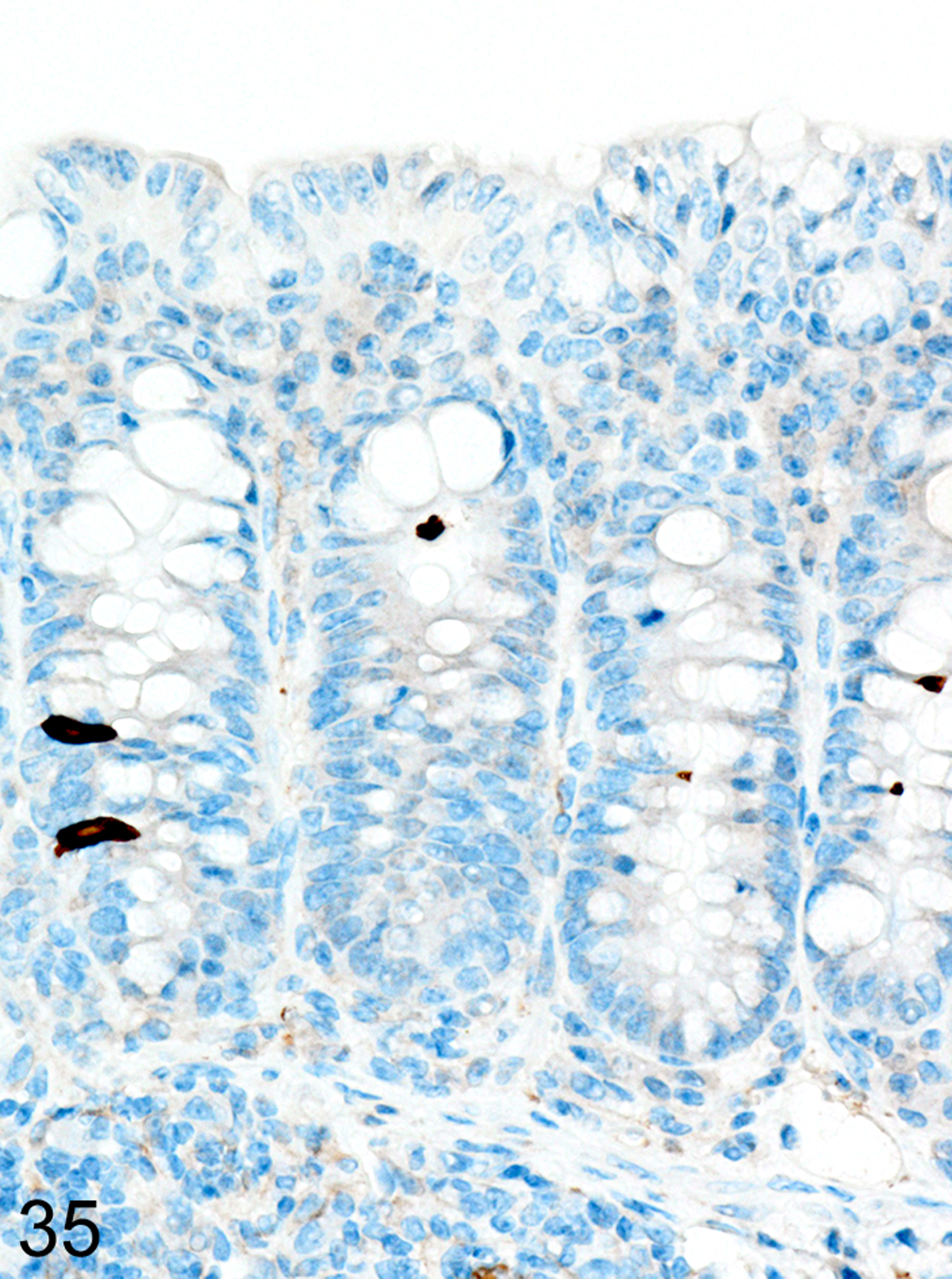
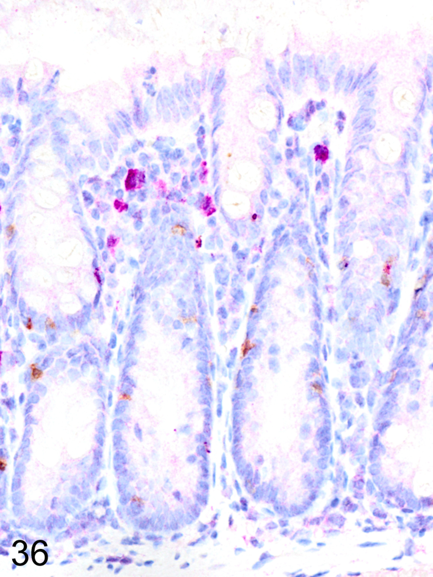
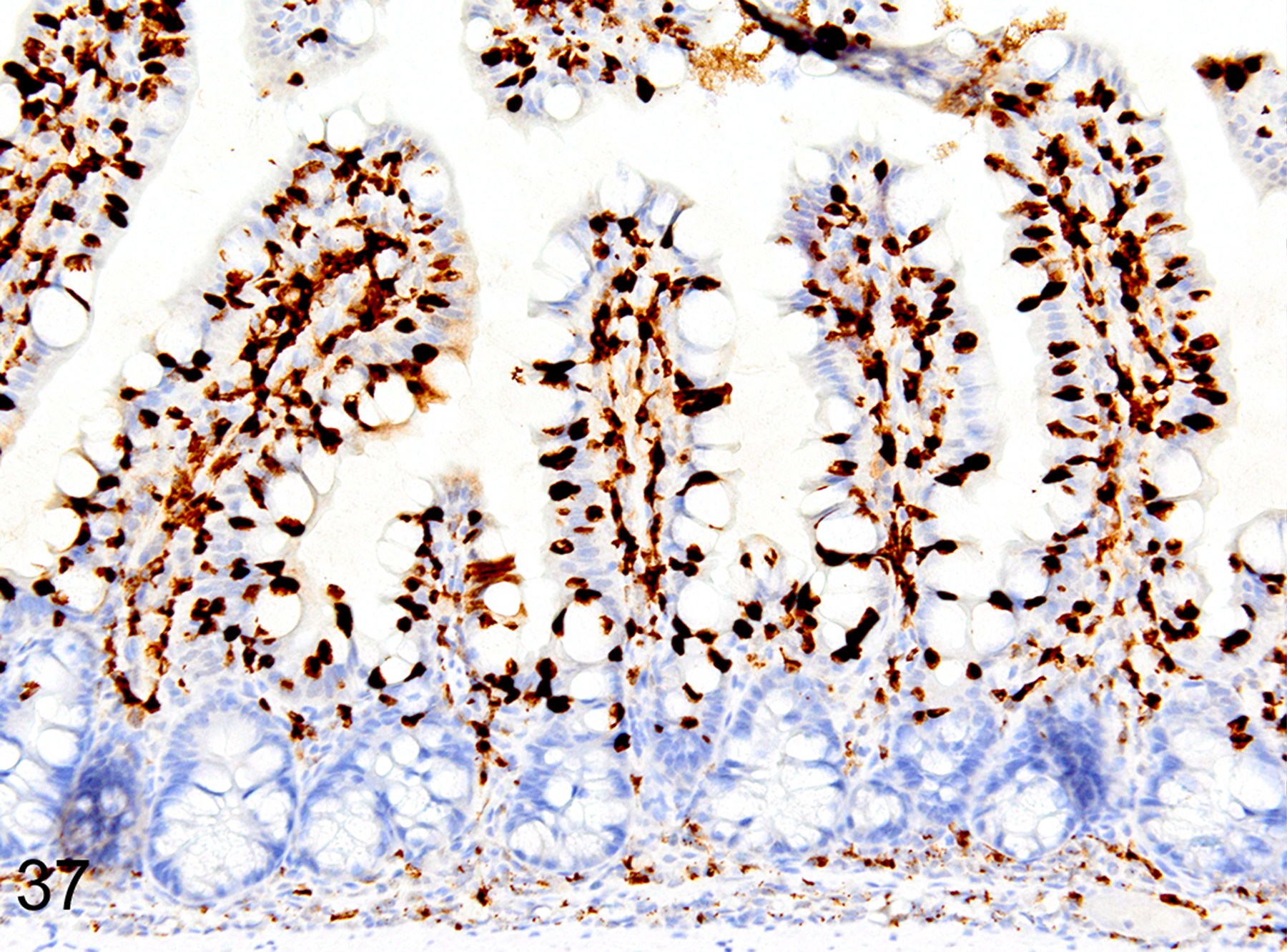
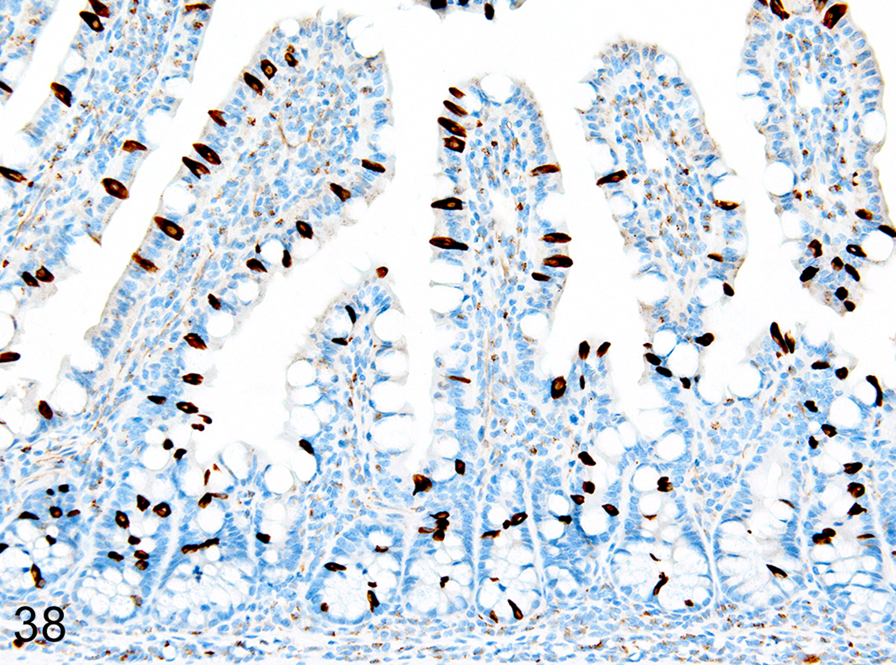
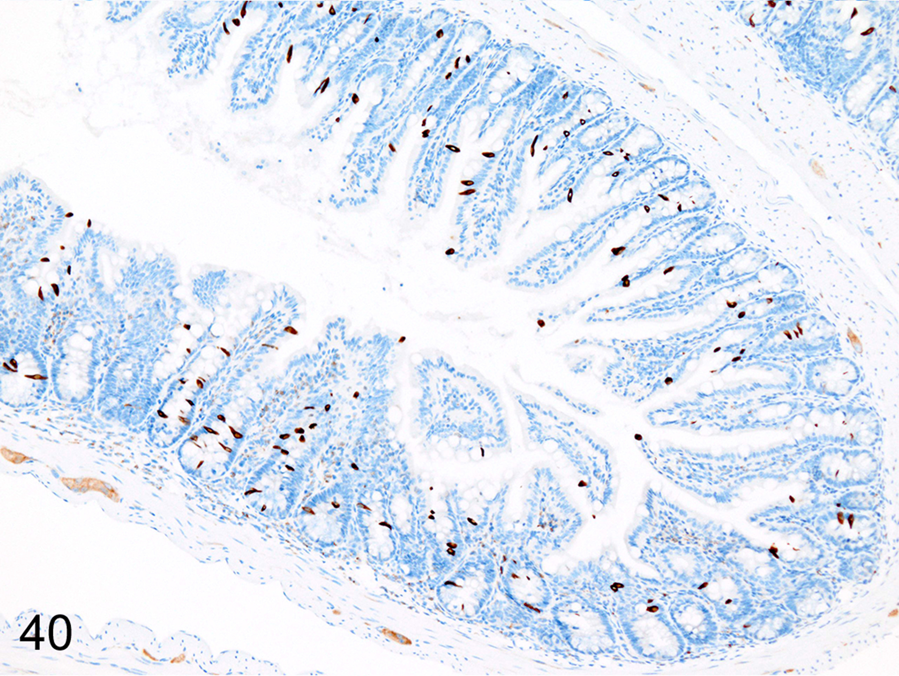
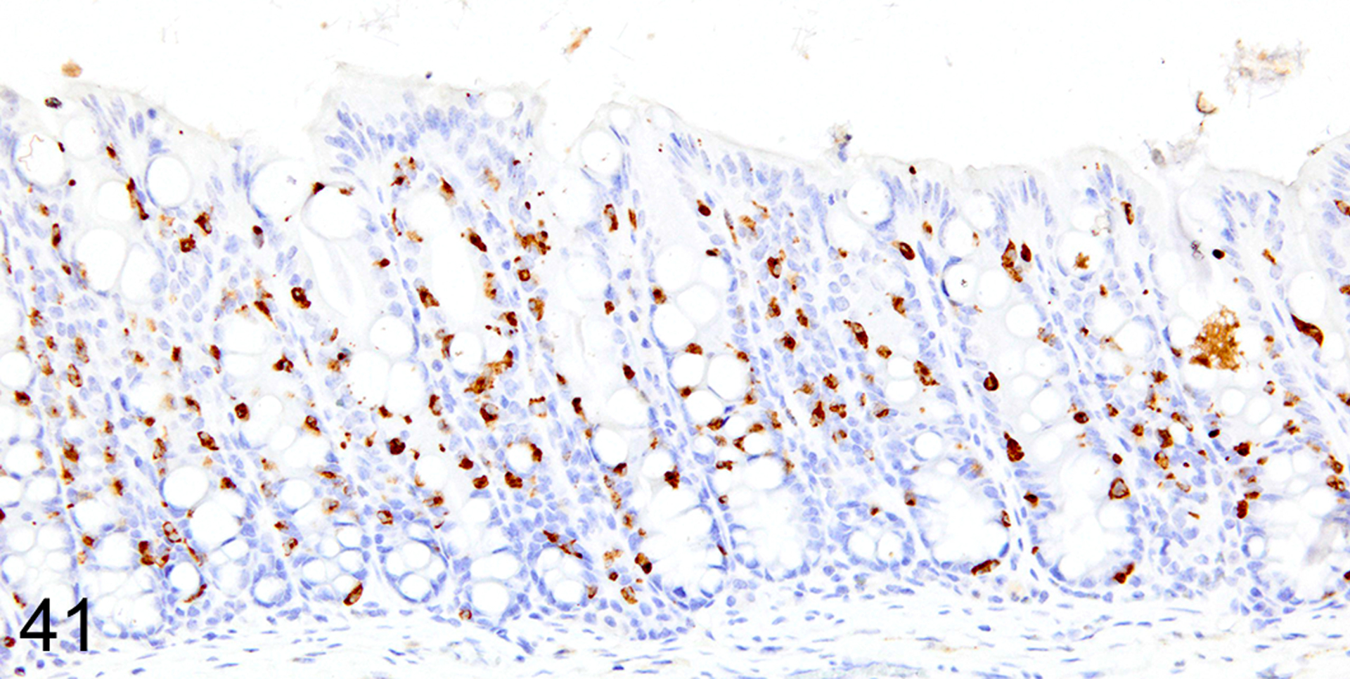
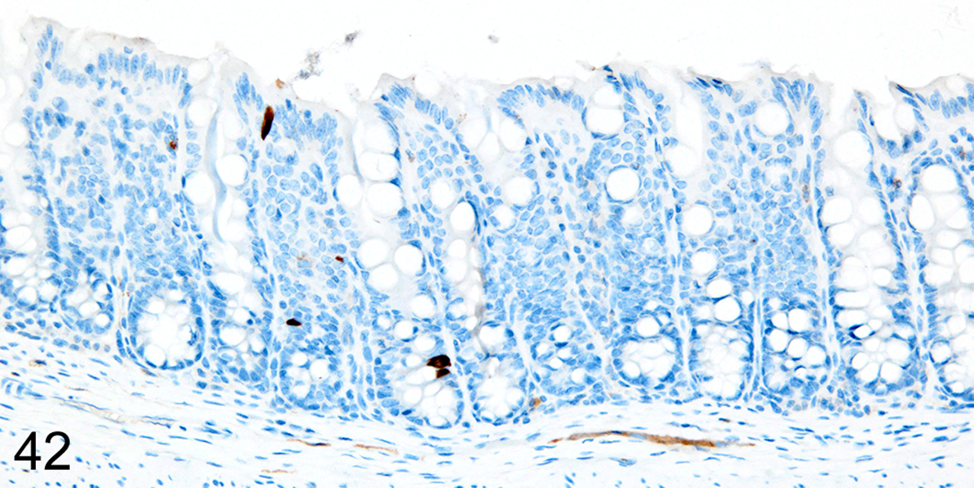
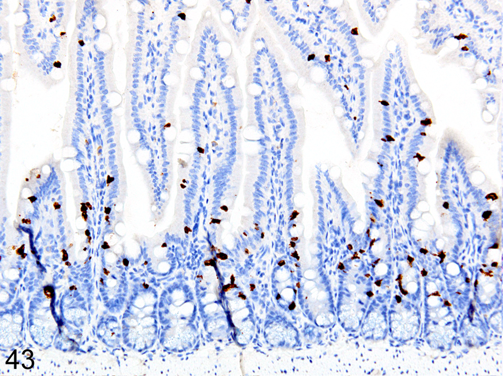
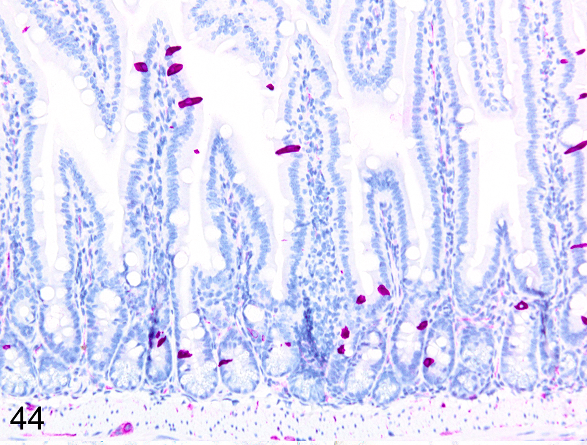
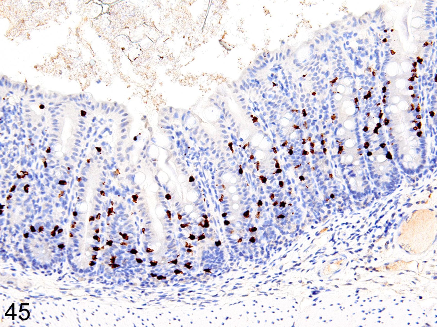
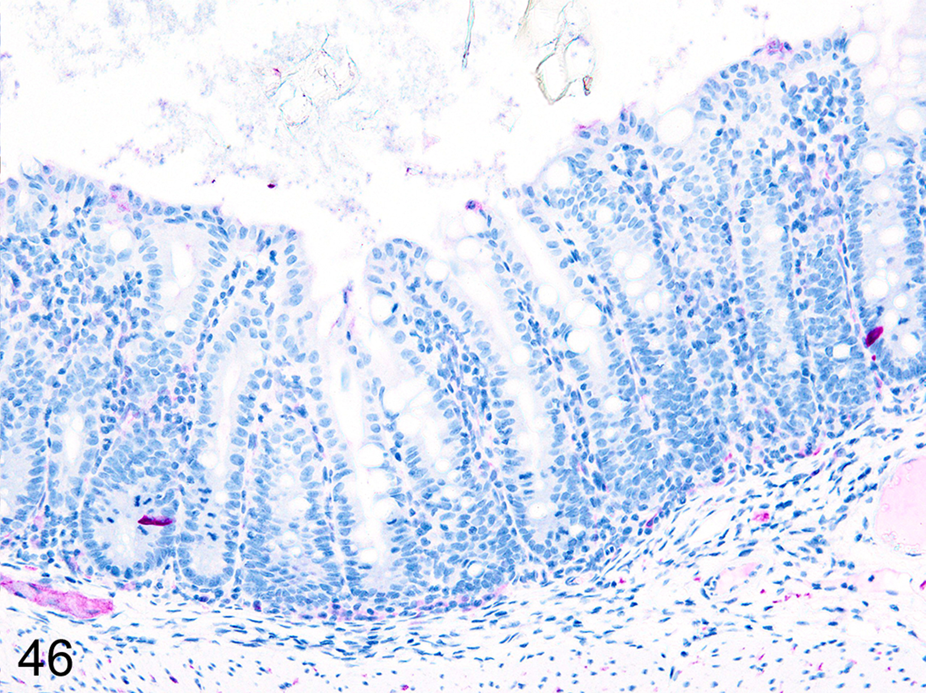
Only 2 major mast cell (MC) subtypes are commonly recognized in the mouse: the large connective tissue mast cells (CTMCs) and the mucosal mast cells (MMCs). Interepithelial mucosal inflammatory cells, most commonly identified as globule leukocytes (GLs), represent a third MC subtype in mice, which we term interepithelial mucosal mast cells (ieMMCs). This term clearly distinguishes ieMMCs from lamina proprial MMCs (lpMMCs) while clearly communicating their common MC lineage. Both lpMMCs and ieMMCs are rare in normal mouse intestinal mucosa, but increased numbers of ieMMCs are seen as part of type 2 immune responses to intestinal helminth infections and in food allergies. Interestingly, we found that increased ieMMCs were consistently associated with decreased mucosal inflammation and damage, suggesting that they might have a role in controlling helminth-induced immunopathology. We also found that ieMMC hyperplasia can develop in the absence of helminth infections, for example, in Treg-deficient mice, Arf null mice, some nude mice, and certain graft-vs-host responses. Since tuft cell hyperplasia plays a critical role in type 2 immune responses to intestinal helminths, we looked for (but did not find) any direct relationship between ieMMC and tuft cell numbers in the intestinal mucosa. Much remains to be learned about the differing functions of ieMMCs and lpMMCs in the intestinal mucosa, but an essential step in deciphering their roles in mucosal immune responses will be to apply immunohistochemistry methods to consistently and accurately identify them in tissue sections.

Keywords: Heligmosomoides bakeri; Treg; Trichuris muris; graft-vs-host; immunohistochemistry; mast cell; mouse.


Figures

Figure 1.
Figure 1.
Dermis, mouse. The prominent dark blue cytoplasmic granules in large connective tissue mast cells (CTMC) are preserved in formalin fixed tissues. Hematoxylin and eosin (HE).
Figure 2.
Figure 2.
Colon, mouse. The red cytoplasmic granules in globule leukocytes/ interepithelial mucosal mast cells are visible in formalin fixed tissues. HE.
Figure 3.
Figure 3.
Small intestine, mouse. CTMCs (arrows) and ieMMCs are visible in formalin fixed tissues, but lamina proprial mast cells (lpMMC) are not. HE.
Figure 4.
Figure 4.
Small intestine, immunohistochemistry (IHC) for MCPT1 and MCPT4, mouse. The small ieMMCs are positive for MCPT1 (brown). MCPT4 (violet) labels both the small lp MMCs and the larger submucosal CTMCs, but they can be distinguished by their differences in location and size.
Figure 5.
Figure 5.
Colon, mouse. Small ieMMCs (arrows) can be difficult to identify and lpMMC are not visible in standard HE stained sections.
Figure 6.
Figure 6.
Colon, (sequential section of Fig. 5), IHC for MCPT4 and MCPT4, mouse. Small ieMMCs (brown) and lpMMCs (violet) are present.
Figures 7.
Figures 7.
Small intestine, IHC for MCPT1, mouse. ieMMCs (arrow) are rare in normal small intestine mucosa.
Figures 8.
Figures 8.
Small intestine, IHC for DCAMKL1, mouse. Tuft cells are scattered diffusely in normal small intestine mucosa.
Figures 9.
Figures 9.
Colon, IHC for MCPT1, mouse. ieMMCs are also rare in normal colon mucosa.
Figures 10.
Figures 10.
Colon, IHC for DCAMKL1, mouse. Tuft cells are rare (arrow) in normal colon mucosa.
Figure 11.
Figure 11.. Trichuris muris infected cecum, mouse.
IHC for MCPT1. There is diffuse ieMMC hyperplasia in the mucosa associated with lymphocytic infiltrates and edema in the mucosa and submucosa. Submucosal lymphatics are dilated by accumulated lymphocytes.
Figure 12.
Figure 12.. Trichuris muris infected cecum, mouse.
IHC for DCAMKL1. There is a diffuse increase in numbers of tuft cells in the cecal mucosa.
Figure 13.
Figure 13.. Trichuris muris infected cecum, mouse.
IHC for MCPT1. The severity of ieMMC hyperplasia is reduced in the immediate vicinity of T. muris attachment sites.
Figure 14.
Figure 14.. Trichuris muris infected cecum, mouse.
The number of tuft cells is also reduced in the general area of T. muris attachment.
Figure 15.
Figure 15.. Trichuris muris infected cecum, mouse.
The anterior end of T. muris attaches to the mucosa by forming tunnels consisting of host cells forming syncytia (arrow). The adjacent mucosa is hyperplastic and lacks goblet cells; the lamina propria contains abundant eosinophils and lymphocytes.
Figures 16.
Figures 16.. Trichuris muris infected cecum, mouse.
Cecum, mouse (high magnification of Fig. 13). IHC for MCPT1. The zone of reduced ieMMCs (brown) is limited to the immediate vicinity of the helminths.
Figures 17.
Figures 17.. Trichuris muris infected cecum, mouse.
Cecum, mouse (high magnification of Fig. 14). IHC for DCAMKL1. A more extensive zone of reduced tuft cells (brown) is often present around helminth attachment sites.
Figure 18.
Figure 18.. Trichuris muris infected cecum, Treg depletion at early time points, mouse.
IHC for MCPT1. There is diffuse ieMMC (brown) hyperplasia in the mucosa.
Figure 19.
Figure 19.. Trichuris muris infected cecum, Treg depletion at early time points, mouse.
IHC for DCAMKL1. Tuft cells (brown) are rare in the cecal mucosa.
Figure 20.
Figure 20.. Trichuris muris infected cecum, Treg depletion at early time points, mouse.
IHC for MCPT4. There are scattered lpMMCs (violet) in the cecal mucosa.
Figure 21.
Figure 21.. Trichuris muris infected cecum, Treg depletion at late time points, mouse.
IHC for MCPT1. ieMMCs (brown) are very rarely present in the diffuse mucosal hyperplasia and more severe inflammatory cell infiltrates.
Figure 22.
Figure 22.. Trichuris muris infected cecum, Treg depletion at late time points, mouse.
IHC for DCAMKL1.Tuft cells (brown) are also very rare in the hyperplastic cecal mucosa.
Figure 23.
Figure 23.. Trichuris muris infected cecum, Treg depletion at late time points, mouse.
IHC for MCPT4. There are increased numbers of lpMMCs (violet) in the lamina propria and submucosa.
Figure 24.
Figure 24.. Heligmosomoides bakeri infected duodenum, mouse.
IHC for MCPT1. Diffuse ieMMC hyperplasia is present in mucosa not in direct contact with helminths.
Figure 25.
Figure 25.. Heligmosomoides bakeri infected duodenum, mouse.
IHC for DCAMKL1. Diffuse tuft cell hyperplasia is present in mucosa not in direct contact with helminths.
Figure 26.
Figure 26.. Heligmosomoides bakeri infected duodenum, mouse.
IHC for MCPT4. Increased numbers of lpMMCs are present in mucosa not in direct contact with helminths.
Figure 27.
Figure 27.. Heligmosomoides bakeri infected duodenum, mouse.
IHC for MCPT1. ieMMCs are absent in the mucosa in direct contact with helminths but MCPT1 is present in helminth digestive tract (arrows).
Figure 28.
Figure 28.. Heligmosomoides bakeri infected duodenum, mouse.
IHC for DCAMKL1. Tuft cells are also absent in the mucosa in direct contact with helminths.
Figure 29.
Figure 29.. Heligmosomoides bakeri infected duodenum, mouse.
IHC for MCPT1. The helminth digestive tracts (arrows) consistently contain MCPT1-positive material.
Figure 30.
Figure 30.. Small intestine and colon, Treg-depleted DEREG mice.
IHC for MCPT1. Diffuse ieMMCs (brown) hyperplasia is present in the small intestine mucosa.
Figure 31.
Figure 31.. Small intestine and colon, Treg-depleted DEREG mice.
IHC for DCAMKL1. Increased tuft cells (brown) are present throughout the small intestine mucosa.
Figure 32.
Figure 32.. Small intestine and colon, Treg-depleted DEREG mice.
IHC for MCPT1. Diffuse ieMMCs (brown) hyperplasia is present in the colon mucosa.
Figure 33.
Figure 33.. Small intestine and colon, Treg-depleted DEREG mice.
IHC for DCAMKL1. In contrast, there is no notable increase in tuft cells (brown) in the same region of colon mucosa.
Figure 34.
Figure 34.. Graft versus host disease (GVHD), colon, mouse.
IHC for MCPT1. Diffuse ieMMCs (brown) hyperplasia is present in the colon mucosa.
Figure 35.
Figure 35.. Graft versus host disease (GVHD), colon, mouse.
IHC for DCAMKL1. In contrast, there is no notable increase in tuft cells (brown) in the colon mucosa.
Figure 36.
Figure 36.. Graft versus host disease (GVHD), colon, mouse.
IHC for and MCPT1 and MCPT4. Scattered ieMMCs (brown) and Clusters of lpMMCs (violet) within the lamina propria are common in GVHD-affected colon mucosa.
Figure 37.
Figure 37.. Small intestine and colon, p19ARF null mouse.
IHC for MCPT1. Severe interepithelial and lamina proprial hyperplasia of ieMMCs (brown) in the small intestine mucosa.
Figure 38.
Figure 38.. Small intestine and colon, p19ARF null mouse.
IHC for DCAMKL1. Tuft cell (brown) hyperplasia can be present in the same areas of mucosa.
Figure 39.
Figure 39.. Small intestine and colon, p19ARF null mouse.
IHC for MCPT1. The ieMMC (brown) hyperplasia in p19ARF null mice often has a patchy distribution.
Figure 40.
Figure 40.. Small intestine and colon, p19ARF null mouse.
IHC for DCAMKL1. This sequential section, illustrates the lack of correlation between tuft cell (brown) numbers and ieMMC hyperplasia.
Figure 41.
Figure 41.. Small intestine and colon, p19ARF null mouse.
IHC for MCPT1. Diffuse ieMMCs (brown) hyperplasia is present in the colon mucosa.
Figure 42.
Figure 42.. Small intestine and colon, p19ARF null mouse.
IHC for DCAMKL1. In contrast, there is no notable increase in tuft cells (brown) in the colon mucosa.
Figure 43.
Figure 43.. Small intestine and colon, CD-1 nude mouse.
IHC for MCPT1. Diffuse ieMMC (brown) hyperplasia in the small intestine mucosa.
Figure 44.
Figure 44.. Small intestine and colon, CD-1 nude mouse.
IHC for DCAMKL1. This sequential section shows tuft cell (violet) hyperplasia is often present in the same areas of small intestine mucosa.
Figure 45.
Figure 45.. Small intestine and colon, CD-1 nude mouse.
IHC for MCPT1. Diffuse ieMMC (brown) hyperplasia in the colon mucosa.
Figure 46.
Figure 46.. Small intestine and colon, CD-1 nude mouse.
IHC for DCAMKL1. This sequential section, illustrates the lack of correlation between tuft cell (violet) numbers and ieMMC hyperplasia in the colon mucosa

Similar articles

Cited by

References

    1. Akpavie SO, Pirie HM. The globule leucocyte: morphology, origin, function and fate, a review. Anatomia, histologia, embryologia 1989;18(1):87–95. - PubMed
    1. Alizadeh H, Murrell KD. The intestinal mast cell response to Trichinella spiralis infection in mast cell-deficient w/wv mice. The Journal of parasitology. 1984;70(5):767–773. - PubMed
    1. Artis D. New weapons in the war on worms: identification of putative mechanisms of immune-mediated expulsion of gastrointestinal nematodes. International journal for parasitology. 2006;36(6):723–733. - PMC - PubMed
    1. Artis D, Villarino A, Silverman M, et al. The IL-27 receptor (WSX-1) is an inhibitor of innate and adaptive elements of type 2 immunity. Journal of immunology. 2004;173(9):5626–5634. - PubMed
    1. Austen KF. The heterogeneity of mast cell populations and products. Hospital practice. 1984;19(9):135–139, 143-136. - PubMed

LinkOut - more resources