A giant squamous cell carcinoma of the skin of the thoracic wall: a case report and review of the literature
- PMID: 28494816
- PMCID: PMC5426016
- DOI: 10.1186/s13256-017-1281-8
A giant squamous cell carcinoma of the skin of the thoracic wall: a case report and review of the literature
Abstract
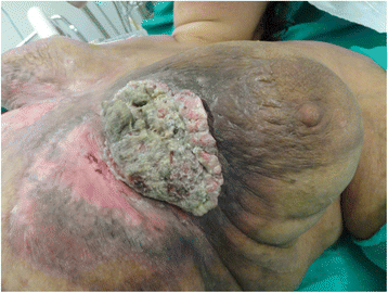
Background: We report a case of a 48-year-old white woman who presented with a huge cutaneous protruding tumor of the thoracic wall below her left breast.
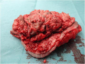
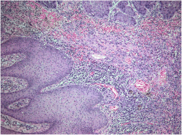
Case presentation: The lesion was excised with clear margins from the adjacent skin, and subcutaneous tissue was left to heal with second intention. A histological examination of the surgical specimen revealed a well-differentiated infiltrative cutaneous squamous cell carcinoma. Our patient neglected to attend our Oncological Department to receive chemotherapy. Today, 12 months after surgery, she is alive and without evidence of disease recurrence.
Conclusions: Cutaneous squamous cell carcinoma can reach a huge size if left untreated. Surgery is the primary mode of treatment, followed by chemotherapy if applicable.
Keywords: Carcinoma; Cutaneous; Invasion; Metastasis; Squamous.
Figures


References
-
- Edge S, Byrd DR, Compton CC, Fritz AG, Greene FL, editors. AJCC Cancer Staging Manual. 7. New York: Springer; 2010.
-
- Miller SJ, Alam M, Andersen J, et al. Basal cell and squamous cell skin cancers. J Natl Compr Canc Netw. 2010;8:836–864. - PubMed
Publication types
MeSH terms
LinkOut - more resources
Full Text Sources
Other Literature Sources
Medical

