. 2017 May 9;19(6):1091-1100.
doi: 10.1016/j.celrep.2017.04.026.

HIV Reprograms Human Airway Basal Stem/Progenitor Cells to Acquire a Tissue-Destructive Phenotype

Affiliations

HIV Reprograms Human Airway Basal Stem/Progenitor Cells to Acquire a Tissue-Destructive Phenotype

Nancy P Y Chung et al. Cell Rep. .

Abstract

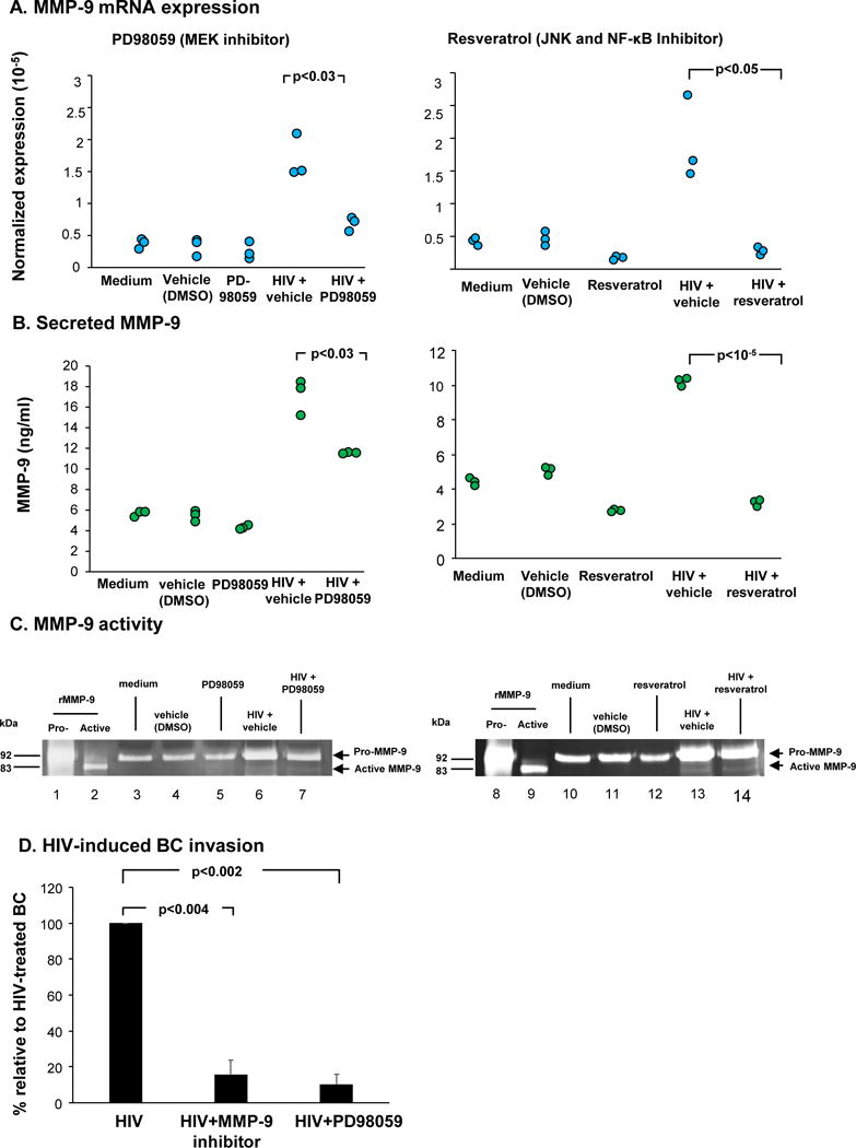
While highly active anti-retroviral therapy has dramatically improved the survival of HIV-infected individuals, there is an increased risk for other co-morbidities, such as COPD, manifesting as emphysema. Given that emphysema originates around the airways and that human airway basal cells (BCs) are adult airway stem/progenitor cells, we hypothesized that HIV reprograms BCs to a distinct phenotype that contributes to the development of emphysema. Our data indicate that HIV binds to but does not replicate in BCs. HIV binding to BCs induces them to acquire an invasive phenotype, mediated by upregulation of MMP-9 expression through activation of MAPK signaling pathways. This HIV-induced "destructive" phenotype may contribute to degradation of extracellular matrix and tissue damage relevant to the development of emphysema commonly seen in HIV+ individuals.

Keywords: HIV; MMP-9; airway basal stem/progenitor cells; matrix metalloproteinase-9; reprogramming.


Figures

Figure 1
Figure 1
Binding of HIV to human airway basal cells. A. Binding of HIV-1NL4-3. BC were exposed to the virus, washed and then lysed in 0.1% Triton-X. Virus binding was measured by HIV-1 p24 levels after 3 and 24 hr. B. Sensitivity of HIV binding to the BC to trypsin. After 3 hr of incubation, cells were incubated with 0.05% trypsin/EDTA for 5 min, washed, lysed and analyzed by HIV-1 p24 levels. C. Flow cytometry assessment of cell surface expression of heparan sulfate proteoglycans (HSPGs) in untreated and trypsin-treated basal cells. The solid line represents staining with monoclonal antibody against the indicated HSPG. The broken line represents staining with corresponding isotype control. Shown are the histograms of heparan sulfate and syndecans 1–4, each from one representative experiment of 3 independent experiments. D. Heparan sulfate inhibition of HIV binding to BC. Pretreatment of HIV with heparan sulfate at different concentrations inhibits HIV binding to airway BC in a dose-dependent manner. Results shown are the average of three independent experiments using cells from 3 different individuals. E. Effect of heparinase III pretreatment of BC on HIV binding. Prior heparinase III treatment on BC significantly abolishes HIV binding. F. HIV binds to, but does not replicate in airway BC. Shown is a time-course of BC levels of p24 following addition of HIV-1 to airway BC. Cell lysates were collected at the indicated time points and used for quantification of HIV-1 p24 levels. *p<0.05, **p<0.01, NL4-3 vs NL4-3 + heparan sulfate.
Figure 2
Figure 2
HIV induced BC to acquire a destructive phenotype. Shown are in vitro assessments of HIV-induced BC morphological changes and cell invasion through connective tissue. A. Morphology of BC after exposure to HIV. At day 5 there are “holes” in the BC culture. B. HIV induces BC invasion through connective tissue. BC were plated onto matrigel-coated chambers. Data shown is the stained migratory cells at the bottom of the chambers in control and HIV-treated culture from one representative of 3 independent experiments. Bar = 100 μm.
Figure 3
Figure 3
Expression of matrix metalloproteinases (MMPs) in mock and HIV-treated BC. BC were exposed to HIVNL4-3 (p24 at 200 ng/ml) for 48 hr, and cell lysates were assessed using a human protease array to detect the expression of MMPs. A. The locations of each MMP on the array membrane are identified. The array images were obtained from 1 min exposure to X-ray film. B. Quantification of the expression of the pixel density of each MMP (in duplicate) was measured. The data represents the average of each MMP after subtraction of the background signal.
Figure 4
Figure 4
HIV modulation of MMP-9 expression in BC. A. HIV induces MMP-9 expression in BC in a dose-dependent manner. BC were exposed to increasing viral input (p24 from 5 to 200 ng/ml) for 2 days and MMP-9 expression quantified by TaqMan PCR. The data is normalized to 18s RNA. B. Assessment of expression of MMP-9 in HIV-treated and HIV + cigarette smoke extract (CSE)-treated BC. C. Quantification of MMP-9 in culture supernatants of untreated, CSE, HIV and HIV+CSE treated BC cultures. MMP-9 levels were quantified by ELISA. D. Gelatin zymography analysis of MMP-9 activity in cylture supernatants from treated BC. Lane 1 - pro-MMP-9 standard; lane 2 - active MMP-9 standard; lane 3 - untreated, lane 4 - HIV-treated BC; lane 5 - heat-inactivated HIV-treated BC; lane 6 - 3% CSE-treated BC; lane 7 - 6% CSE-treated BC; lane 8 - HIV+ 3% CSE-treated BC; lane 9 - HIV+ 6% CSE-treated BC.
Figure 5
Figure 5
MEK inhibitor (PD98059) and resveratrol suppress HIV-induced MMP-9 expression in BC. BC were treated with PD98059 (20 M) or resveratrol (75 M) for 1 hr prior to virus exposure. BC were collected and analyzed. A. MMP-9 mRNA expression by Taqman PCR. B. Level of secreted MMP-9 in culture supernatants assessed by ELISA from BC culture treated with HIV in the absence and presence of inhibitors. C. MMP-9 activity in culture supernatants assessed by gelatin zymography analysis. Lanes 1, 8 - pro-MMP-9 standard; lane 2, 9 - active MMP-9 standard; lanes 3, 10 - untreated BC; lanes 4, 11 - vehicle control; lane 5 - PD98059 alone; lanes 6, 13 - HIV+vehicle; lane 7 - HIV+PD98059; lane 12- resveratrol; lane 14 - HIV+resveratrol. D. Suppression of invasion of HIV-bound airway BC through connective tissue by MEK and MMP-9 inhibitors. The number of migratory cells in HIV-treated in the absence and presence of inhibitors were quantified. Data is presented as the percentage of cell invasion relative to HIV-treated BC from three independent experiments.

Similar articles

Cited by

References

    1. Atkinson JJ, Senior RM. Matrix metalloproteinase-9 in lung remodeling. Am J Respir Cell Mol Biol. 2003;28:12–24. - PubMed
    1. Beck JM, Rosen MJ, Peavy HH. Pulmonary complications of HIV infection. Report of the Fourth NHLBI Workshop. Am J Respir Crit Care Med. 2001;164:2120–2126. - PubMed
    1. Beeh KM, Beier J, Kornmann O, Buhl R. Sputum matrix metalloproteinase-9, tissue inhibitor of metalloprotinease-1, and their molar ratio in patients with chronic obstructive pulmonary disease, idiopathic pulmonary fibrosis and healthy subjects. Respir Med. 2003;97:634–639. - PubMed
    1. Bhatia NS, Chow FC. Neurologic Complications in Treated HIV-1 Infection. Curr Neurol Neurosci Rep. 2016;16:62. - PubMed
    1. Bhatia R, Ryscavage P, Taiwo B. Accelerated aging and human immunodeficiency virus infection: emerging challenges of growing older in the era of successful antiretroviral therapy. J Neurovirol. 2012;18:247–255. - PubMed

Publication types

Substances