. 2017 May 9;112(9):1997-2010.
doi: 10.1016/j.bpj.2017.03.022.

Rate-Dependent Role of IKur in Human Atrial Repolarization and Atrial Fibrillation Maintenance

Affiliations

Rate-Dependent Role of IKur in Human Atrial Repolarization and Atrial Fibrillation Maintenance

Martin Aguilar et al. Biophys J. .

Abstract

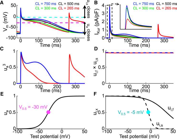
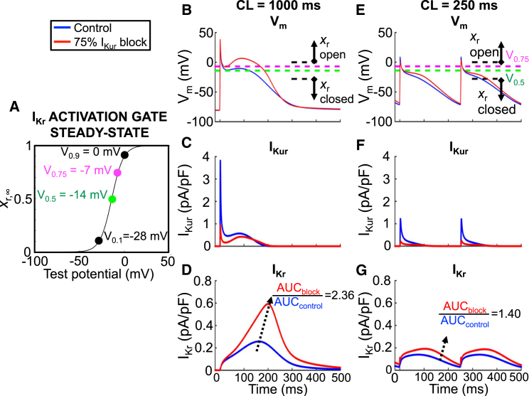
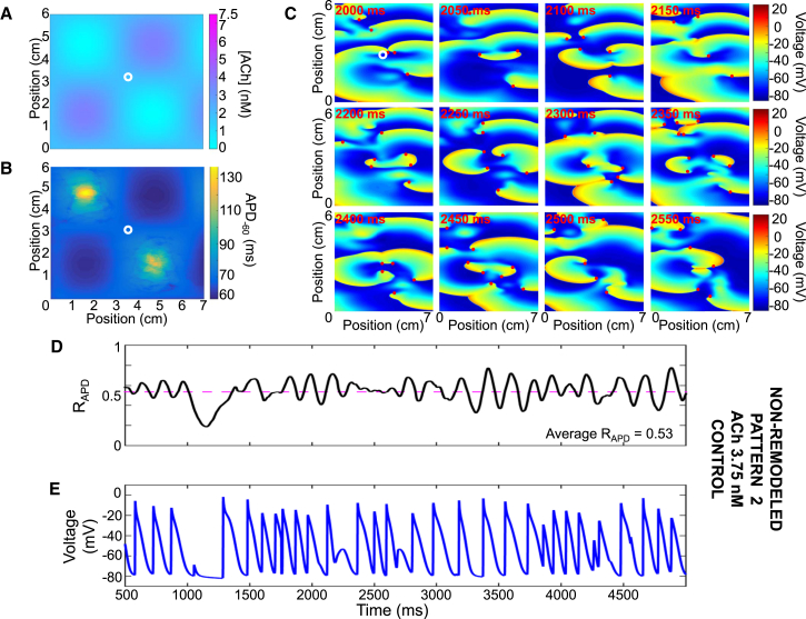
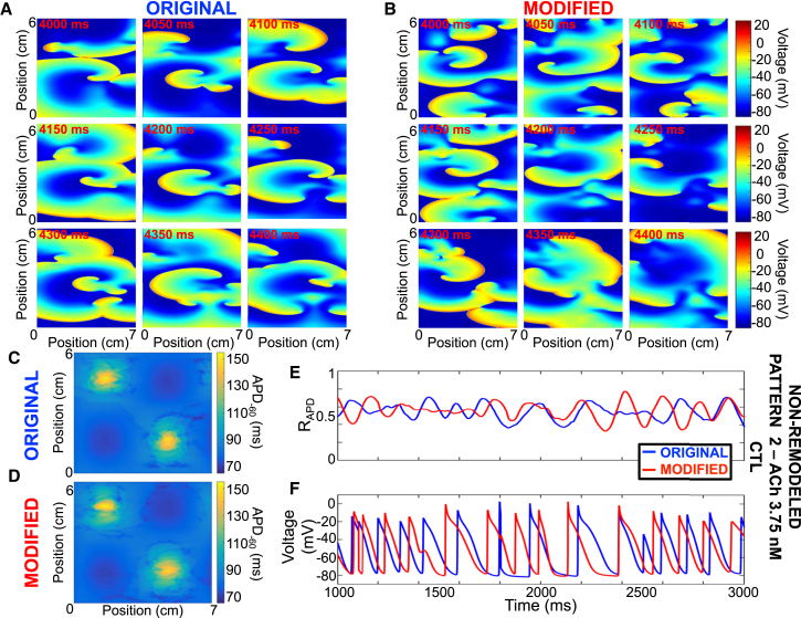
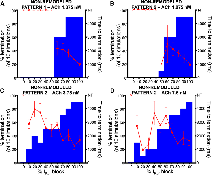
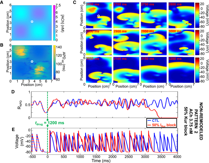
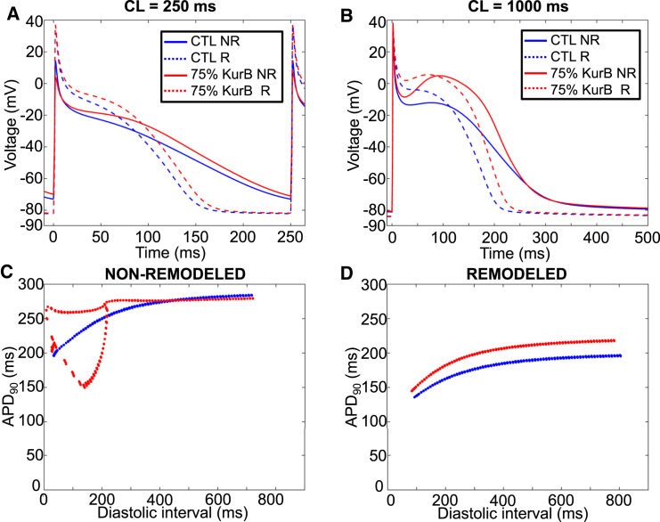
The atrial-specific ultrarapid delayed rectifier K+ current (IKur) inactivates slowly but completely at depolarized voltages. The consequences for IKur rate-dependence have not been analyzed in detail and currently available mathematical action-potential (AP) models do not take into account experimentally observed IKur inactivation dynamics. Here, we developed an updated formulation of IKur inactivation that accurately reproduces time-, voltage-, and frequency-dependent inactivation. We then modified the human atrial cardiomyocyte Courtemanche AP model to incorporate realistic IKur inactivation properties. Despite markedly different inactivation dynamics, there was no difference in AP parameters across a wide range of stimulation frequencies between the original and updated models. Using the updated model, we showed that, under physiological stimulation conditions, IKur does not inactivate significantly even at high atrial rates because the transmembrane potential spends little time at voltages associated with inactivation. Thus, channel dynamics are determined principally by activation kinetics. IKur magnitude decreases at higher rates because of AP changes that reduce IKur activation. Nevertheless, the relative contribution of IKur to AP repolarization increases at higher frequencies because of reduced activation of the rapid delayed-rectifier current IKr. Consequently, IKur block produces dose-dependent termination of simulated atrial fibrillation (AF) in the absence of AF-induced electrical remodeling. The inclusion of AF-related ionic remodeling stabilizes simulated AF and greatly reduces the predicted antiarrhythmic efficacy of IKur block. Our results explain a range of experimental observations, including recently reported positive rate-dependent IKur-blocking effects on human atrial APs, and provide insights relevant to the potential value of IKur as an antiarrhythmic target for the treatment of AF.


Figures

Figure 1
Figure 1
Time-, voltage-, and frequency dependence of IKur inactivation. (A) Shown here is the normalized IKur current during a 1000-ms test pulse (TP) at +40 mV, 5 ms after a 100-ms prepulse (PP), to inactivate Ito (inset) as recorded from the experimental preparation (black) and the original Courtemanche human atrial model (blue). (B) Normalized IKur current using a similar protocol, but with test pulse duration of 50 s as recorded from the experimental preparation (black) and the original and modified Courtemanche model (blue and red, respectively), is given. The experimental and modified model time constants are τui,f,exp = 702 ± 7 ms, τui,s,exp = 5688 ± 26 ms and τui,f,model = 713 ± 18 ms, τui,s,model = 5848 ± 188 ms, respectively. (C) Normalized fast (τui,f) and slow (τui,s) time constants as a function of test pulse potential for the experimental preparation and the modified Courtemanche model at 37°C are shown. (D) Shown here is the normalized IKur current using a CPD to +40 mV, followed by a 100-ms prepulse (PP) to +40 mV to inactivate Ito preceding a 100-ms test pulse (TP) to +40 mV (inset), as recorded from the experimental preparation and the original and modified models; the half-inactivation CPD (CPD1/2) was 1100 ms. (E) Shown here is the normalized IKur current using a 50 s-conditioning pulse (CP) to various voltages followed by a 240-ms test pulse to +50 mV (inset); the half-inactivation voltage (V1/2) was –6.5 mV in the model and –7.5 ± 0.6 mV experimentally. (F) Shown here is the normalized IKur current obtained by applying 100 pretest stimuli of 100 ms duration at +40 mV at various frequencies, followed by a 100-ms conditioning pulse to +40 mV to inactivate Ito and a 140-ms test pulse to +40 mV (inset); the normalized current at 4 Hz was 16% of its value at 0.1 Hz. For all panels, experimental data are in black; original and modified models are in blue and red, respectively.
Figure 2
Figure 2
Mechanism of IKur rate dependence. (A) Action potentials at stimulation cycle lengths from 265 ms (red), 300 ms (green), 500 ms (black), and 750 ms (blue) are shown. The purple and teal dashed lines correspond to the activation and inactivation gating variable 50% (V0.5,ua = −30 mV and V0.5,uis = −5 mV) opening potentials, respectively. (B) Shown here are corresponding IKur tracings; there is a rate-dependent decrease in IKur during phase 2 of the AP. (C) Shown here is the activation gate open probability (ua3) and (D) inactivation gate open probability (ui,f × ui,s) as a function of time for CLs of 265 ms (red) and 750 ms (blue). The activation open probability is rate dependent and the inactivation open probability is rate independent. (E) Shown here is the activation gating variable (ua) as a function of transmembrane potential; the purple dot corresponds to activation gating variable V0.5 as transposed on (A). (F) Fast (ui,f; black solid) and slow (ui,s; black dashed) inactivation gating variables as a function of transmembrane potential are given; the teal dot corresponds to the slow inactivation gating variable V0.5 as transposed on (A). The inactivation open probability is rate independent because the action potential spends very little time positive to V0.5,uis (−5 mV; teal).
Figure 3
Figure 3
Rate-dependent effects of IKur block on the AP. (A) Shown here is the IKr activation gate steady state (xr,∞) as a function of test potential. V0.1, V0.5, V0.75, and V0.9 are marked and transposed onto (B) and (E) as dashed lines. (B) Shown here are APs obtained at a cycle length of 1000 ms under control (blue) and with 75% IKur blockade (red); the AP duration at −60 mV (APD−60) was 255 ms for both. (C and D) Shown here are corresponding IKur and IKr tracings; the ratio of IKr with 75% IKur block to control was 2.36. (E) Shown here are APs obtained at a cycle length of 250 ms under control (blue) and with 75% IKur blockade (red); the APD−60 for control and 75% IKur block was 199 and 209 ms, respectively. (F and G) Shown here are corresponding IKur and IKr tracings; the ratio of IKr with 75% IKur block to control was 1.40.
Figure 4
Figure 4
Representative example of simulated vagotonic AF using pattern #2 with a peak ACh concentration of 3.75 nM and the non-remodeled cardiomyocyte model. (A) Shown here is ACh distribution with peak concentration of 3.75 nM and (B) a corresponding APD−60 distribution. (C) Shown here is transmembrane potential over time at 50-ms intervals; reentry is maintained by multiple short-lived spiral waves. (D) Shown here is the ratio of depolarized cells (ratio of cells with a voltage positive to −60 mV to the total number of cells) and (E) transmembrane potential over time for the cardiomyocyte marked with a white circle in (A) and (B).
Figure 5
Figure 5
Representative example comparing reentry dynamics in the original and modified models. (A and B) Transmembrane potential snapshots over time at 50-ms intervals for the original and modified models are shown. (C and D) APD−60 values for the original and modified models are given. (E and F) Shown here is the ratio of depolarized cells (ratio of cells with a voltage positive to −60 mV to the total number of cells) and transmembrane potential for the original (blue) and modified (red) models. Non-remodeled cardiomyocyte model with ACh pattern #2 with peak concentration of 3.75 nM is given.
Figure 6
Figure 6
Dose-response (bar-graphs) and average time to termination (red data) by IKur block using the non-remodeled cardiomyocyte model for (A) ACh pattern #1 with peak ACh concentration of 1.875 nM. (BD) Shown here are the ACh pattern #2 and peak ACh concentrations of 1.875, 3.75, and 7.5 nM, respectively.
Figure 7
Figure 7
Representative example of reentry termination by 50% IKur block using ACh pattern #2 with peak ACh concentration of 3.75 nM and the non-remodeled cardiomyocyte model. (A)Shown here is the ACh distribution with peak concentration of 3.75 nM and (B) its corresponding APD−60 distribution. (C) Transmembrane potential snapshots over time at 50 ms intervals are given; 50% IKur block was introduced at tdrug = 1200 ms. (D) Shown here is the ratio of depolarized cells (ratio of cells with a voltage positive to −60 mV to the total number of cells) and (E) transmembrane potential over time for the cardiomyocyte marked with a white circle in (A) and (B) for control (blue) and 50% IKur block (red).
Figure 8
Figure 8
IKur blocking effects in remodeled cardiomyocytes. (A) Shown here is a single cell AP at a stimulation CL of 250 ms for a non-remodeled (NR; solid) and remodeled (R; dashed) cardiomyocyte without drug (blue) and with 75% IKur block (red); (B) same as in (A), but at CL of 1000 ms. (C) Shown here is an AP at 90% repolarization (APD90) as a function of diastolic interval without drug (blue) and with 75% IKur block (red) for a non-remodeled cardiomyocyte; (D) same as in (C), but for a remodeled cardiomyocyte.

References

    1. Wang Z., Fermini B., Nattel S. Sustained depolarization-induced outward current in human atrial myocytes. Evidence for a novel delayed rectifier K+ current similar to Kv1.5 cloned channel currents. Circ. Res. 1993;73:1061–1076. - PubMed
    1. Feng J., Xu D., Nattel S. Ultrarapid delayed rectifier current inactivation in human atrial myocytes: properties and consequences. Am. J. Physiol. 1998;275:H1717–H1725. - PubMed
    1. Ford J., Milnes J., Ravens U. The positive frequency-dependent electrophysiological effects of the IKur inhibitor XEN-D0103 are desirable for the treatment of atrial fibrillation. Heart Rhythm. 2016;13:555–564. - PMC - PubMed
    1. Courtemanche M., Ramirez R.J., Nattel S. Ionic mechanisms underlying human atrial action potential properties: insights from a mathematical model. Am. J. Physiol. 1998;275:H301–H321. - PubMed
    1. Kneller J., Zou R., Nattel S. Cholinergic atrial fibrillation in a computer model of a two-dimensional sheet of canine atrial cells with realistic ionic properties. Circ. Res. 2002;90:E73–E87. - PubMed

MeSH terms