. 2017 Jul:72:70-75.e1.
doi: 10.1016/j.pediatrneurol.2017.03.015. Epub 2017 Apr 1.

Neonatal Vein of Labbé Infarction Size is Associated With Long-Term Language Outcomes

Affiliations

Neonatal Vein of Labbé Infarction Size is Associated With Long-Term Language Outcomes

Kristen L Benninger et al. Pediatr Neurol. 2017 Jul.

Abstract

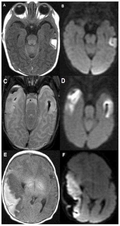
Background: The vein of Labbé is a superficial cortical vein, which drains the lateral surface of the temporal lobe. Thrombosis of the vein of Labbé can occur in the neonatal period. The developmental outcomes of infants who had vein of Labbé thrombosis are unknown as few studies of outcomes exist.

Methods: We completed a retrospective review of infants born ≥34 weeks of gestation, diagnosed with vein of Labbé thrombosis, and/or infarction on neuroimaging during the first 30 days of life. Size of each temporal lobe infarction was estimated based on the number of temporal lobe segments involved. Primary outcomes were the presence of major neurodevelopmental impairments in childhood and Bayley scores at two years.

Results: Our cohort of 19 infants had a median gestational age of 38 weeks (interquartile range 36 to 39) and mean birth weight 2892 ± 920 grams. The most common presenting symptoms of vein of Labbé thrombosis and infarction of surrounding tissue were seizures, apnea, lethargy, and either hypertonia or hypotonia. At the latest clinical follow-up appointment documented in the electronic medical record (mean 4.4 ± 3.08 years), 44% had major neurodevelopmental impairment. Patients with large vein of Labbé infarctions had significantly worse average Bayley scores than those with small to moderate lesions, and differences in language composite were statistically significant (72.7 vs 107.8, P = 0.017).

Conclusions: Neonates with large vein of Labbé infarctions are more likely to have poor language outcomes. This finding suggests a need for targeted surveillance to ensure early identification of deficits and referral for intervention.

Keywords: cerebral sinovenous thrombosis; neonate; neurodevelopment; outcome; vein of Labbé.


Conflict of interest statement

Conflicts of interest: none

Figures

Figure 1
Figure 1
A. and B. 8 day old infant with small left VOL hemorrhagic venous infarction (HVI). A. Axial T1 weighted image demonstrates the typical appearance of a small mid left temporal lobe infarct with peripheral hemorrhage. B. Axial diffusion weighted image demonstrates a medial band of bright signal consistent with restricted diffusion (infarction) within the cortex of the VOL HVI. C. and D. 3 day old infant with moderate right VOL hemorrhagic venous infarction (HVI) and small left VOL HVI. C. Axial T2 weighted image demonstrates peripheral hemorrhage in the moderate right VOL HVI as well as T2 bright edema in the involved right temporal lobe cortex and underlying white matter. The small left VOL HVI shows focal cortical and white matter edema with deeper T2 dark hemorrhage. D. Axial diffusion weighted image demonstrates bright signal consistent with restricted diffusion (infarction) in both moderate and small VOL HVIs. E. and F. 1 day old infant with large right VOL hemorrhagic venous infarction (HVI) who presented with apneic events at 8 hours of life, confirmed as electrographic seizures. He was discharged on anti-epilepsy medication and was ultimately diagnosed with Lennox Gastaut Syndrome at age 3 years. He has marked language delays. E. Axial T1 weighted image demonstrates peripheral T1 bright hemorrhage along the large right temporal VOL HVI with low T1 signal edema in the underlying infarcting cortex and subcortical white matter. F. Axial diffusion weighted image demonstrates bright signal consistent with restricted diffusion (infarction) within the cortex and subcortical white matter of the entire right temporal lobe. The peripheral hemorrhage lateral to the infarcting right temporal lobe has dark signal.

References

    1. deVeber G, Andrew M, Adams C, et al. Canadian Pediatric Ischemic Stroke Study Group. Cerebral sinovenous thrombosis in children. N Engl J Med. 2001;345:417–423. doi: 10.1056/NEJM200108093450604. - DOI - PubMed
    1. Moharir MD, Shroff M, Pontigon AM, Askalan R, Yau I, Macgregor D, et al. A prospective outcome study of neonatal cerebral sinovenous thrombosis. J Child Neurol. 2011;26(9):1137–44. doi: 10.1177/0883073811408094. - DOI - PMC - PubMed
    1. Yang JY, Chan AK, Callen DJ, Paes BA. Neonatal cerebral sinovenous thrombosis: sifting the evidence for a diagnostic plan and treatment strategy. Pediatrics. 2010;126:e693700. doi: 10.1542/peds.2010-1035. - DOI - PubMed
    1. Coutinho JM, Gerritsma JJ, Zuurbier SM, Stam J. Isolated cortical vein thrombosis: systematic review of case reports and case series. Stroke. 2014;45:1836–1838. doi: 10.1161/strokeaha.113.004414. - DOI - PubMed
    1. Singh R, Cope WP, Zhou Z, De Witt ME, Boockvar JA, Tsiouris AJ. Isolated cortical vein thrombosis: case series. J Neurosurg. 2015;123:427–433. doi: 10.3171/2014.9.jns141813. - DOI - PubMed